NAD+ levels do fall in the ageing and failing heart — that part is well documented. Whether that fall drives cardiovascular disease or merely accompanies it is the open question, and it is the one that decides whether topping NAD+ back up could ever help. This article separates the two. What depletes cardiac NAD+: over-activation of PARP enzymes after accumulated DNA damage, reduced expression of NAMPT (the main NAD+ biosynthetic enzyme) in failing human hearts, plus ageing and metabolic disease. What follows downstream: impaired fatty-acid oxidation, mitochondrial dysfunction and lower ATP output. And what the human trials have actually delivered so far — including NAD-HF, in which 30 patients with stable heart failure received nicotinamide riboside at up to 1,000 mg twice daily.
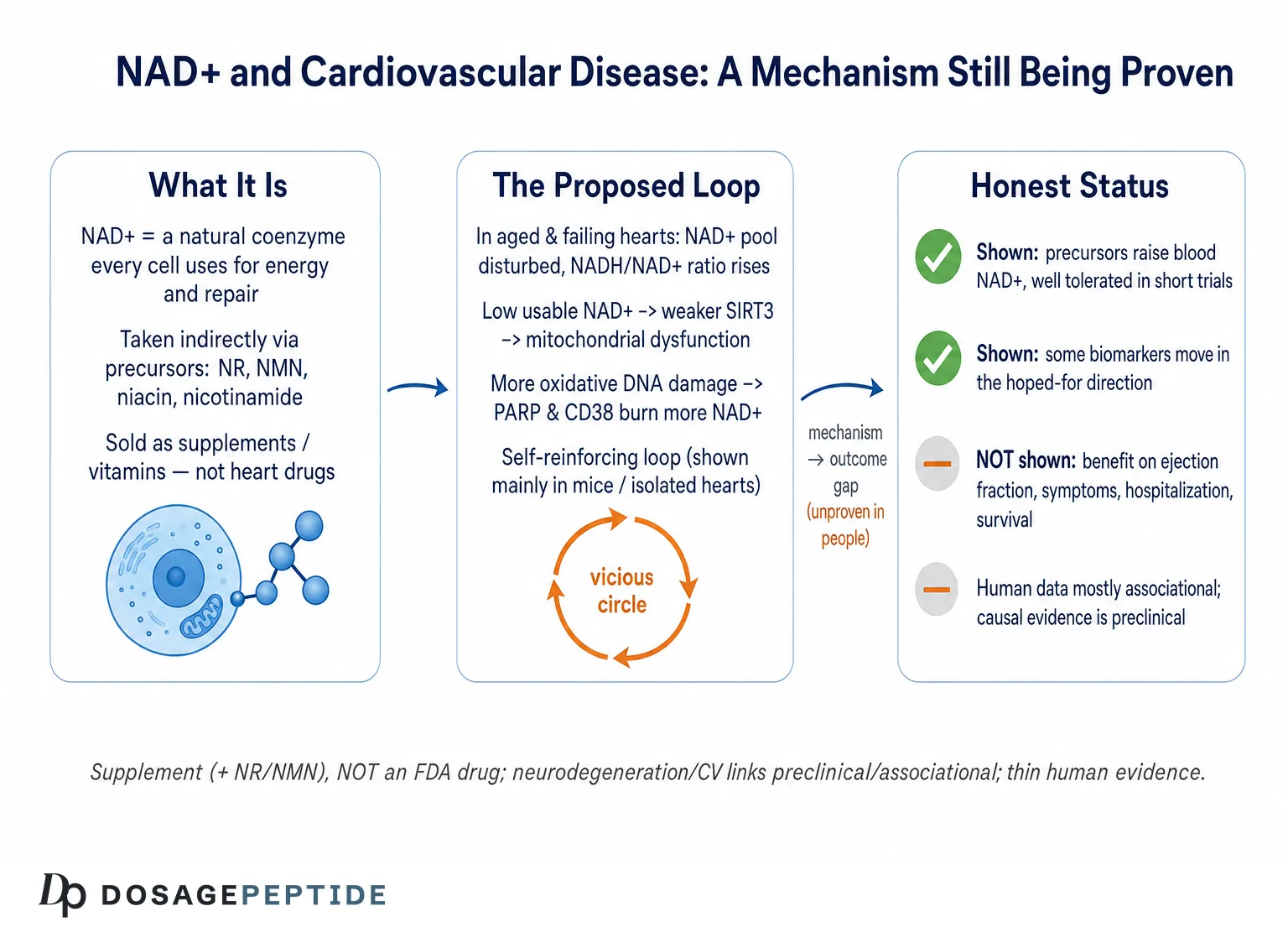
Here is the state of play as of mid-2026. There is strong, reproducible evidence that tissue NAD+ and the NAD+/NADH redox ratio are disturbed in failing hearts and in aged blood vessels, and that the enzymes governing NAD+ turnover shift in a characteristic direction with age and stress.12 There is compelling preclinical evidence — in mice, rats, and isolated hearts — that restoring NAD+ with precursors blunts hypertrophy, protects against ischemic injury, and improves contractile function.39 And there is a small but growing set of early human trials showing that NAD+ precursors do raise blood NAD+ and are well tolerated, with scattered signals on blood pressure and inflammation but no demonstrated effect on hard cardiovascular outcomes.57 What does not yet exist is a large, outcome-powered clinical trial proving that raising NAD+ slows or reverses cardiovascular disease in people.
So the correct reading of the title question is not “explain the established therapy” but “examine an active, mechanistically rich, but clinically unproven hypothesis.” NAD+ itself is a naturally occurring coenzyme; the compounds people actually take to raise it — nicotinamide riboside (NR), nicotinamide mononucleotide (NMN), niacin, and nicotinamide — are marketed as dietary supplements, not as approved drugs for heart disease. None is FDA-approved to treat, prevent, or slow any cardiovascular condition. This piece maps what the biology says, where the human evidence stops, and how to think clearly about the gap between an elegant mechanism and a proven treatment. That gap is the recurring theme of everything below, and keeping it in view is the single most useful habit for evaluating any confident claim about NAD+ and the heart. Readers interested in the parallel story in the brain can compare this with the site’s discussion of the link between NAD+ deficiency and Parkinson’s progression, which raises the same causation-versus-correlation problem in a different organ.
What NAD+ Is and Why the Failing Heart Runs Low
Nicotinamide adenine dinucleotide (NAD+) is one of the most heavily trafficked small molecules in the cell. It exists in two interconverting forms — oxidized (NAD+) and reduced (NADH) — and the ratio between them is the cell’s central redox currency. Every time a fuel molecule is oxidized in glycolysis, the tricarboxylic acid cycle, or fatty-acid oxidation, electrons are handed to NAD+ to make NADH; those electrons are then delivered to the mitochondrial electron transport chain to drive ATP synthesis, regenerating NAD+ in the process.12 The heart is the most energy-hungry organ in the body by mass, cycling its entire ATP pool many times per minute, so it depends on this shuttle more acutely than almost any other tissue.
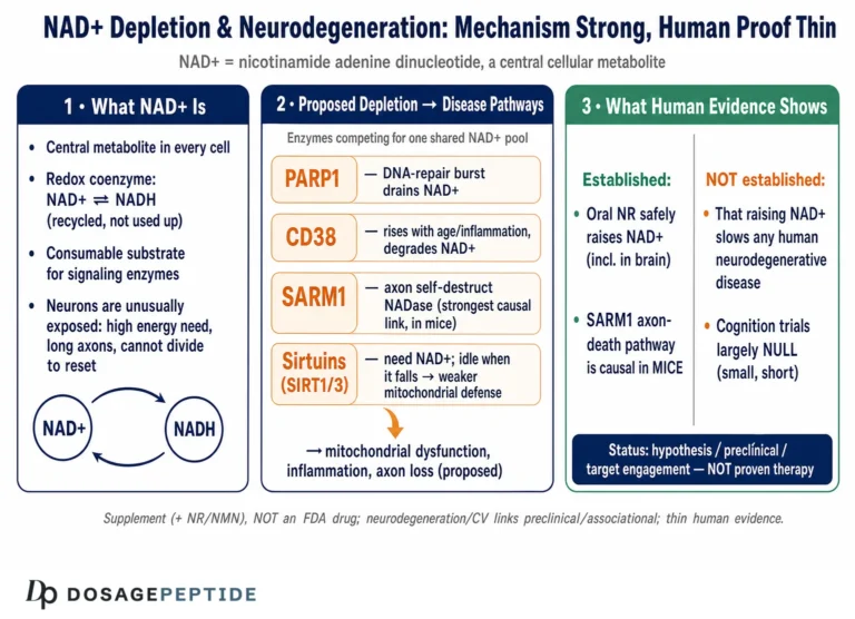
Beyond its redox role, NAD+ is also a consumable substrate. Three families of enzymes cleave NAD+ and use its nicotinamide moiety as a signaling or repair currency: the sirtuins (NAD+-dependent deacylases such as SIRT1 and SIRT3), the poly(ADP-ribose) polymerases (PARPs, which respond to DNA damage), and the NAD+ glycohydrolases CD38 and CD157.1012 Because these enzymes consume NAD+ rather than merely borrowing its electrons, their activity directly lowers the free NAD+ pool. This dual identity — coenzyme and substrate — is the key to understanding depletion: the heart can run short of NAD+ either because it makes less or because it burns through more.
The evidence that the failing heart is NAD+-poor comes from several directions. Analyses of failing human myocardium and of mouse hearts with pathological hypertrophy show an elevated NADH/NAD+ ratio and widespread protein hyperacetylation, the biochemical fingerprint of insufficient sirtuin activity relative to demand.2 Elderly patients hospitalized with decompensated heart failure have significantly lower whole-blood NAD+ than healthy controls.4 And in the vasculature, NAD+-dependent SIRT1 activity declines with age, tracking the onset of endothelial dysfunction and arterial stiffening.8 The direction of these findings is consistent, which is why the depletion hypothesis has gained traction. What the findings cannot settle on their own is whether low NAD+ is pushing the disease forward or simply reflecting a sick, energy-starved tissue — a distinction we return to in detail later.
It helps to hold a simple mental model: NAD+ is both the wire that carries electrons for energy production and the fuel that powers a set of protective repair-and-maintenance enzymes. When the pool shrinks, both jobs suffer at once — the heart makes energy less efficiently and loses some of its capacity to defend and repair itself. That double hit is what makes the depletion hypothesis mechanistically attractive.
A further point of nuance separates the sophisticated version of this hypothesis from the marketing caricature. What the heart most likely suffers from is not simply a lower absolute quantity of NAD+ but a disturbed redox ratio and a disturbed compartmental distribution. In the failing heart, the reduced form NADH tends to accumulate relative to oxidized NAD+ because electrons are handed off to the respiratory chain less efficiently; the pool can even appear numerically preserved while being functionally “stuck” in the reduced state, so that sirtuins and other NAD+-consuming enzymes are starved of the oxidized co-substrate they require.2 This is why the elevated NADH/NAD+ ratio, rather than total NAD+ alone, is the more informative biochemical marker in cardiac tissue — and why crude blood measurements of “total NAD” can mislead. The distinction matters for interpreting every claim that follows: “depletion” in this article means a functional shortfall of usable, oxidized NAD+ where and when the heart needs it, not merely a smaller number on an assay.
The Three Levers of NAD+ Depletion: Synthesis, Consumption, and CD38
NAD+ levels in any tissue reflect a balance between synthesis and consumption, and cardiovascular stress appears to tilt that balance in three converging ways. Understanding these three levers is essential, because they are the actual mechanistic content behind the word “depletion,” and each is a candidate point of intervention.
Lever one: reduced salvage synthesis. Most NAD+ in mammalian cells is not made from scratch but recycled through the salvage pathway, in which nicotinamide is reconverted to NAD+. The rate-limiting enzyme of that pathway is nicotinamide phosphoribosyltransferase (NAMPT). NAMPT expression and activity fall in stressed and aging cardiac tissue, throttling the heart’s ability to regenerate NAD+ from the nicotinamide liberated by sirtuins and PARPs.1 Because the salvage pathway is the dominant source, a drop in NAMPT has an outsized effect on the steady-state pool.
Lever two: increased PARP consumption. DNA damage — from oxidative stress, ischemia, or mechanical strain — activates PARP1, which consumes large quantities of NAD+ to build poly(ADP-ribose) chains as part of the repair response. In acute or sustained stress this becomes a major NAD+ sink, and hyperactivated PARP1 can drive the pool low enough to compromise energy metabolism.112 The DNA-repair role of NAD+ is a genuine double-edged sword, a tension the site explores further in its piece on the mechanisms linking NAD+ to DNA repair and cancer prevention.
Lever three: rising CD38 activity. Perhaps the most influential single finding in this field is that the NADase CD38 increases with age and inflammation and is a principal driver of the age-related NAD+ decline. In a landmark study, CD38-knockout mice were protected from the age-associated fall in tissue NAD+ and from the accompanying mitochondrial dysfunction, and this protection operated through preserved SIRT3 activity.1 CD38 is expressed on immune cells and is upregulated by the inflammatory milieu that accompanies heart failure and atherosclerosis, which ties chronic inflammation directly to NAD+ depletion.
These levers are not independent. Inflammation raises CD38; ischemia activates PARP; aging lowers NAMPT. In a chronically stressed heart, all three can operate simultaneously, which is why NAD+ depletion in cardiovascular disease is best understood as a convergent endpoint of several pathways rather than a single lesion. It also explains why interventions have been tried at multiple points: precursor supplementation to boost synthesis, PARP inhibitors to reduce consumption, and CD38 inhibitors to plug the largest leak. Only the precursor approach has reached human cardiovascular testing in any meaningful way.
It is worth pausing on why the three-lever framing is more than academic bookkeeping. Each lever implies a different, and potentially conflicting, therapeutic logic. If depletion is driven mainly by falling NAMPT-mediated synthesis, then flooding the salvage pathway with precursors such as NR or NMN is the rational move. If it is driven mainly by PARP over-consumption in the setting of DNA damage, then a precursor alone may be like pouring water into a leaking bucket — the more targeted intervention would be to reduce the damage or restrain PARP. And if CD38 is the dominant sink, as the aging literature suggests, then CD38 inhibition might do more than any amount of precursor, because CD38 will also degrade the NMN that supplementation provides before it can be used.1 This last point is genuinely awkward for the popular NMN narrative: the very enzyme most responsible for age-related NAD+ decline is also an efficient consumer of the precursor people take to counter that decline. Which lever dominates almost certainly varies by patient, tissue, and disease stage, and no human study has resolved this, which is one reason the “just take a precursor” framing oversimplifies a genuinely unsettled biology.
The Depletion–Dysfunction Loop: PARP, SIRT3, and Mitochondrial Hyperacetylation
The reason NAD+ depletion is thought to drive progression, rather than merely mark it, is that the leading mechanistic account describes a self-reinforcing loop — a vicious circle in which low NAD+ begets mitochondrial dysfunction, which begets more DNA damage and inflammation, which further lowers NAD+. Following this loop carefully is worthwhile because it is the strongest theoretical argument for the depletion hypothesis, and also because its individual steps are where the evidence is firmest.
Start with the sirtuins. SIRT3 is the principal mitochondrial deacetylase, and it depends absolutely on NAD+ as a co-substrate. When NAD+ falls, SIRT3 activity falls with it, and the acetylation marks that SIRT3 would normally remove accumulate on mitochondrial proteins. This is not a cosmetic change: hyperacetylation of tricarboxylic-acid-cycle enzymes and of components of the malate-aspartate shuttle impairs substrate oxidation and the transfer of reducing equivalents into the mitochondria, degrading the very machinery that regenerates NAD+ and produces ATP.2 Proteomic and acetylome analyses of failing hearts show exactly this signature — a subpopulation of mitochondrial proteins whose acetylation status is sensitive to the NADH/NAD+ ratio.2
SIRT3 has a second job that closes part of the loop: it deacetylates cyclophilin D, a regulator of the mitochondrial permeability transition pore (mPTP). When SIRT3 activity falls, cyclophilin D is hyperacetylated, the mPTP opens more readily, and mitochondria swell and become leaky — a change linked in animal models to age-related cardiac hypertrophy and fibrosis.1 A leaky, dysfunctional mitochondrion produces more reactive oxygen species and more DNA damage, which reactivates PARP1, which consumes still more NAD+.
The result is the pathogenic loop that anchors the depletion hypothesis: elevated PARP activity depletes NAD+, low NAD+ impairs SIRT3, impaired SIRT3 worsens mitochondrial function and increases oxidative DNA damage, and that damage drives PARP activity higher still.12 Because the loop is self-amplifying, even a modest initial perturbation could, in principle, ratchet a heart toward progressive dysfunction over time. This is the mechanistic heart of the argument that NAD+ depletion contributes to progression and not merely to a snapshot of illness.
Interestingly, the therapeutic benefit of raising NAD+ may not be entirely explained by restoring sirtuin-driven deacetylation. A 2023 study found that raising NAD+ levels alleviated heart failure partly by stimulating short-chain dehydrogenase/reductase proteins, independent of mitochondrial protein deacetylation — a reminder that even the mechanism of any observed benefit is still being worked out.2 The loop is a useful organizing model, not a closed case.
Two honest caveats belong here. First, most of the loop has been mapped in rodents and isolated systems; the human data are largely correlational snapshots of hyperacetylation and low NAD+, not demonstrations that breaking the loop pharmacologically changes the human disease course. Second, a loop that is real in principle can still be a minor contributor in practice, dwarfed by hemodynamic overload, fibrosis, or neurohormonal activation. The loop is plausible and partly proven; it is not established as the dominant engine of human heart failure. Cardiovascular progression is multifactorial, and it would be a mistake to promote one biochemical circuit — however elegant — to the status of master switch when decades of cardiology point to pressure, volume, ischemia, and fibrosis as the heavyweight drivers. The most defensible position is that NAD+ depletion is one contributing thread in a thick rope, potentially more prominent in some phenotypes (for example, mitochondrial or age-dominant heart failure) than in others.
Mapping Depletion Onto Specific Cardiovascular Diseases
“Cardiovascular disease” is an umbrella, and NAD+ biology intersects each condition under it slightly differently. Separating them prevents the common error of treating a single mechanism as if it explained everything from atherosclerosis to arrhythmia.
Heart failure. This is where the depletion story is most developed. Failing human and hypertrophic mouse hearts show elevated NADH/NAD+ and mitochondrial hyperacetylation, and stimulating the salvage pathway in mice suppresses that hyperacetylation and improves function under stress.2 In a dilated-cardiomyopathy mouse model, nicotinamide riboside preserved cardiac function and raised myocardial NAD+.3 The mechanistic case here is the strongest of any cardiovascular indication.
Vascular aging and atherosclerosis. Here the central player is endothelial SIRT1, whose NAD+-dependent activity declines with age. Reduced SIRT1 lowers nitric-oxide bioavailability, permits NADPH-oxidase-driven oxidative stress, and promotes the inflammation, foam-cell formation, and senescence that underlie atherosclerosis.8 In aged mice, supplying the NAD+ precursor NMN restored SIRT1 activity and reversed measures of arterial dysfunction and oxidative stress.6 The link is mechanistically coherent but rests heavily on animal work. It is also the arm of the story with the most direct human echo: the arterial-stiffness and blood-pressure signals seen in the small NR pilots are exactly what this endothelial-SIRT1 mechanism would predict, which is encouraging — but those pilots are small and their positive findings live mostly in subgroups and secondary analyses, so the vascular case remains suggestive rather than settled.7
Ischemia–reperfusion injury. Ischemia collapses NAD+ synthesis while reperfusion floods the tissue with oxidative damage that activates PARP. Restoring NAD+ — with NMN given before ischemia or at reperfusion — preserved cardiac NAD+ and reduced infarct size in mouse hearts, and NAMPT is required for the protection conferred by ischemic preconditioning.9 This is an acute, high-turnover setting where the depletion logic is especially clean.
Arrhythmia and sudden cardiac death. Cardiac NAD+ depletion in mice can promote hypertrophic remodeling and arrhythmias, and the pattern of reduced NAMPT with increased CD38 and PARP activity has been proposed as a substrate for the electrical instability associated with sudden cardiac death.1 This remains the most speculative of the four.
| Condition | Proposed NAD+-related mechanism | Strongest evidence level |
|---|---|---|
| Heart failure | ↑NADH/NAD+, mitochondrial hyperacetylation, impaired SIRT32 | Human tissue + rodent intervention |
| Vascular aging / atherosclerosis | ↓SIRT1, ↓nitric oxide, oxidative stress, senescence8 | Mostly rodent + human observational |
| Ischemia–reperfusion injury | NAD+ collapse + PARP over-activation; NAMPT-dependent protection9 | Rodent / isolated heart only |
| Arrhythmia / sudden death | ↓NAMPT, ↑CD38/PARP as remodeling substrate1 | Rodent / hypothesis-generating |
The table makes the honest structure of the field visible at a glance: the mechanistic reasoning is consistent across conditions, but the evidence is overwhelmingly preclinical, thinning to pure hypothesis at the arrhythmia end. For a related look at how a different compound class is scrutinized against cardiovascular endpoints, see the site’s analysis of how retatrutide influences cardiovascular risk factors, which illustrates the higher evidentiary bar that approved-track therapies are held to.
Correlation or Cause? Examining the Premise Honestly
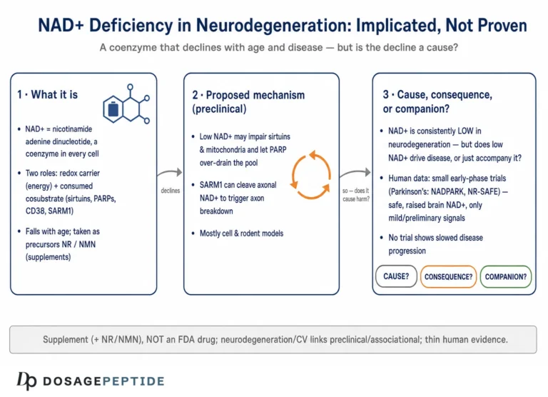
This is the section the title most needs, because the word “linked” is doing a lot of quiet work. A link can be causal (low NAD+ drives disease), reverse-causal (disease lowers NAD+), or confounded (a third factor, such as inflammation or aging, lowers both NAD+ and cardiac function independently). Distinguishing these is not pedantry; it determines whether raising NAD+ could plausibly help at all.
The case for causation rests on three pillars. First, the mechanistic loop described earlier gives a plausible physical route from low NAD+ to worse function.12 Second, intervention studies in animals — where NAD+ is deliberately raised or lowered and the heart’s response is observed — consistently show that raising NAD+ protects and that genetically crippling NAD+ synthesis harms.39 Intervention evidence is far stronger than correlation because it manipulates the proposed cause. Third, the direction is reproducible across labs, models, and species.
The case for caution is equally real. Almost all of the causal intervention evidence is preclinical. Rodent hearts differ from human hearts in heart rate, metabolic substrate preference, and the time course of disease; compounds that rescue a mouse model routinely fail to translate. The human data are dominated by associations — low blood NAD+ in heart-failure patients, declining SIRT1 with age — and associations cannot distinguish cause from consequence.48 A failing heart is a catabolic, inflamed, energy-starved organ; it would be surprising if its NAD+ metabolism were not disturbed, regardless of whether that disturbance is driving anything.
There is also a specific reason for humility unique to this field. NAD+ metabolism is homeostatically defended and highly compartmentalized — the concentration in the mitochondrion, the cytosol, the nucleus, and the bloodstream are regulated somewhat separately. Whole-blood NAD+, which is what most human studies measure, may not faithfully report myocardial or endothelial NAD+.5 A drop measured in blood is a useful biomarker but an uncertain proxy for what matters at the tissue level. This is exactly the kind of measurement gap that keeps a mechanistically attractive hypothesis from graduating to established fact.
One more subtlety deserves airtime because it is routinely ignored. Even if NAD+ depletion is genuinely causal, it does not automatically follow that supplementation reverses the damage. Causation and reversibility are different claims. A nutrient deficiency can cause a disease that supplementation nonetheless fails to undo once structural remodeling — fibrosis, cardiomyocyte loss, vascular calcification — has set in. The window in which restoring NAD+ helps might be early and preventive rather than late and restorative, which is precisely the kind of question that only long, well-designed human trials can answer, and precisely the kind of nuance that supplement marketing erases when it implies benefit at any stage. The animal successes largely involve intervening early or concurrently with the insult; whether a precursor helps a heart already remodeled over years is a separate, unproven proposition.
The intellectually honest summary is that NAD+ depletion is a credible contributing mechanism with strong preclinical causal support and consistent human associational support, but that its causal role in human cardiovascular progression — and therefore its value as a treatment target — remains unproven. The same open-question posture applies to NAD+’s proposed protective role elsewhere, as discussed in the site’s review of the scientific evidence for NAD+ preserving neurons during stroke.
What the Human Evidence Actually Shows
Because the mechanistic story is so appealing, it is important to be precise about how thin the human clinical evidence actually is. The trials that exist are small, short, and focused on safety and biomarkers rather than on outcomes that matter to patients.
The most directly relevant cardiac study is the NAD-HF trial: 30 patients with clinically stable heart failure with reduced ejection fraction received nicotinamide riboside at up to 1,000 mg twice daily. NR was safe and well tolerated and roughly doubled whole-blood NAD+. Within-subject increases in NAD+ correlated with improved mitochondrial respiration and reduced pro-inflammatory expression in peripheral blood mononuclear cells, but the trial was far too small to detect any effect on clinical endpoints, and none was found.5 A companion mechanistic study showed that boosting NAD+ suppressed the inflammatory activation of blood immune cells from heart-failure patients — a biomarker result, not a clinical one.4
On the vascular side, a randomized, double-blind, placebo-controlled trial in midlife and older adults with above-normal systolic blood pressure tested NR at 500 mg twice daily. A secondary analysis reported that in the subgroup with stage-1-range systolic pressure, NR was associated with a reduction in systolic blood pressure and a suggestion of reduced aortic stiffness; the overall primary comparison was more modest, and the study was a pilot.7 An earlier trial had already established that chronic NR raises NAD+ and is well tolerated in healthy middle-aged and older adults.11 NMN, meanwhile, has been tested mostly for metabolic and exercise endpoints; dedicated, adequately powered cardiovascular-outcome data in humans are essentially absent.
| Human study | Design | Cardiovascular-relevant finding |
|---|---|---|
| NAD-HF (NR in HFrEF) | Randomized, ~30 patients, safety-focused5 | Safe; NAD+ ~doubled; ↑PBMC respiration, ↓inflammation; no clinical-endpoint effect |
| PBMC inflammation study | Mechanistic sub-study, HF patients4 | NR suppressed inflammatory activation of immune cells (biomarker only) |
| NR for systolic BP / stiffness | Randomized, double-blind, pilot, adults ≥507 | Signal toward lower systolic BP/stiffness in a subgroup; pilot scale |
| NR tolerability (healthy adults) | Randomized crossover, healthy middle-aged/older11 | Raised NAD+; well tolerated; no CV outcomes assessed |
The pattern is unambiguous and worth stating plainly: NAD+ precursors reliably raise blood NAD+ and are well tolerated over weeks to a few months, and they move some mechanistic biomarkers in the hypothesized direction. What they have not done is demonstrate improvement in ejection fraction, exercise capacity, hospitalization, or survival in any adequately powered cardiovascular trial. Anyone who tells you NAD+ supplementation is proven to slow heart disease is describing the mouse literature, not the human one. Readers can situate these precursor doses against general reconstitution and measurement practice using the site’s peptide and compound glossary.
The Precursors: NR, NMN, Niacin, and Nicotinamide
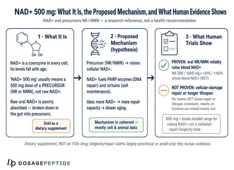
People do not administer NAD+ itself very often — the intact molecule is large, charged, and poorly cell-permeable, and its cellular uptake is inefficient. Instead, the practical question is which precursor to use, and this is a source of endless marketing confusion that is worth clearing up honestly.
All roads lead to the salvage pathway. Niacin (nicotinic acid) is the classic vitamin B3; it raises NAD+ efficiently but causes flushing via a prostaglandin-mediated receptor and, at the high doses historically used for lipids, has a complicated cardiovascular record. Nicotinamide (niacinamide) is flush-free but at high doses can inhibit sirtuins, which is self-defeating for a strategy meant to fuel them. Nicotinamide riboside (NR) and nicotinamide mononucleotide (NMN) are the two precursors that have attracted the most research money and hype; both feed the salvage pathway, both reliably raise blood NAD+ in humans, and both have been reasonably well tolerated in short trials.1011 NMN is generally converted to NR before crossing the cell membrane in many tissues, though a direct NMN transporter has been described, and the NR-versus-NMN debate remains more commercially heated than scientifically settled.12
| Precursor | Notable feature | Regulatory / evidence status |
|---|---|---|
| Niacin (nicotinic acid) | Efficient NAD+ raise; causes flushing | Approved as a vitamin/lipid agent; high-dose CV benefit unproven for outcomes |
| Nicotinamide | Flush-free; may inhibit sirtuins at high dose | Vitamin/supplement; not a CV drug |
| Nicotinamide riboside (NR) | Raises blood NAD+; most human CV pilot data57 | Marketed as a supplement; not FDA-approved for CV disease |
| Nicotinamide mononucleotide (NMN) | Raises NAD+; strong rodent vascular data6 | Supplement; U.S. status contested; no CV-outcome trials |
The essential honest point is that none of these is an approved cardiovascular medicine. NR and NMN are sold as dietary supplements; niacin and nicotinamide are vitamins. Their ability to raise NAD+ is real and measurable, but raising a biomarker is not the same as treating a disease. The gap between “this raises NAD+” and “this helps your heart” is precisely the gap the human trials have not yet closed. The same reasoning — a plausible mechanism outrunning the outcome evidence — recurs across the vascular literature, for instance in work on GLP-1 signaling mechanisms in arterial stiffness.
Research Models and Methodology
How the NAD+–cardiovascular link has been studied determines what the data can and cannot support, so a brief tour of the methodology is clarifying. The evidence architecture falls into four tiers, and the confidence we can place in a claim tracks the tier it comes from.
Biochemical and cell work. Isolated cardiomyocytes, endothelial cells, and mitochondrial preparations are used to measure NAD+/NADH ratios, sirtuin and PARP activity, protein acetylation, and mitochondrial respiration. These systems established the SIRT3-hyperacetylation mechanism and the CD38/PARP consumption pathways.12 They are excellent for mechanism but say nothing about whole-organism outcomes.
Genetic mouse models. The most persuasive causal evidence comes from animals in which a single NAD+-handling gene is deleted or overexpressed — CD38-knockout mice protected from age-related NAD+ decline, NAMPT-manipulated hearts, and models with altered salvage flux.1 These isolate the contribution of one lever, which is exactly what correlation studies cannot do.
Disease-model interventions. Pressure-overload and dilated-cardiomyopathy models, aged-animal vascular studies, and ischemia–reperfusion preparations test whether raising NAD+ changes the disease course. This tier produced the headline preclinical results: NR preserving function in dilated cardiomyopathy, NMN reversing vascular dysfunction in aged mice, and NMN limiting infarct size in ischemia–reperfusion.369
Human trials. The clinical tier is the thinnest. It consists of small, short randomized trials with biomarker and safety endpoints — NAD-HF in heart failure, the NR blood-pressure pilot, tolerability studies — none powered for clinical outcomes.5711 A recurring methodological limitation across the human work is reliance on whole-blood NAD+ as the readout, which may not reflect cardiac or vascular tissue NAD+.5
A specific methodological trap recurs across this literature and is worth naming so readers can spot it. Because NAD+ precursors reliably move an easy-to-measure biomarker — blood NAD+ — studies frequently report that biomarker success as though it were the endpoint of interest. It is not. Raising blood NAD+ is the exposure, the thing the intervention obviously does; the endpoints that would justify calling NAD+ depletion a driver of disease are downstream and clinical: cardiac structure, function, symptoms, events, survival. Conflating “the drug did what it mechanically should” with “the drug helped the patient” is one of the most common ways promising mechanisms are oversold, and the NAD+ field is unusually prone to it because the biomarker is so responsive and so marketable. A disciplined reader asks not “did NAD+ go up?” but “did anything the patient would notice or that predicts their prognosis change?”
The methodological bottom line mirrors the evidence hierarchy in most translational fields: mechanism is well characterized, animal causation is reasonably strong, and human outcome data are largely missing. A definitive answer to the title question would require large, long, outcome-powered randomized trials in defined cardiovascular populations, ideally with tissue-level or validated surrogate NAD+ measurements rather than blood alone. Until then, the strength of any claim should be calibrated to the tier of evidence it rests on.
Safety, Tolerability, and Research-Context Handling
Short-term safety is one of the more reassuring aspects of the NAD+-precursor literature, but it must be framed carefully: “well tolerated in small trials of weeks to months” is a much narrower claim than “safe for indefinite use in people with heart disease.”
In the trials conducted so far, NR at doses up to 1,000–2,000 mg per day and NMN at comparable ranges have generally been well tolerated, without the endocrine or hemodynamic red flags that would halt development.511 Niacin is the exception with a longer and more cautionary history: at the high doses once used for cholesterol, large outcome trials failed to show cardiovascular benefit and raised concerns about adverse effects, a reminder that a compound that raises a favorable biomarker can still fail or harm at the level of outcomes. Several important caveats apply across the class:
- Population. Safety has been assessed largely in stable, relatively healthy or stable-heart-failure participants, not in acutely decompensated, frail, or polypharmacy-heavy patients where drug handling differs.
- Duration. Trials run weeks to months; cardiovascular disease is a decades-long process, and the long-term safety of sustained NAD+ elevation is uncharacterized.
- The NAD+ paradox. Because NAD+ fuels PARP and can be co-opted by proliferating and inflammatory cells, there is legitimate theoretical debate about whether chronically raising NAD+ is universally beneficial — a nuance the site addresses in its coverage of NAD+, DNA repair, and cancer prevention.
- Product quality. As unregulated supplements, real-world NR and NMN products vary in purity and actual content; label claims are not guaranteed.
On handling: NAD+ and its precursors are most often encountered by researchers as powders or, in the case of injectable NAD+, as solutions requiring cold-chain storage. Standard laboratory practice applies — protection from heat, light, and repeated freeze-thaw; reconstitution with appropriate sterile diluent using gentle technique; and refrigerated storage of reconstituted material for a limited window. General reconstitution arithmetic and storage conventions are cataloged in the site’s reference glossary. None of this handling guidance implies a recommendation for human use; NAD+ and its precursors are not approved to treat cardiovascular disease, and meticulous technique changes nothing about that evidentiary status.
Limitations and the Human-Evidence Gap
Drawing the threads together, the limitations bearing on the title question are substantial and, importantly, they compound rather than cancel.
The blood-versus-tissue problem. Most human evidence rests on whole-blood NAD+, which is an accessible but imperfect proxy for the myocardial and endothelial pools that actually matter.5 A precursor that doubles blood NAD+ may raise cardiac NAD+ far less, or differently across compartments.
The correlation-causation problem. Human data linking low NAD+ to cardiovascular disease are overwhelmingly associational; the causal intervention evidence is preclinical.34 A sick heart lowering its own NAD+ is at least as plausible as low NAD+ sickening the heart, and both can be true at once.
The translation problem. The history of cardiovascular therapeutics is littered with mechanisms that were elegant in mice and inert or harmful in humans. NAD+ precursors have not yet run the gauntlet of a large outcome trial, and the niacin experience is a cautionary precedent for assuming biomarker success predicts clinical benefit.
The dose-and-form problem. Which precursor, at what dose, for how long, in which patients, remains entirely unsettled. The optimal strategy for a young athlete, an aging vasculature, and a failing ventricle need not be the same, and no trial has compared them head-to-head for outcomes.
The human-evidence gap here is not a technicality to be waved away with mechanism talk and rodent extrapolation. It is the crux. NAD+ depletion is a genuine, reproducible feature of the aging and failing cardiovascular system, and the mechanistic loop connecting it to dysfunction is real and partly proven. But “linked to progression” should be read, honestly, as “mechanistically implicated and preclinically causal, clinically unproven.” For readers tracking how this evidence base matures, the site’s ongoing coverage includes a look at how NAD+ influences cellular damage repair and longevity in human studies.
Regulatory Status
Precision about regulatory status matters because it is routinely blurred in marketing that leans on the mechanistic story to imply an approved therapy.
No cardiovascular drug approval. Neither NAD+ nor any of its precursors — NR, NMN, niacin, or nicotinamide — is approved by the FDA, EMA, or comparable regulators to treat, prevent, or slow the progression of heart failure, atherosclerosis, arrhythmia, or any other cardiovascular disease. There is no such approved indication anywhere.
Supplement and vitamin framing. NR and NMN are marketed in the United States and elsewhere as dietary supplements. That framing addresses commerce and a baseline of ingredient safety at supplement-level exposure; it is emphatically not a finding of efficacy for any disease and not drug approval. Niacin and nicotinamide are recognized vitamins with defined nutritional roles; high-dose niacin has a regulated history as a lipid-modifying agent, but its cardiovascular-outcome record is, at best, disappointing.
NMN’s contested U.S. status. NMN’s regulatory position in the United States has been unsettled, with regulatory questions about whether it can lawfully be sold as a dietary supplement given its investigation as a drug candidate. This ambiguity underscores that NMN’s place in commerce is not a statement about its clinical value.
The regulatory synthesis is straightforward: NAD+ precursors occupy the supplement-and-vitamin space, not the approved-drug space, for cardiovascular disease. Any legitimate exploration of NAD+ repletion as a cardiovascular therapy belongs in properly designed and authorized clinical trials, not in off-label or informal use. The honest one-line answer to a patient asking whether they should take NMN or NR to protect their heart is that the evidence does not currently support doing so as a treatment, and that established, proven measures — blood-pressure control, lipid management, exercise, and guideline-directed therapy — remain the foundation.
Frequently Asked Questions
Does NAD+ decline actually cause cardiovascular disease, or just accompany it?
The honest answer is that both may be true and the causal share is unproven in humans. There is strong preclinical evidence that deliberately lowering NAD+ harms the heart and raising it protects it, which is genuine causal evidence — but almost entirely in animals.139 Human data are largely associational: failing and aged cardiovascular tissue shows low NAD+ and reduced sirtuin activity, but a sick, inflamed, energy-starved heart could lower its own NAD+ as a consequence.248 NAD+ depletion is best described as a credible contributing mechanism, not a proven cause of human disease progression.
Will taking NMN or NR reverse or slow my heart disease?
There is no clinical evidence that it will. NR and NMN reliably raise blood NAD+ and are well tolerated in short trials, and they nudge some biomarkers in a favorable direction, but no adequately powered trial has shown improvement in ejection fraction, exercise capacity, hospitalization, or survival.57 They are marketed as supplements, not approved as cardiovascular drugs. Proven measures — blood-pressure and lipid control, exercise, and guideline-directed medical therapy — remain the foundation of cardiovascular care.
What is the strongest evidence that NAD+ depletion drives heart disease?
The single strongest strand is the mechanistic loop confirmed across biochemical and rodent studies: low NAD+ impairs the mitochondrial deacetylase SIRT3, causing hyperacetylation of energy-metabolism proteins and mitochondrial dysfunction, which increases oxidative DNA damage and PARP activity, further consuming NAD+.12 Failing human hearts show the predicted hyperacetylation signature, and CD38-knockout mice are protected from age-related NAD+ decline via preserved SIRT3.1
Why does NAD+ fall with age and heart disease in the first place?
Three converging changes: the salvage-synthesis enzyme NAMPT declines, DNA-damage-driven PARP1 activity rises and consumes NAD+, and the NADase CD38 increases with age and inflammation, becoming a major NAD+ sink.112 In a chronically stressed, inflamed heart, all three can operate at once, which is why depletion is a convergent endpoint rather than a single defect.
Is NAD+ the same thing as NR or NMN?
No. NAD+ is the working coenzyme; NR (nicotinamide riboside) and NMN (nicotinamide mononucleotide) are precursors that cells convert into NAD+ through the salvage pathway. People rarely administer NAD+ directly because the intact molecule crosses cell membranes poorly, so precursors are used to raise NAD+ indirectly.1012
Are NAD+ precursors safe?
In short trials of weeks to a few months, NR and NMN have generally been well tolerated at studied doses, without major safety signals.511 That is not the same as proven long-term safety in people with cardiovascular disease. Long-term data are lacking, the studied populations were relatively stable, product quality varies among unregulated supplements, and there is legitimate theoretical debate about whether chronically raising NAD+ is universally beneficial.
Does whole-blood NAD+ reflect what is happening in my heart?
Not necessarily. NAD+ is compartmentalized and regulated somewhat separately in blood, cytosol, nucleus, mitochondria, and different tissues. Most human studies measure whole-blood NAD+ because it is accessible, but it is an imperfect proxy for myocardial or endothelial NAD+, which is a key limitation of the current human evidence.5
Could NAD+ repletion ever become an approved cardiovascular therapy?
It cannot be ruled out, and the mechanism is strong enough to justify continued trials. But it would require large, long, outcome-powered randomized studies in defined cardiovascular populations — ideally with tissue-level or validated surrogate NAD+ measurement — and it would have to overcome the field’s poor track record of translating elegant mouse mechanisms into human benefit. As of 2026, that evidence does not exist.57
References
- Camacho-Pereira J, Tarragó MG, Chini CCS, et al. CD38 Dictates Age-Related NAD Decline and Mitochondrial Dysfunction through an SIRT3-Dependent Mechanism. Cell Metab. 2016;23(6):1127-1139. PMID: 27304511. PMCID: PMC4911708. https://pmc.ncbi.nlm.nih.gov/articles/PMC4911708/
- Lee CF, Chavez JD, Garcia-Menendez L, et al. Normalization of NAD+ Redox Balance as a Therapy for Heart Failure. Circulation. 2016;134(12):883-894. PMID: 27489254. https://pubmed.ncbi.nlm.nih.gov/27489254/
- Diguet N, Trammell SAJ, Tannous C, et al. Nicotinamide Riboside Preserves Cardiac Function in a Mouse Model of Dilated Cardiomyopathy. Circulation. 2018;137(21):2256-2273. PMID: 29217642. PMCID: PMC6954688. https://pmc.ncbi.nlm.nih.gov/articles/PMC6954688/
- Zhou B, Wang DD, Qiu Y, et al. Boosting NAD level suppresses inflammatory activation of PBMCs in heart failure. J Clin Invest. 2020;130(11):6054-6063. PMID: 32790648. PMCID: PMC7598081. https://pmc.ncbi.nlm.nih.gov/articles/PMC7598081/
- Wang DD, Airhart SE, Zhou B, et al. Safety and Tolerability of Nicotinamide Riboside in Heart Failure With Reduced Ejection Fraction. JACC Basic Transl Sci. 2022;7(12):1183-1196. PMCID: PMC9831861. https://pmc.ncbi.nlm.nih.gov/articles/PMC9831861/
- de Picciotto NE, Gano LB, Johnson LC, et al. Nicotinamide mononucleotide supplementation reverses vascular dysfunction and oxidative stress with aging in mice. Aging Cell. 2016;15(3):522-530. PMID: 26970090. PMCID: PMC4854911. https://pubmed.ncbi.nlm.nih.gov/26970090/
- Freeberg KA, Craighead DH, Martens CR, You Z, Chonchol M, Seals DR. Nicotinamide Riboside Supplementation for Treating Elevated Systolic Blood Pressure and Arterial Stiffness in Midlife and Older Adults. Front Cardiovasc Med. 2022;9:881703. PMID: 35620522. PMCID: PMC9127073. https://pmc.ncbi.nlm.nih.gov/articles/PMC9127073/
- Kane AE, Sinclair DA. Sirtuins and NAD+ in the Development and Treatment of Metabolic and Cardiovascular Diseases. Circ Res. 2018;123(7):868-885. PMID: 30355077. PMCID: PMC6206916. https://pmc.ncbi.nlm.nih.gov/articles/PMC6206916/
- Yamamoto T, Byun J, Zhai P, et al. Nicotinamide Mononucleotide, an Intermediate of NAD+ Synthesis, Protects the Heart from Ischemia and Reperfusion. PLoS One. 2014;9(6):e98972. PMID: 24905194. PMCID: PMC4048236. https://pmc.ncbi.nlm.nih.gov/articles/PMC4048236/
- Rajman L, Chwalek K, Sinclair DA. Therapeutic Potential of NAD-Boosting Molecules: The In Vivo Evidence. Cell Metab. 2018;27(3):529-547. PMID: 29514064. PMCID: PMC6342515. https://pmc.ncbi.nlm.nih.gov/articles/PMC6342515/
- Martens CR, Denman BA, Mazzo MR, et al. Chronic nicotinamide riboside supplementation is well-tolerated and elevates NAD+ in healthy middle-aged and older adults. Nat Commun. 2018;9(1):1286. PMID: 29599478. PMCID: PMC5876407. https://pmc.ncbi.nlm.nih.gov/articles/PMC5876407/
- Abdellatif M, Sedej S, Kroemer G. NAD+ Metabolism in Cardiac Health, Aging, and Disease. Circulation. 2021;144(22):1795-1817. PMID: 34843394. https://pubmed.ncbi.nlm.nih.gov/34843394/
- Covarrubias AJ, Perrone R, Grozio A, Verdin E. NAD+ metabolism and its roles in cellular processes during ageing. Nat Rev Mol Cell Biol. 2021;22(2):119-141. PMID: 33353981. PMCID: PMC7963035. https://pmc.ncbi.nlm.nih.gov/articles/PMC7963035/
Educational and research-use disclaimer: This article is provided solely for scientific and educational purposes. NAD+ is a naturally occurring coenzyme, and its precursors — nicotinamide riboside (NR), nicotinamide mononucleotide (NMN), niacin, and nicotinamide — are marketed as dietary supplements or vitamins, not as approved drugs. None is approved by the FDA, EMA, or any comparable regulator to treat, cure, prevent, or slow the progression of heart failure, atherosclerosis, arrhythmia, or any other cardiovascular disease, and no adequately powered human trial has demonstrated such a benefit. The link between NAD+ depletion and cardiovascular disease progression is mechanistically plausible and supported by preclinical and associational human data, but it remains an open research question rather than an established clinical fact. Nothing here is medical advice or a recommendation for human use. Anyone with cardiovascular disease should rely on proven, guideline-directed care and consult qualified healthcare professionals before making any decisions.