Two corrections before the evidence: NAD+ is not a peptide, and it has never been shown to protect neurons in a human stroke trial. NAD+ (nicotinamide adenine dinucleotide) and its precursors are pyridine-nucleotide coenzymes and small-molecule vitamers derived from vitamin B3, and the neuroprotection claim rests almost entirely on cell cultures and rodent models with no validated human clinical endpoint behind it.1 What the literature does support is narrower and still interesting: ischemia collapses NAD+ in brain tissue, and restoring it improves neuronal survival in animal models. Below we set out that evidence, how the precursors differ, and where the human gap begins. Dosing conventions used in research are summarised in our NAD+ 500 mg vial protocol.
That distinction is not pedantic. Stroke is the second leading cause of death worldwide, and the history of neuroprotection research is littered with compounds that reduced infarct size in rodents and then failed in humans. NAD+ precursors — principally nicotinamide riboside (NR), nicotinamide mononucleotide (NMN), and nicotinamide itself — sit squarely in that preclinical-promise category. There is a coherent, mechanistically grounded rationale for why raising NAD+ availability might matter in the injured brain, and there are numerous animal studies reporting benefit. What there is not, as of 2026, is a single completed randomized controlled trial demonstrating that any NAD+ precursor improves neurological outcome in human stroke patients.11
The sections below walk through the biochemistry, the proposed mechanisms, the actual level of evidence, the comparisons between precursors, how these studies are done, safety data from unrelated human supplement trials, laboratory handling in a research context, the specific gaps that separate a rodent result from a clinical claim, and the current regulatory reality. Throughout, the intention is to be evidence-cautious: to describe what has been observed without inflating it into a therapy that does not yet exist.
What NAD+ and Its Precursors Are, and Where the Stroke Question Comes From
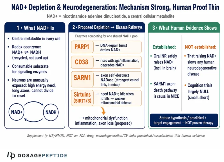
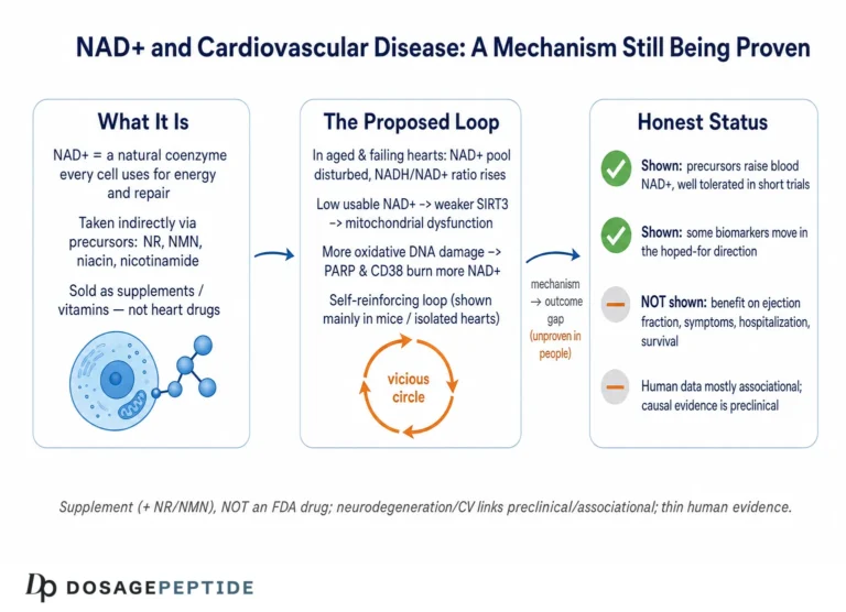
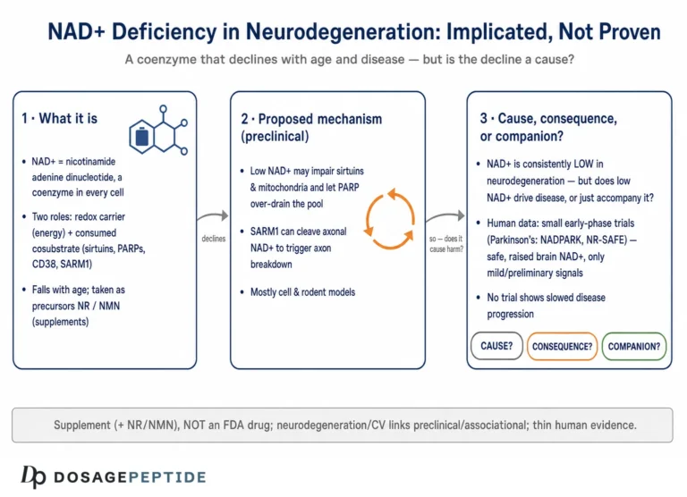
NAD+ is one of the most abundant and essential small molecules in living cells. It functions as a redox cofactor, cycling between its oxidized (NAD+) and reduced (NADH) forms to shuttle electrons through glycolysis, the tricarboxylic acid cycle, and oxidative phosphorylation. Every cell in the brain depends on this cycling to generate ATP. Beyond its redox role, NAD+ is a consumable substrate for three families of signaling enzymes: the sirtuins (NAD+-dependent deacylases, of which SIRT1 and SIRT3 are the most studied in the brain), the poly(ADP-ribose) polymerases (PARPs), and the cyclic ADP-ribose synthases such as CD38 and SARM1.9 Because these enzymes cleave NAD+ during their reactions, cellular NAD+ must be continuously resynthesized, and the rate of that resynthesis becomes rate-limiting under metabolic stress.
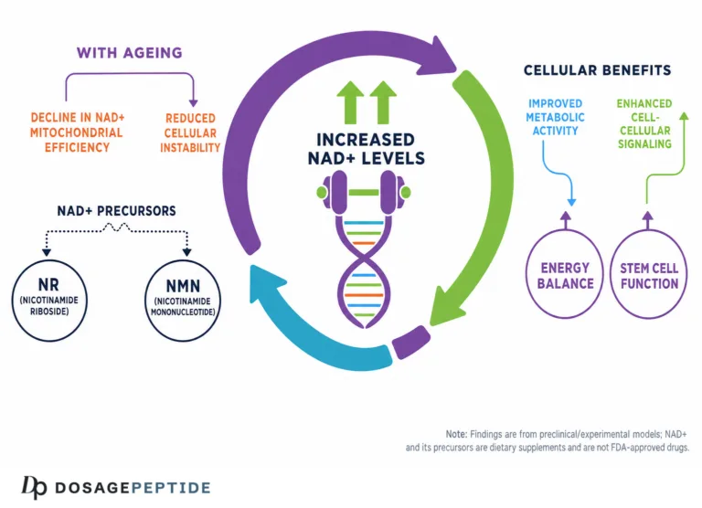
Cells build NAD+ through several routes: de novo synthesis from the amino acid tryptophan via the kynurenine pathway, and salvage pathways that recycle nicotinamide, nicotinic acid, nicotinamide riboside, and nicotinamide mononucleotide back into NAD+. The salvage pathway is the workhorse in most tissues. In it, nicotinamide is converted by nicotinamide phosphoribosyltransferase (NAMPT) into NMN, and NMN is then adenylylated by the nicotinamide mononucleotide adenylyltransferases (NMNAT1, 2, and 3) into NAD+. Nicotinamide riboside enters this pathway one step earlier, being phosphorylated by nicotinamide riboside kinases into NMN. This is why NR and NMN are described as NAD+ “precursors” or “boosters” — they feed the salvage machinery.1
The stroke question emerged from a convergence of observations. Researchers noticed that NAD+ levels fall sharply in ischemic brain tissue, that the enzymes consuming NAD+ (especially PARP-1) are hyperactivated by the oxidative and DNA-damaging conditions of ischemia-reperfusion, and that manipulating NAD+ availability changed the amount of tissue that died in animal models.8 A parallel line of work in axon biology showed that the NAD+ metabolome is a master regulator of whether an injured axon survives or self-destructs.9 Together these findings made NAD+ metabolism an attractive target, and NR and NMN — orally bioavailable, generally well-tolerated in supplement studies — became the practical tools for testing the idea.
| Molecule | Class | Position in NAD+ pathway | Typical research form |
|---|---|---|---|
| NAD+ | Dinucleotide coenzyme | End product / active cofactor | Lyophilized powder; IV or SC in studies |
| NMN | Mononucleotide | Immediate precursor (one enzyme step from NAD+) | Oral capsule; injectable in animal work |
| NR | Nucleoside (vitamin B3 vitamer) | Two steps upstream (via NRK → NMN) | Oral chloride salt (e.g., NIAGEN) |
| Nicotinamide | Vitamin B3 amide | Salvage entry via NAMPT | Oral; long history as vitamin |
It is worth stressing that none of these are peptides, and that the “NAD+” sometimes sold for injection in research-chemical contexts is the intact coenzyme, whose ability to cross cell membranes and the blood-brain barrier is itself debated. Educational reference material on the compound, including reconstitution parameters used in research settings, is catalogued in the NAD+ 500 mg/10 mL vial protocol and the broader dosage index. These references describe handling, not clinical treatment, and none of them should be read as evidence that NAD+ preserves neurons in human stroke.
Why Stroke Depletes NAD+: The Molecular Rationale
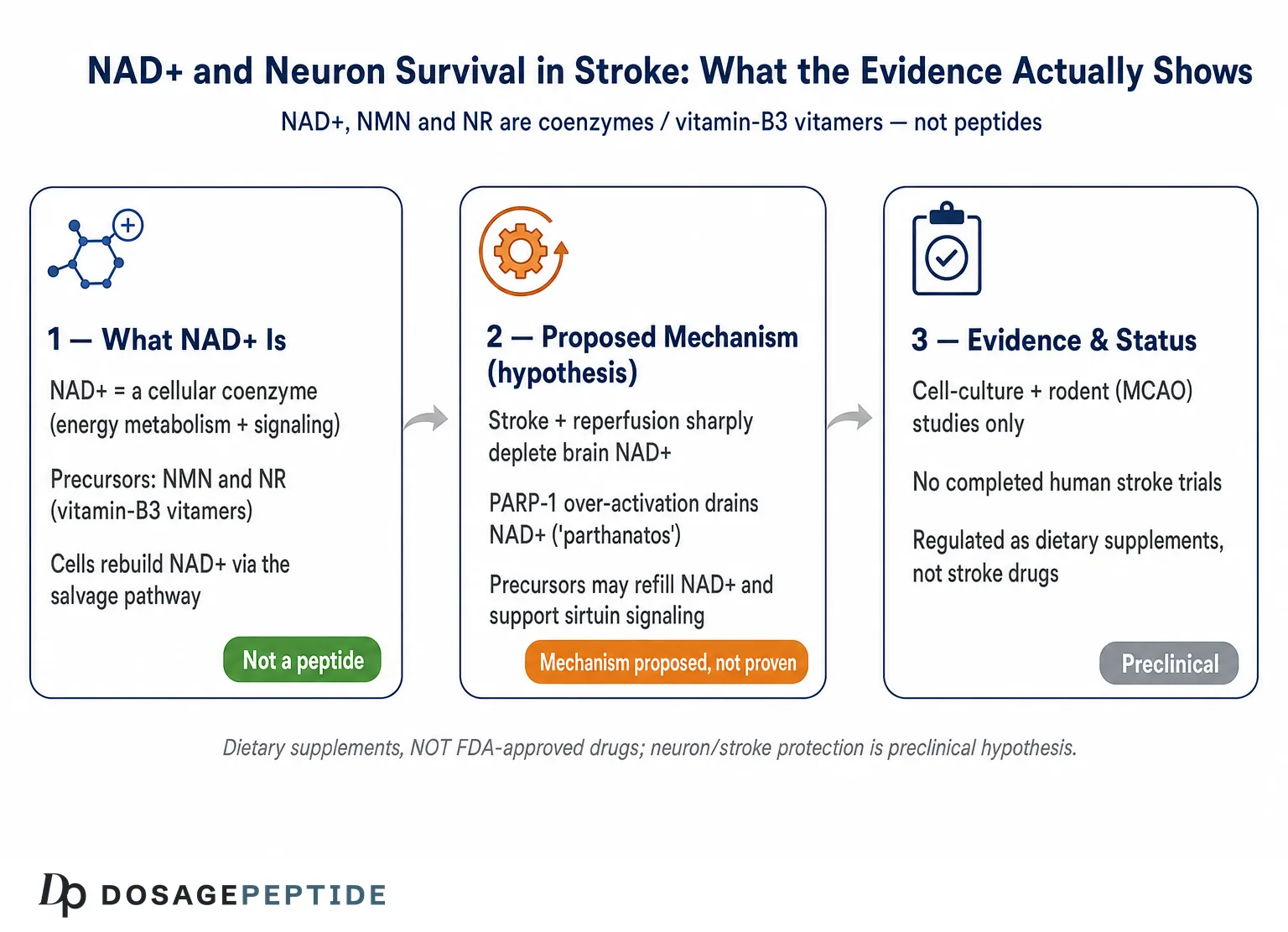
To understand why NAD+ became a candidate at all, one has to look at what happens to the coenzyme during an ischemic insult. When cerebral blood flow is interrupted, neurons lose their oxygen and glucose supply within seconds. ATP production collapses, ion gradients fail, and a cascade of excitotoxicity, calcium overload, and reactive oxygen species production begins. When blood flow is restored — reperfusion, whether spontaneous or via thrombolysis or thrombectomy — the sudden reintroduction of oxygen produces a burst of oxidative stress and extensive DNA strand breaks.8
DNA damage is the trigger for the central NAD+-depleting event in ischemic neuronal death. PARP-1, a nuclear DNA-repair enzyme, detects the strand breaks and becomes hyperactivated. Each catalytic cycle consumes NAD+ to build poly(ADP-ribose) chains on target proteins. Under the massive DNA damage of reperfusion, PARP-1 runs unchecked and can drain the cellular NAD+ pool. Because NAD+ is required for glycolysis and oxidative phosphorylation, its depletion produces a secondary energetic catastrophe on top of the primary ischemic one. This PARP-1-driven cell death program has its own name, parthanatos, distinguished by poly(ADP-ribose) accumulation, mitochondrial release of apoptosis-inducing factor (AIF), and AIF partnering with the nuclease MIF to fragment the genome.8
Reviews of ischemic stroke pathophysiology describe parthanatos as a meaningful contributor to the death of neurons in the ischemic penumbra — the at-risk tissue surrounding the irreversibly damaged core, and the tissue that neuroprotective strategies aim to salvage.8 The logic that follows is straightforward, if unproven in humans: if NAD+ depletion is part of what kills penumbral neurons, then replenishing NAD+, or supplying precursors that speed its resynthesis, might blunt the energetic collapse and give those neurons a better chance of surviving until reperfusion is achieved. It is important to note the nuance emphasized in the primary literature: some studies suggest PARP-mediated bioenergetic failure operates partly through inhibition of glycolysis rather than through NAD+ depletion alone, so “just add NAD+” is likely an oversimplification of a multi-node problem.8
The temporal dynamics of NAD+ loss also complicate the therapeutic picture. NAD+ does not simply vanish uniformly; it falls fastest in the ischemic core, where cell death is already largely committed, and more gradually in the penumbra, where intervention could in principle still matter. The window during which raising NAD+ might rescue penumbral tissue is therefore narrow and moving, expanding outward as the infarct matures. This spatial and temporal heterogeneity is one reason a blood-level increase in NAD+ metabolites does not straightforwardly translate into salvaged neurons: the coenzyme has to reach the right cells at the right moment and in a form they can use. PARP inhibitors, which block NAD+ consumption rather than boosting its supply, have themselves been studied extensively in experimental stroke with mixed clinical translation, underscoring that targeting this pathway — from either the supply or the demand side — has repeatedly looked promising preclinically without yet yielding an approved human therapy.8
A second, distinct axis of the rationale comes from axon biology. The enzyme SARM1 is a latent NAD+-cleaving enzyme that, when activated, executes a self-destruction program in injured axons (the molecular engine of Wallerian degeneration). SARM1 is held in check by NMNAT2, an NAD+-synthesizing enzyme that must be continuously delivered to the axon. When the ratio of NMN to NAD+ rises — as it does when NAD+ synthesis falters — SARM1 switches on and demolishes the axon.9 Ischemic and traumatic injuries can perturb this balance, so preserving axonal NAD+ has been proposed as a way to limit the axonal, not just somatic, component of brain injury. These two rationales — PARP/parthanatos in the cell body and SARM1/NMNAT2 in the axon — are the biochemical foundation on which the entire NAD+-and-stroke hypothesis rests.
How NAD+ Precursors Are Proposed to Protect Neurons
Assuming a precursor can raise NAD+ in the relevant brain compartment, several downstream mechanisms have been invoked to explain the neuroprotection reported in animal studies. These are the mechanistic hypotheses; each is supported by some experimental data and none should be taken as proven in the human ischemic brain.
Sirtuin activation. SIRT1 is an NAD+-dependent deacetylase concentrated in neurons that regulates stress resistance, mitochondrial biogenesis (via PGC-1α), autophagy, and inflammation. Because SIRT1 activity depends on NAD+ availability, raising NAD+ is thought to enhance SIRT1 signaling. Studies of nicotinamide phosphoribosyltransferase (NAMPT) in ischemic stroke reported that it protects neurons through a SIRT1-dependent activation of AMP-activated protein kinase (AMPK), linking NAD+ synthesis to cellular energy sensing and survival.6 SIRT3, a mitochondrial sirtuin, has been implicated in limiting mitochondrial reactive oxygen species in ischemia-reperfusion models.1
Blood-brain barrier preservation. One consequence of stroke is breakdown of the blood-brain barrier, which worsens edema and secondary injury. Work on NMNAT1 — the nuclear NAD+-synthesizing enzyme — reported that boosting it reduced blood-brain barrier permeability after experimental ischemic stroke, an effect correlated with a higher NAD+/NADH ratio and elevated SIRT1 in brain microvascular endothelial cells.4 This positions NAD+ metabolism as relevant not only to neurons but to the neurovascular unit as a whole.
Autophagy and mitochondrial quality control. In an aged-rat model of transient cerebral ischemia, overexpression of NMNAT1 reduced ischemic injury with involvement of autophagy, apparently through a SIRT1/mTOR axis.5 Restoring the capacity of stressed neurons to clear damaged mitochondria and protein aggregates is a plausible route by which NAD+ restoration could improve survival, and it dovetails with the sirtuin story.
Neuroinflammation and immune metabolism. A more recent and mechanistically interesting line of evidence moved outside the neuron entirely. A 2025 study reported that NAD+ is critically depleted in the blood monocytes of aged mice after stroke, that this depletion drives a hyperinflammatory monocyte phenotype and accumulation of the neurotoxic kynurenine-pathway metabolite quinolinic acid, and that pretreatment with nicotinamide riboside restored monocyte NAD+, reduced systemic and brain inflammation, and improved stroke outcomes in the aged animals.3 This suggests that at least part of any benefit may be immunometabolic — acting on the peripheral immune cells that traffic into the injured brain — rather than a direct rescue of neurons.
Curbing PARP-driven collapse. Finally, and most directly tied to the depletion rationale, supplying precursors is hypothesized to help the salvage pathway keep pace with PARP-1 consumption, sustaining the NAD+ needed for glycolysis and mitochondrial ATP production during the critical early window. In practice, the animal literature more often reports the net outcome (smaller infarcts, less apoptosis, better behavior) than it dissects which of these mechanisms dominates, and different studies emphasize different nodes.2 The honest reading is that NAD+ precursors are pleiotropic: they touch energy metabolism, sirtuin signaling, vascular integrity, autophagy, and inflammation simultaneously, which is both their appeal and a reason it is hard to attribute any observed benefit to a single clean mechanism.
This pleiotropy carries a double edge that is easy to overlook. On one hand, a stroke injures the brain through many parallel routes — excitotoxicity, oxidative stress, inflammation, apoptosis, vascular breakdown — and a single agent that nudges several of them at once is theoretically attractive compared with a narrowly targeted drug. On the other hand, pleiotropy makes dose-finding and mechanism attribution genuinely hard: the concentration that optimally supports sirtuin signaling may differ from the one that best restrains inflammatory monocytes or protects endothelium, and the same NAD+ pool feeds enzymes with opposing consequences (sirtuins that tend to protect, and PARPs and SARM1 that can destroy). Raising total NAD+ does not guarantee that the protective consumers, rather than the destructive ones, capture the extra substrate. Untangling which mechanism drives a given result requires genetic and pharmacological dissection — knocking out SIRT1, inhibiting PARP-1, or manipulating NMNAT/SARM1 — that most efficacy studies do not fully perform, which is why the field still debates the relative contribution of each node.
What the Evidence Actually Shows — and Its Honest Level
This is the section where enthusiasm must be tempered by rigor. The evidence that NAD+ precursors preserve neurons in stroke is preclinical: it comes from cultured cells and from rodent models of cerebral ischemia. It is real, it is peer-reviewed, and it is also, categorically, not clinical proof of benefit in humans.
Among the more concrete results: in a mouse model of ischemic injury, acute treatment with nicotinamide riboside chloride (300 mg/kg administered about 20 minutes after reperfusion) reduced hippocampal infarct volume, decreased neuronal loss and apoptosis in the hippocampus, and improved learning and memory performance on the Morris water maze relative to untreated controls.2 Studies on NMNAT enzymes reported reduced infarct volume and improved functional scores after experimental stroke, with the blood-brain barrier and autophagy effects described above.4,5 In neonatal hypoxic-ischemic brain injury models, NMN administration has been reported to be protective as well.7 Beyond ischemic stroke proper, NR reduced brain injury, oxidative stress, and neuroinflammation in a mouse model of intracerebral hemorrhage, and NAD+ precursors have shown protection in related insults such as traumatic brain injury.14
What does “honest level of evidence” mean here? A useful hierarchy runs from mechanistic cell-culture work, to small-animal efficacy studies, to large-animal studies, to human phase I safety trials, to phase II/III efficacy trials with clinical endpoints. NAD+ precursors for stroke sit at the second rung. They have abundant cell and rodent data; they have human safety data from unrelated indications (metabolic health, Parkinson’s disease); but they have zero completed human trials in stroke demonstrating improved neurological outcome. No regulatory agency has evaluated, let alone approved, any NAD+ precursor for stroke prevention or treatment.13
| Evidence tier | Status for NAD+ / precursors in stroke |
|---|---|
| In-vitro / mechanistic | Substantial — PARP, SIRT1, SARM1/NMNAT pathways characterized |
| Rodent efficacy | Multiple positive reports (NR, NMN, NMNAT overexpression) |
| Large-animal efficacy | Very limited / essentially absent for stroke |
| Human safety (any indication) | Available for NR and NMN; generally favorable |
| Human stroke efficacy (clinical endpoints) | None — no completed RCT |
| Regulatory approval for stroke | None |
Several caveats within the preclinical literature further temper the picture. Effect sizes vary widely; timing of administration relative to the insult matters enormously (many positive studies dose before or immediately after the injury, which is difficult to translate to real-world stroke, where treatment necessarily begins after symptom onset); young healthy animals dominate the datasets even though human stroke overwhelmingly affects older, comorbid patients; and publication bias almost certainly inflates the apparent consistency of benefit. The 2025 monocyte study is notable partly because it used aged animals and explicitly addressed the age dimension.3 The overall verdict a cautious reader should reach is: biologically plausible, preclinically supported, clinically unproven.
Comparing the Precursors: NAD+, NMN, NR, and Nicotinamide
“NAD+” is often used loosely to mean any member of a family of related molecules that raise cellular NAD+. In research they behave quite differently, and the distinctions matter for interpreting the stroke literature.
Intact NAD+. The coenzyme itself is large, charged, and does not freely cross cell membranes or the blood-brain barrier. When NAD+ is administered (typically intravenously in human wellness settings, or by injection in animal work), its central-nervous-system effects are thought to depend heavily on extracellular breakdown to precursors such as nicotinamide riboside and nicotinamide, which are then taken up and re-synthesized into NAD+ inside cells. A retrospective tolerability pilot compared intravenous NAD+ against NR in a real-world setting, reflecting how the intact molecule is used clinically for symptom-oriented, non-approved indications rather than stroke.17 The poor membrane permeability of intact NAD+ is one reason precursors are favored for research aiming at intracellular NAD+.
Nicotinamide mononucleotide (NMN). NMN is one enzymatic step from NAD+ (NMNAT converts it directly). It is the precursor most often used in the cerebral and cardiac ischemia animal literature and has documented protective effects in myocardial ischemia-reperfusion models, where it preserved cardiac NAD+ and reduced injury.15 In humans, NMN has been studied mainly for metabolic and performance endpoints; a systematic review and meta-analysis of randomized controlled trials found no significant benefit on glucose or lipid parameters, with no stroke data.12
Nicotinamide riboside (NR). NR is two steps upstream, entering via nicotinamide riboside kinases. It has the most robust human safety dataset of the boosters, having been through multiple randomized, placebo-controlled trials that consistently show it raises the blood NAD+ metabolome.10,11 Much of the recent stroke-relevant rodent work — the hippocampal ischemia study and the aged-monocyte study — used NR, partly because of this safety pedigree.2,3 Notably, in a rat middle-cerebral-artery occlusion model tested under clinically relevant conditions, NR was the only NAD+ precursor of those compared that reduced infarct size, underscoring that the different boosters are not interchangeable in ischemia research.16
Nicotinamide (niacinamide). The simplest B3 vitamer, nicotinamide, also raises NAD+ but is a feedback inhibitor of sirtuins and PARPs at high concentrations, which complicates its interpretation. It has a very long history of human use as a vitamin and has been studied in various neurological contexts, though again not with definitive stroke-outcome data.
A comparative review of NMN and NR concluded that both raise NAD+ and share overlapping mechanisms, but differ in pharmacokinetics, tissue distribution, and the specific transporters and enzymes they engage; the review stopped short of declaring either superior for any disease, and certainly not for stroke.1 For the purposes of the stroke question, the practical takeaway is that positive results with one precursor in one model should not be generalized into a claim about “NAD+ therapy” broadly — the molecules, doses, routes, and timing all differ. Readers comparing compounds may also encounter adjacent NAD+-modulating research chemicals such as the NNMT inhibitor 5-Amino-1MQ, referenced in the 5-Amino-1MQ protocol; these act on NAD+ metabolism by a different route and are equally preclinical with respect to stroke.
Research Models and Methodology
Understanding how these studies are conducted is essential to judging their weight. The dominant model of ischemic stroke in NAD+ research is middle cerebral artery occlusion (MCAO) in rodents. A filament is advanced to block the middle cerebral artery, cutting off blood flow to a defined territory of the brain, and then withdrawn after a set interval (commonly 60 to 120 minutes) to allow reperfusion. Investigators then measure infarct volume (often by triphenyltetrazolium chloride staining), neuronal apoptosis, blood-brain barrier permeability, biochemical markers, and functional recovery using behavioral batteries such as neurological deficit scores, rotarod, and the Morris water maze.2
Several methodological features recur and each carries an interpretive caveat:
Timing of administration. In many positive studies the precursor is given before the occlusion (prophylactic pretreatment) or within minutes of reperfusion.2,3 Human stroke does not announce itself in advance, and even the fastest emergency systems begin treatment tens of minutes to hours after onset. A compound that only works when given before the insult has limited translational value as an acute treatment, though it might inform prevention research in high-risk populations — a distinct and still-unproven proposition.
Dose and route. Rodent doses are frequently high on a milligram-per-kilogram basis (the hippocampal study used 300 mg/kg NR chloride) and are often delivered by intraperitoneal or intravenous injection rather than the oral route used in human supplementation.2 Allometric scaling from a 25-gram mouse to a human is not a simple multiplication, and the pharmacokinetics of getting a precursor into brain tissue differ across species and routes.
Animal age and comorbidity. Historically most stroke models used young, healthy male rodents, whereas human stroke patients are typically older and carry hypertension, diabetes, and atherosclerosis — conditions that themselves alter NAD+ metabolism. The field has been criticized for this mismatch, and the emergence of aged-animal studies represents a partial correction.3
Outcome measures. Infarct volume at 24 to 72 hours is a common primary readout, but it is a surrogate. Long-term functional recovery, the outcome that matters to patients, is measured less often and over shorter windows than a human trial would require. The Stroke Therapy Academic Industry Roundtable (STAIR) criteria were developed precisely because so many neuroprotectants looked good on these surrogates and then failed clinically. Among the STAIR recommendations relevant here are testing in both sexes, in aged and comorbid animals, across independent laboratories, with blinded and randomized designs, and with functional outcomes assessed weeks rather than hours after the insult. Most published NAD+ stroke studies satisfy only a subset of these criteria, which is a fair and specific critique rather than a dismissal.
Cell-culture models add their own layer of methodology and limitation. Oxygen-glucose deprivation (OGD) of cultured neurons is the standard in-vitro surrogate for ischemia, and it is where much of the mechanistic dissection of NAD+, PARP-1, and sirtuins is performed. OGD offers precise control and the ability to knock genes in or out, but a dish of isolated neurons lacks the vasculature, immune cells, and three-dimensional architecture of real brain tissue — the very features that the 2025 monocyte study showed can dominate the outcome.3 A compound that rescues neurons in a dish may fail in an animal because the limiting factor in vivo was inflammation or blood-brain barrier breakdown rather than intrinsic neuronal energy failure, and conversely an agent acting mainly on peripheral immune cells might do little to a pure neuronal culture. Reading the literature well means noting which model generated each claim and resisting the temptation to stitch results from different systems into a single tidy narrative.
These methodological realities do not invalidate the preclinical findings, but they explain why a smaller infarct in a pretreated young mouse cannot be reported as evidence that NAD+ “preserves neurons during stroke” in people. The methods generate hypotheses; they do not generate clinical proof. A rigorous reader treats each positive MCAO result as one data point in an early-stage program, weighted by how closely its design mimics the human clinical scenario — and by that standard, most current studies are still quite far from it.
Safety and Tolerability
Because NAD+ precursors are widely sold as dietary supplements, their human safety has been examined more thoroughly than their efficacy for any specific disease — but that safety data comes from healthy volunteers and non-stroke patient populations, not from stroke patients.
The most informative safety studies involve nicotinamide riboside. In a randomized, double-blind, placebo-controlled trial of long-term NIAGEN (NR chloride) in healthy overweight adults, NR was well tolerated, did not increase adverse events versus placebo, did not elevate LDL cholesterol, and did not dysregulate one-carbon metabolism; it dose-dependently raised the blood NAD+ metabolome.10 Dose-ranging work showed 100, 300, and 1000 mg of NR increased whole-blood NAD+ by roughly 22%, 51%, and 142% respectively within two weeks, with the increases sustained.10
At the higher end of the dose range, the NR-SAFE trial — a randomized, double-blind, placebo-controlled phase I study — gave people with Parkinson’s disease 1500 mg of NR twice daily (3000 mg/day) for four weeks. All participants completed the study; there were no moderate or severe adverse events attributed to NR; mild adverse events occurred at similar rates in the NR and placebo arms; and the high dose produced a pronounced systemic increase in the NAD+ metabolome without methyl-donor depletion. The authors concluded the data supported extending NR to 3000 mg/day in future phase II trials with appropriate monitoring.11
For NMN, human studies (mostly in metabolic and performance contexts) have generally reported good tolerability at doses up to several hundred milligrams per day, with a meta-analysis finding no serious safety signals though also modest and inconsistent efficacy on the metabolic endpoints examined.12 Intact NAD+ given intravenously has been used in wellness settings; infusion-related effects (chest tightness, nausea, flushing when infused rapidly) are described anecdotally and in tolerability pilots, and slowing the infusion mitigates them.17
| Compound | Human safety context | General tolerability signal |
|---|---|---|
| NR | RCTs to 3000 mg/day (healthy, Parkinson’s) | No serious AEs vs placebo; raises NAD+ |
| NMN | RCTs, metabolic/performance endpoints | Generally well tolerated; efficacy modest |
| Nicotinamide | Long vitamin history; various trials | Well tolerated at usual doses; hepatic caution at very high doses |
| Intact NAD+ (IV) | Wellness/pilot use, not approved | Infusion-rate-dependent effects |
Two honesty points must accompany this table. First, “well tolerated in healthy volunteers” is not the same as “safe and beneficial in acute stroke,” a setting with altered hemodynamics, blood-brain barrier disruption, and concurrent thrombolytics or anticoagulants — interactions that have never been formally studied. Second, favorable safety is sometimes misused to imply efficacy (“it can’t hurt, so it must help”); that inference is invalid. A safe compound that does not work is still a compound that does not work. Nothing in the safety literature bears on whether NAD+ precursors improve human stroke outcomes.
Handling and Reconstitution in a Research Context
Because this site catalogues laboratory-handling information, a brief, strictly research-oriented note on the physical properties of NAD+ preparations is warranted — with the explicit caveat that describing how a substance is stored says nothing about whether it treats any disease, and that none of the following is medical guidance or an endorsement of human self-administration.
NAD+ supplied for research is typically a lyophilized (freeze-dried) powder that is hygroscopic and sensitive to heat, light, and oxidation. General laboratory practice for such nucleotide preparations is to store the unopened lyophilized material cold and dry — commonly at −20 °C or below, with −80 °C preferred for long-term storage — protected from light and moisture. Once reconstituted in bacteriostatic or sterile water, the solution is generally refrigerated at 2–8 °C and used within a limited window (often cited as roughly two weeks) because NAD+ in solution degrades over time; repeated freeze-thaw cycles are avoided. These parameters are described for the intact coenzyme in the NAD+ vial reference, and general aseptic reconstitution technique is covered in the reconstitution guide.
Reconstitution math in a research setting follows the same logic as for any lyophilized compound: the powder mass divided by the diluent volume gives the concentration, and the concentration determines the volume corresponding to any nominal amount. For example, a 500 mg quantity brought up in 3.0 mL of diluent yields approximately 166.7 mg/mL. Tools that automate this arithmetic are described in general reconstitution references. None of this arithmetic implies a therapeutic dose, a route of administration for humans, or any claim of clinical benefit; it is purely the bookkeeping of preparing a solution at a known concentration for in-vitro or animal research.
It also bears repeating that the oral NR and NMN used in the human safety trials are chemically and pharmacokinetically different from injectable intact NAD+. The rodent neuroprotection studies most often used injected precursors at high mg/kg doses, the human safety data come from oral capsules, and the intact-NAD+ handling parameters above describe yet another preparation. Conflating these — treating storage information about an injectable coenzyme as if it validated an oral supplement’s effect on stroke, or vice versa — is exactly the kind of category error this article is written to prevent. Handling knowledge is logistics, not evidence of efficacy. Anyone using such references should recognize that the compounds are sold for laboratory research, that self-experimentation carries real and unquantified risk, and that appropriate professional and ethical oversight is non-negotiable.
Limitations and the Human-Evidence Gap
The gap between the animal literature and a human stroke claim is not a minor caveat to be waved away; it is the central fact of this topic. Several specific limitations define that gap.
No completed human stroke trials. The single most important limitation is the absence of any completed randomized controlled trial testing an NAD+ precursor in human stroke patients with neurological or functional outcomes. Every claim of neuroprotection rests on cells and rodents. Open-label studies of NAD+ in other contexts exist (for example, a registered open-label study assessing increasing NAD+ levels), but these are not stroke-efficacy trials and do not fill the gap.18
The translational graveyard. Stroke neuroprotection has one of the worst translational track records in medicine. Dozens of compounds — NXY-059, various NMDA antagonists, free-radical scavengers — reduced infarct size in animals and then failed in phase III. The base rate for a promising preclinical neuroprotectant reaching clinical utility is very low. NAD+ precursors have not yet been tested at the stage where most candidates fail, so their apparent promise has not been stress-tested by the trials that historically break such candidates.
Delivery to the injured brain. Whether an orally dosed precursor raises NAD+ in the specific neurons of the ischemic penumbra, at the right time, and to a degree sufficient to change cell fate, is largely unestablished in humans. Blood NAD+ rising by 142% (as with 1000 mg NR) does not guarantee a corresponding, timely rise in penumbral neuronal NAD+.10
Timing and the therapeutic window. Positive rodent results skew toward pretreatment or immediate post-reperfusion dosing.2 Real strokes are treated after a delay; whether a precursor helps when started hours after onset is unknown.
Comorbidity and interactions. Human stroke patients are older, sicker, and on multiple medications (antithrombotics, antihypertensives, statins). Interactions with these, and with reperfusion therapies, have not been characterized. Aged-animal studies begin to address the biology but not the pharmacology of polypharmacy.3
Heterogeneity of “NAD+ therapy.” The literature spans intact NAD+, NMN, NR, nicotinamide, and enzyme-overexpression approaches (NMNAT1), across different doses, routes, and injury models. Pooling these into a single “NAD+ preserves neurons” claim obscures that we do not know which molecule, dose, route, or timing — if any — would work in a human.
The intellectually honest summary is that NAD+ metabolism is a genuinely interesting target with a coherent mechanistic story and encouraging early data, and simultaneously that there is currently no scientific basis for telling a person that taking NAD+, NMN, or NR will preserve their neurons if they have a stroke. Both statements are true at once, and responsible communication holds them together rather than collapsing into either hype or dismissal.
Regulatory Status
The regulatory picture reinforces the evidence picture: no NAD+ precursor is an approved drug for stroke, or for any neurological disease, anywhere.
In the United States, NAD+ precursors are regulated as dietary supplements or ingredients, not as drugs. That classification specifically means they may be marketed to support general health but may not be marketed to diagnose, treat, cure, or prevent any disease — including stroke — and no manufacturer may make such a claim legally. Nicotinamide riboside chloride (NIAGEN) has been the subject of new dietary ingredient notifications and generally-recognized-as-safe determinations that concern its use as a food ingredient, not its efficacy against disease.
Nicotinamide mononucleotide had a more turbulent regulatory history. The FDA initially took the position (in 2022) that NMN was excluded from the dietary-supplement definition because it had been authorized for investigation as a new drug. After petitions from industry associations, the FDA reversed course, and by late 2025 confirmed that beta-NMN is lawful for use in dietary supplements, while noting that NMN remains a new dietary ingredient subject to premarket notification requirements.13 Crucially, none of this back-and-forth concerns stroke: it is about whether NMN may be sold as a supplement, not whether it works for any medical condition. Being “lawful in supplements” is a marketing-status determination, not an efficacy endorsement.
Intact NAD+ administered intravenously occupies a grayer zone. It is used in some wellness and functional-medicine settings for non-approved, symptom-oriented indications, but it is not an FDA-approved therapy for stroke or neuroprotection, and such use is off-label and unproven. Compounds sold as “research chemicals,” including injectable NAD+ vials, are explicitly not for human consumption and are not evaluated by regulators for safety or efficacy in people.
Internationally, the picture is similar: NAD+ precursors circulate as supplements or novel foods under various food-safety frameworks, and no major regulator has approved any of them as a medicine for cerebrovascular disease. There is no European Medicines Agency or FDA drug approval, no stroke indication on any label, and no clinical guideline that recommends NAD+ precursors for stroke prevention or treatment. Any product or source implying otherwise is making a claim unsupported by both the evidence and the regulatory record. Readers exploring the broader category can consult general educational overviews such as the compound reference index and combination reference material, keeping in mind that these are educational resources about research compounds, not clinical recommendations.
Frequently Asked Questions
Is NAD+ a peptide?
No. NAD+ (nicotinamide adenine dinucleotide) is a dinucleotide coenzyme, and its common precursors — nicotinamide mononucleotide (NMN) and nicotinamide riboside (NR) — are nucleotides and nucleosides derived from vitamin B3. None of them are peptides (chains of amino acids). The term is sometimes used loosely in research-chemical marketing, but chemically NAD+ and its precursors belong to a completely different molecular class than peptides.
Does the evidence show NAD+ preserves neurons during a human stroke?
No. The evidence that NAD+ or its precursors preserve neurons comes from cell cultures and rodent models of cerebral ischemia.2,3,4 There are no completed randomized controlled trials in human stroke patients showing improved neurological outcomes. The idea is a preclinical hypothesis, not a demonstrated human treatment, and no regulator has approved any NAD+ precursor for stroke.
Why do scientists think NAD+ might matter in stroke at all?
Because NAD+ levels fall sharply in ischemic brain tissue. DNA damage from reperfusion hyperactivates the enzyme PARP-1, which consumes NAD+ and can trigger a cell-death program called parthanatos.8 Since NAD+ is also required for energy production and for protective sirtuin signaling, researchers hypothesized that replenishing it might help at-risk neurons survive. That rationale is coherent but remains unproven in humans.
What is the difference between NAD+, NMN, and NR?
They are related molecules at different points in the same pathway. Intact NAD+ is the active coenzyme but crosses cell membranes poorly. NMN is one enzymatic step from NAD+ and is common in animal ischemia studies.15 NR is two steps upstream and has the strongest human safety dataset among the boosters.10,11 A comparative review found overlapping mechanisms but different pharmacokinetics, and did not declare any one superior for disease.1
Are NAD+ precursors safe?
In healthy volunteers and some patient groups, oral NR and NMN have generally been well tolerated in randomized trials, including NR at up to 3000 mg/day for four weeks with no serious adverse events versus placebo.11 However, favorable safety in those settings does not mean the compounds are safe or beneficial in acute stroke, where hemodynamics, blood-brain barrier integrity, and concurrent medications differ and have never been formally studied.
Can I take NMN or NR to prevent a stroke?
There is no evidence supporting that use. NAD+ precursors are regulated as dietary supplements, which by law may not be marketed to prevent or treat any disease, including stroke.13 No clinical guideline recommends them for stroke prevention. Anyone concerned about stroke risk should discuss evidence-based measures — blood pressure control, atrial fibrillation management, lifestyle — with a qualified clinician rather than relying on unproven supplements.
What is the regulatory status of NMN and NAD+?
NR and NMN are regulated as dietary supplements/ingredients in the United States. After earlier uncertainty, the FDA confirmed by late 2025 that beta-NMN is lawful in dietary supplements while remaining a new dietary ingredient subject to notification requirements.13 None of this concerns disease efficacy: no NAD+ precursor is an approved drug for stroke or any neurological condition, and intact injectable NAD+ sold as a research chemical is not for human consumption.
What would it take to actually prove NAD+ helps in stroke?
Well-designed human trials: randomized, placebo-controlled studies in stroke patients, using aged and comorbid populations that reflect real patients, dosing within a realistic therapeutic window after symptom onset, and measuring meaningful clinical outcomes (disability, recovery) rather than only surrogate markers.3 Until such trials are completed and replicated, the neuroprotection claim remains a hypothesis generated by preclinical work.
References
- Yang et al. An Updated Review on the Mechanisms, Pre-Clinical and Clinical Comparisons of Nicotinamide Mononucleotide (NMN) and Nicotinamide Riboside (NR). Food Frontiers, 2025. https://iadns.onlinelibrary.wiley.com/doi/10.1002/fft2.511
- Acute Treatment with Nicotinamide Riboside Chloride Reduces Hippocampal Damage and Preserves the Cognitive Function of Mice with Ischemic Injury. PubMed 35585298, 2022. https://pubmed.ncbi.nlm.nih.gov/35585298/
- NAD+ depletion drives age-related monocyte hyperinflammation after stroke and is reversed by nicotinamide riboside. Journal of Neuroinflammation, 2025. https://link.springer.com/article/10.1186/s12974-025-03638-6
- Nicotinamide Mononucleotide Adenylyltransferase 1 Regulates Cerebral Ischemia-Induced Blood-Brain Barrier Disruption Through NAD+/SIRT1 Signaling Pathway. Molecular Neurobiology, 2022. https://link.springer.com/article/10.1007/s12035-022-02903-6
- Neuroprotection by NMNAT1 with involvement of autophagy in an aged rat model of transient cerebral ischemia and reperfusion. Brain Research (ScienceDirect). https://www.sciencedirect.com/science/article/abs/pii/S0006899319304457
- Nicotinamide phosphoribosyltransferase protects against ischemic stroke through SIRT1-dependent AMPK pathway. PubMed 21246601. https://pubmed.ncbi.nlm.nih.gov/21246601/
- Therapeutic Effect of Nicotinamide Mononucleotide for Hypoxic-Ischemic Brain Injury in Neonatal Mice. ASN Neuro, 2023. https://www.tandfonline.com/doi/full/10.1177/17590914231198983
- Liu et al. Emerging role of PARP-1 and PARthanatos in ischemic stroke. Journal of Neurochemistry, 2022. https://pmc.ncbi.nlm.nih.gov/articles/PMC9010225/
- The SARM1 Axon Degeneration Pathway: Control of the NAD+ Metabolome Regulates Axon Survival in Health and Disease. PMC7483800. https://pmc.ncbi.nlm.nih.gov/articles/PMC7483800/
- Safety and Metabolism of Long-term Administration of NIAGEN (Nicotinamide Riboside Chloride) in a Randomized, Double-Blind, Placebo-controlled Clinical Trial of Healthy Overweight Adults. PMC6611812. https://pmc.ncbi.nlm.nih.gov/articles/PMC6611812/
- NR-SAFE: a randomized, double-blind safety trial of high dose nicotinamide riboside in Parkinson’s disease. Nature Communications, 2023. https://www.nature.com/articles/s41467-023-43514-6
- Effects of Nicotinamide Mononucleotide on Glucose and Lipid Metabolism in Adults: A Systematic Review and Meta-analysis of Randomised Controlled Trials. PMC11557618. https://www.ncbi.nlm.nih.gov/pmc/articles/PMC11557618/
- FDA Declares Nicotinamide Mononucleotide Is a Dietary Supplement. Venable LLP / NutraIngredients, 2025. https://www.venable.com/insights/publications/2025/10/fda-declares-nicotinamide-mononucleotide-is
- Nicotinamide riboside restores NAD+ levels and alleviates brain injury by inhibiting oxidative stress and neuroinflammation in a mouse model of intracerebral hemorrhage. PMC11772386. https://pmc.ncbi.nlm.nih.gov/articles/PMC11772386/
- Nicotinamide Mononucleotide, an Intermediate of NAD+ Synthesis, Protects the Heart from Ischemia and Reperfusion. PMC4048236. https://www.ncbi.nlm.nih.gov/pmc/articles/PMC4048236/
- Only the NAD Precursor Nicotinamide Riboside Reduces Infarct Size in a Rat Ischemia-Reperfusion Model Under Clinically Relevant Conditions. PMC8435675. https://www.ncbi.nlm.nih.gov/pmc/articles/PMC8435675/
- Intravenous infusion of NAD+ versus nicotinamide riboside (NR): a retrospective tolerability pilot study in a real-world setting. PMC12907335. https://www.ncbi.nlm.nih.gov/pmc/articles/PMC12907335/
- Open-label Study to Assess Increasing Levels of NAD+ (Nicotinamide Adenine Dinucleotide). ClinicalTrials.gov NCT03707652. https://clinicaltrials.gov/study/NCT03707652
Educational and research-use disclaimer: This article is provided solely for scientific education and to summarize the current state of preclinical research. It is not medical advice and does not describe an approved therapy. NAD+, NMN, and NR are not FDA-approved drugs for stroke or any neurological disease; where sold, they are dietary supplements or research chemicals and may not lawfully be marketed to diagnose, treat, cure, or prevent any disease. The neuroprotection discussed here is a hypothesis supported only by cell and animal studies and has never been demonstrated in human stroke trials. Nothing herein should be interpreted as a recommendation to use any compound to preserve neurons, treat, or prevent stroke. Injectable research-chemical preparations are not for human consumption. Anyone with concerns about stroke risk or any medical condition should consult a qualified, licensed healthcare professional and rely on evidence-based, guideline-recommended care.