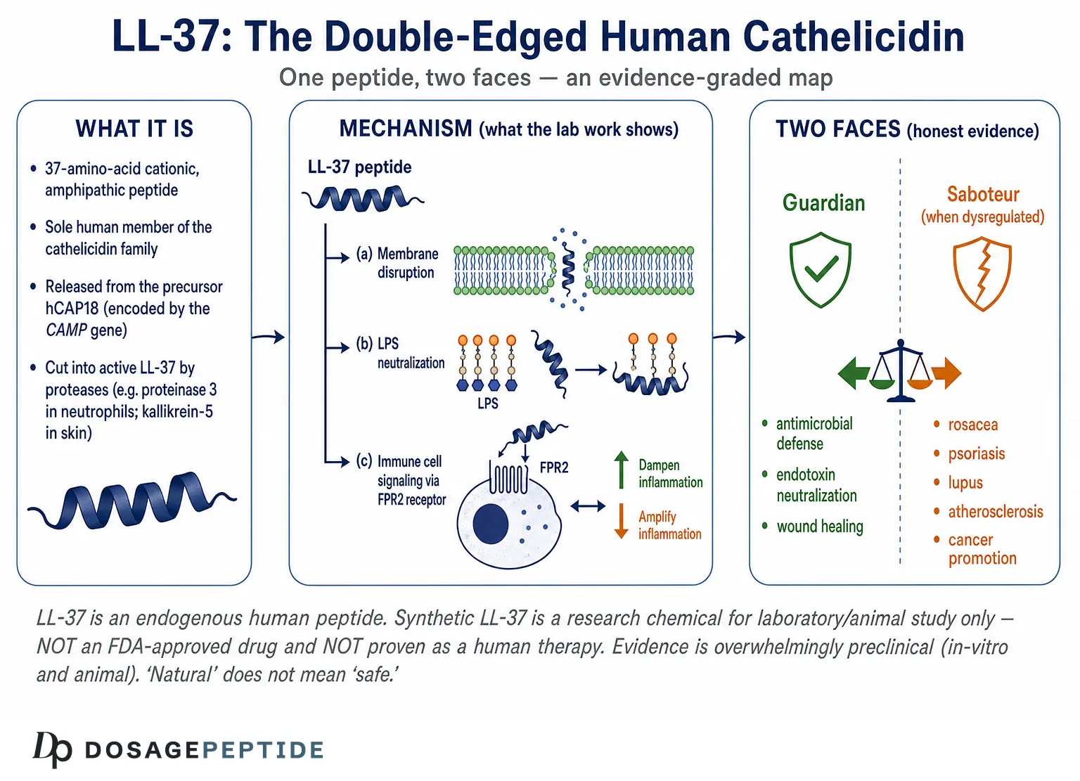
LL-37 is the only cathelicidin the human body makes. It is a 37-amino-acid peptide cut from a precursor called hCAP18, released by skin, gut and airway lining cells and by neutrophils, and it does two jobs at once. It disrupts bacterial membranes directly, and it tells the immune system what to do next — recruiting white cells, shaping macrophage and dendritic-cell behaviour, and pushing wound repair along. It also has documented antiviral and anti-biofilm activity.
The complication, and the reason researchers call it double-edged, is that the same peptide can be pro-inflammatory or anti-inflammatory depending on context. In psoriasis and lupus it appears to be part of the problem rather than the defence. Vitamin D controls how much of it your cells produce. This article covers what LL-37 does, what laboratory and animal work has actually shown, how far the drug-development attempts have got, and what is still unknown.
Framing note: LL-37 is an endogenous peptide your own body makes. Synthetic LL-37 sold to laboratories is a research chemical for in-vitro and animal study only. It is not an approved drug, and nothing here is medical advice or a dosing recommendation. The overwhelming majority of the findings below come from cell-culture and animal experiments; human therapeutic use of synthetic LL-37 remains early and experimental.
What is LL-37, and why is it called the human cathelicidin?
LL-37 is a 37-amino-acid, cationic, amphipathic peptide that constitutes the antimicrobial business end of the human cathelicidin protein. Its name is literal: the mature peptide begins with two leucine residues (“LL”) and is 37 residues long. Cathelicidins are an ancient class of host-defense (antimicrobial) peptides defined not by their active fragment but by a shared, highly conserved N-terminal pro-region called the cathelin domain — a cathepsin-L-inhibitor-like sequence. Different species carry very different C-terminal antimicrobial payloads attached to this conserved cassette, but humans carry exactly one.[1]
The cathelicidin family across species
Cathelicidins are found across mammals, birds, fish, and reptiles, and they are strikingly diverse in their mature-peptide sequences: pigs make protegrins and PR-39, cattle make the disulfide-bridged bactenecins and indolicidin, and mice and rats make CRAMP (cathelicidin-related antimicrobial peptide). What unites them is the cathelin pro-domain that keeps the peptide inactive while it is stored, plus the general theme that the released C-terminal peptide is cationic and membrane-active. This evolutionary conservation of a stored-then-cleaved antimicrobial strategy is one reason researchers treat cathelicidins as a core component of innate immunity rather than a curiosity.[1]
One gene, one peptide: CAMP and hCAP18
In humans, a single gene — CAMP (cathelicidin antimicrobial peptide), on chromosome 3 — encodes a preproprotein. After the signal sequence is removed, the circulating precursor is an 18-kDa protein called hCAP18 (human cationic antimicrobial protein, 18 kDa). hCAP18 is the storage and transport form; it comprises the conserved cathelin domain followed by the C-terminal 37 residues that become LL-37 only after proteolytic release. Because there is exactly one human cathelicidin gene producing exactly one canonical antimicrobial peptide, LL-37 is often described as “the only human cathelicidin” — a phrase that captures both its uniqueness and the outsized amount of biology packed into a single short sequence.[1][2] Readers new to the vocabulary of peptide research may find our peptide research glossary useful for terms such as amphipathic, cationic, and host-defense peptide.
How is LL-37 generated from hCAP18?
A central and often-misunderstood point is that LL-37 does not exist pre-formed inside resting cells. Cells store the inactive precursor hCAP18 and only liberate the active LL-37 peptide by cutting it at the right moment with the right protease. This “stored precursor, on-demand activation” design is what allows a potentially membrane-damaging peptide to be kept safely sequestered until it is needed — and it means the enzymes doing the cutting are as biologically important as the peptide itself.
Proteinase 3 in neutrophils
Neutrophils are the body’s largest reservoir of cathelicidin. They store hCAP18 in their specific (secondary) granules. When neutrophils are activated and degranulate, hCAP18 is exocytosed and then cleaved extracellularly by proteinase 3, a serine protease released from the neutrophil’s azurophil (primary) granules. Classic work established that, of the azurophil serine proteases, proteinase 3 is the one responsible for converting exocytosed hCAP18 into mature LL-37 — a neat spatial arrangement in which two different granule populations must both be released for the active peptide to appear.[3]
Kallikrein-5 and the skin
Skin uses a different set of scissors. In keratinocytes, hCAP18 is processed by kallikrein-related peptidases, principally kallikrein-5 (also historically called stratum corneum tryptic enzyme, SCTE), and kallikrein-7. Crucially, the skin proteases can generate not only canonical LL-37 but a family of alternative, shorter cathelicidin fragments. As we will see in the section on rosacea, the identity of those fragments — not just the total amount of cathelicidin — determines whether the result is orderly defense or destructive inflammation.[7] Other tissues use still other proteases; in seminal plasma, for example, gastricsin cleaves hCAP18 into a longer 38-residue peptide (ALL-38) at low pH, illustrating how the same precursor is tuned to its local environment.[2]
Where is LL-37 expressed in the body?
One reason LL-37 is described as a “factotum” — a jack-of-all-trades — is that it is made almost everywhere the body meets the outside world. Beyond neutrophils, hCAP18/LL-37 is produced by epithelial cells of the skin, airways, gastrointestinal tract, and urogenital system, and by monocytes, mast cells, natural killer cells, B cells, and other leukocytes. It is secreted into wound fluid, airway surface liquid, saliva, sweat, breast milk, and other secretions, positioning it as a first-line sentinel at barrier surfaces.[2]
| Site / source | Cell type | Notes on the research |
|---|---|---|
| Blood / pus | Neutrophils (specific granules) | Largest store of hCAP18; released and cleaved by proteinase 3 on degranulation |
| Skin | Keratinocytes, sweat & sebaceous glands | Induced by injury/infection; processed by kallikrein-5/7 into LL-37 and variants |
| Airway | Bronchial/alveolar epithelium | Present in airway surface liquid; studied in cystic fibrosis and respiratory infection |
| Gut | Colonic epithelium | Contributes to mucosal barrier defense; interfaces with the microbiome |
| Urogenital | Epithelium; seminal plasma | Seminal hCAP18 cleaved to ALL-38 by gastricsin at low pH |
| Secretions | Multiple | Detected in saliva, sweat, breast milk, wound and airway fluid |
This broad distribution is why LL-37 is studied not as a niche molecule but as a hub connecting barrier immunity, inflammation, and tissue repair. It also foreshadows the double-edged theme: a peptide present in so many compartments has many opportunities to help — and many opportunities to be dysregulated.
What does LL-37 look like? Structure and biophysics
To understand both the protective and pathogenic sides of LL-37, you have to start with its physical shape, because nearly every activity flows from how this peptide folds and how it interacts with membranes.
The amphipathic α-helix
In water at physiological salt, LL-37 is largely disordered, but when it encounters an anionic (negatively charged) membrane or a membrane-mimicking micelle, it folds into an amphipathic α-helix. “Amphipathic” means the helix has two faces: one hydrophobic (water-avoiding) face studded with leucine, phenylalanine, and other nonpolar residues, and one cationic hydrophilic face bearing the peptide’s many positively charged arginines and lysines. High-resolution NMR structures show that in detergent micelles the peptide forms a curved helix-bend-helix motif spanning roughly residues 2–31, followed by a disordered C-terminal tail, with the bend located near residues 14–16.[4] That same structural work identified a minimal antimicrobial core — the 12-residue fragment KR-12 (residues 18–29) — which retains membrane activity and has become an important template for engineered mimetics.[4]
Why LL-37 targets microbial membranes and (mostly) spares host cells
The selectivity of LL-37 for microbes over host cells is largely an electrostatic story. Bacterial membranes present abundant negatively charged lipids (phosphatidylglycerol, cardiolipin, and in Gram-negative bacteria the anionic lipopolysaccharide of the outer leaflet), and bacterial surfaces carry other anionic polymers such as teichoic acids. The cationic face of LL-37 is drawn to these negative charges, concentrating the peptide at the microbial surface. Mammalian plasma membranes, by contrast, keep their outer leaflet largely zwitterionic (net-neutral phosphatidylcholine and sphingomyelin) and enrich it with cholesterol, which rigidifies the bilayer and blunts peptide insertion. The result is a preferential attack on microbial membranes at concentrations that host cells can better tolerate.[1] This selectivity is relative, not absolute — at high concentrations LL-37 can damage host cells too, a fact that becomes central to its toxicity and its dark side.
Aggregation, oligomerization, and environmental sensitivity
LL-37 is unusual among antimicrobial peptides in that it tends to oligomerize in solution, and this self-association influences both its stability against proteolysis and how it engages membranes. Its behavior is also highly sensitive to the local environment: physiological salt concentrations, divalent cations, serum proteins, and pH all modulate its folding and activity. This environmental sensitivity is a recurring caveat throughout the literature — a concentration or condition that produces crisp bacterial killing in a low-salt buffer may produce weak or entirely different effects in serum-rich, physiological conditions. Keeping that in mind is essential when interpreting in-vitro potency claims.[1]
How does LL-37 kill microbes?
The original and best-characterized function of LL-37 is direct antimicrobial activity. But even here, “kill” is an oversimplification: LL-37 disrupts membranes, neutralizes microbial toxins, and interferes with biofilms, and its net effect depends heavily on the model system.
Membrane disruption models
Once concentrated at a microbial surface, LL-37 is thought to disrupt the membrane by mechanisms usually described with the carpet and toroidal-pore models. In the carpet model, peptide molecules accumulate parallel to the membrane surface until, above a threshold density, they act like a detergent and dissolve the bilayer into fragments and micelles. In the toroidal-pore model, peptides insert and induce the lipid head groups to curve inward, forming transient aqueous pores lined by both peptide and lipid. Either way, the barrier function of the membrane collapses, ions and metabolites leak, and the microbe dies. LL-37’s tendency to lie flat on and carpet membranes fits the surface-active picture rather than a discrete, stable channel.[1]
Bacteria and biofilms
In vitro, LL-37 shows broad-spectrum activity against Gram-positive and Gram-negative bacteria, including species such as Staphylococcus aureus, Escherichia coli, and Pseudomonas aeruginosa, though potency varies widely with salt, serum, and strain. A particularly active area of research is LL-37’s effect on biofilms — structured microbial communities encased in a self-produced matrix that are notoriously tolerant of conventional antibiotics. Studies report that LL-37 and its truncated mimetics (such as KR-12 derivatives) can inhibit biofilm formation and, in some systems, disrupt established biofilms of organisms including P. aeruginosa and S. aureus, sometimes at concentrations below those needed to kill planktonic cells. This anti-biofilm potential is a major reason engineered LL-37 fragments are studied for device- and wound-associated infections.[2]
Viruses and fungi
LL-37’s activity is not limited to bacteria. In cell-culture and biochemical assays it shows antifungal effects against Candida albicans and antiviral effects against several enveloped viruses, where it can disrupt the viral envelope or interfere with entry and replication steps. These findings are consistently in-vitro or animal-model in nature; they establish biological plausibility for a broad host-defense role but do not demonstrate that administering LL-37 treats fungal or viral disease in people.[2]
An important subtlety is that LL-37 does not indiscriminately sterilize the surfaces where it is expressed. At barrier sites such as the gut and skin, it operates within a resident microbial community, and its concentrations are usually well below those that would wipe out that community. Rather than acting as a blunt disinfectant, endogenous LL-37 appears to help shape which organisms colonize and to reinforce the barrier against opportunistic overgrowth — a role that is closer to gardening than to scorched earth. This is another reason researchers increasingly emphasize LL-37’s regulatory and immunomodulatory functions over raw microbicidal potency: in the living organism, the peptide’s job seems to be tuning the host-microbe relationship as much as killing.[2]
| Target class | Representative activity reported in vitro | Key caveat |
|---|---|---|
| Gram-positive bacteria | Membrane disruption of S. aureus and others | Potency drops sharply in physiological salt/serum |
| Gram-negative bacteria | Activity vs E. coli, P. aeruginosa; LPS binding | Strain- and condition-dependent |
| Biofilms | Inhibition/disruption at sub-MIC levels in some models | Mostly in-vitro; device and wound models |
| Fungi | Anti-Candida effects | Preclinical only |
| Enveloped viruses | Envelope disruption / entry interference | In-vitro; not proven antiviral therapy |
| Endotoxin (LPS) | Binding and neutralization | Immunomodulatory, not “killing” |
LPS and endotoxin neutralization
Perhaps the most therapeutically intriguing direct effect is LL-37’s ability to bind and neutralize lipopolysaccharide (LPS), the endotoxin of Gram-negative bacterial outer membranes that drives septic inflammation through Toll-like receptor 4. By binding LPS directly, LL-37 can prevent it from engaging its receptor, dampening the downstream cytokine storm. Multiple studies characterize LL-37 as an LPS-neutralizing, pleiotropic peptide, and this activity underpins interest in cathelicidin-derived molecules for sepsis and endotoxemia.[8] Notably, endotoxin neutralization blurs the line between “antimicrobial” and “immunomodulatory” — the peptide is not killing anything here; it is editing the host’s inflammatory reaction. That editing function is where the story gets genuinely complex.
Beyond killing microbes: how does LL-37 modulate the immune system?
A pivotal shift in cathelicidin research was the recognition that, at the concentrations typically present in tissue, LL-37’s direct microbicidal effect is often modest, while its ability to reshape the host immune response is profound. This reframed LL-37 from a simple “natural antibiotic” into a signaling molecule that helps orchestrate defense — for better and for worse.
Chemotaxis via FPR2
One of the foundational immunomodulatory findings is that LL-37 is a chemoattractant. It recruits human neutrophils, monocytes, and T cells by acting as an agonist at the G-protein-coupled receptor formyl peptide receptor-like 1 (FPRL1, now called FPR2/ALX). Through FPR2, LL-37 triggers calcium mobilization and directed cell migration, effectively summoning immune cells to sites where the peptide is released. This receptor-mediated recruitment is a genuine signaling function, entirely separate from membrane poking, and it places LL-37 upstream of the cellular inflammatory response rather than merely alongside it.[5]
Effects on monocytes, macrophages, and dendritic cells
LL-37 influences the differentiation and behavior of the innate immune cells it recruits. It can shape how monocytes mature into macrophages or dendritic cells, alter the surface receptors those cells express, and modulate their responsiveness to microbial signals. In some settings it enhances phagocytosis and pathogen clearance; in others it tempers cellular activation. These effects, together with its chemotactic activity, are why LL-37 is grouped with a small set of “host-defense peptides” that function as endogenous immune modulators. This modulatory theme is shared with other immunologically active peptides that researchers study for immune balance, such as thymosin alpha-1 and its immune-modulation research.[2]
Cytokine modulation: dampening and amplifying
The single most important nuance in LL-37 biology is that its effect on inflammation is bidirectional and context-dependent. In classic experiments using human monocytic cells, low, physiologically relevant concentrations of LL-37 (on the order of 1 µg/mL or less) suppressed the LPS-induced release of the pro-inflammatory cytokine TNF-α and down-regulated a large set of NF-κB-driven pro-inflammatory genes, while leaving certain chemokine responses intact. In other words, LL-37 can act as a brake on the endotoxin response, biasing the reaction toward controlled recruitment rather than runaway inflammation.[6] Yet in different cellular contexts — and especially when LL-37 forms complexes with nucleic acids — the very same peptide amplifies inflammation dramatically. This Jekyll-and-Hyde behavior is not a contradiction in the data; it is the defining feature of the molecule, and it is why blanket claims that LL-37 is “anti-inflammatory” are misleading.
Bridging innate and adaptive immunity
Through chemotaxis, dendritic-cell modulation, and its effects on antigen sensing, LL-37 helps connect the immediate innate response to the slower, specific adaptive response. It can influence which T-cell programs are favored and, as later sections describe, can even become a target of adaptive immunity itself in autoimmune disease. The peptide thus operates as a node where innate detection is translated into adaptive consequences — a powerful position that also makes its dysregulation consequential.[2]
Does LL-37 help wounds heal?
Wound healing is one of the most studied “beneficial” functions of LL-37, and it ties together several of the activities already described. LL-37 is secreted into wound fluid, where it both limits infection and appears to actively promote repair. A landmark study demonstrated that LL-37 is angiogenic — it directly activates endothelial cells, increasing their proliferation and driving the formation of new vessel-like structures, an effect mediated through FPR2. In that work, LL-37 promoted neovascularization in a chick chorioallantoic membrane assay and in a rabbit model of hind-limb ischemia, linking the peptide to the blood-vessel growth that healing tissue requires.[11]
Beyond angiogenesis, LL-37 promotes the migration and proliferation of keratinocytes, supporting re-epithelialization of the wound surface. Reduced or dysfunctional cathelicidin has been associated with impaired healing in some models, and topical LL-37 has been explored in early-phase human wound studies — for example in hard-to-heal venous leg ulcers — though such trials remain small, early, and far from establishing routine therapy. The honest summary is that LL-37 is a genuine participant in the wound-repair program, best documented in preclinical systems, with human data still preliminary. Readers interested in how other peptides are studied for tissue protection and repair may compare this with research on BPC-157 and gastrointestinal ulcer protection.[11]
How does vitamin D control LL-37? The CAMP axis
Of all the regulatory inputs to LL-37, the vitamin D connection is the best-supported at a mechanistic level, and it is one of the more remarkable stories in innate immunology. The human CAMP gene carries a functional vitamin D response element (VDRE) in its promoter, so the active hormone 1,25-dihydroxyvitamin D3, working through the vitamin D receptor, directly up-regulates cathelicidin transcription.[10]
The evolutionary twist is that this VDRE was inserted by an ancient Alu short interspersed nuclear element (SINE) that is present in primates but absent in mice, rats, and dogs. As a result, vitamin D’s control of cathelicidin is essentially a primate-specific innovation — a caution flag for anyone extrapolating from rodent cathelicidin (CRAMP) biology to human LL-37.[10] The functional importance of this axis was shown in a now-classic study of tuberculosis: activation of Toll-like receptors on human macrophages up-regulated the vitamin D receptor and the vitamin-D-activating enzyme, leading to cathelicidin induction and enhanced killing of intracellular Mycobacterium tuberculosis. Strikingly, serum from individuals with low 25-hydroxyvitamin D supported cathelicidin induction poorly, offering a molecular rationale for the long-observed link between vitamin D status and infection susceptibility.[9]
It is important to state the boundary of this evidence carefully. The vitamin D → CAMP → LL-37 pathway is a genuinely well-supported molecular axis. That does not mean vitamin D supplementation is a proven way to treat or prevent infection through LL-37 in the general population; the clinical trial literature on vitamin D and infection outcomes is mixed and beyond the scope of this peptide-focused article. The mechanism is real; the clinical translation is unsettled.
Why is LL-37 called “double-edged”?
Everything above describes a useful peptide. But the defining feature of modern LL-37 research — and the reason this article exists — is that the same molecule that defends and repairs can, when dysregulated, drive disease. The switch usually depends on context: how much LL-37 is present, which fragments are produced, what it complexes with, and which cells respond. Below are the best-characterized examples of LL-37’s pathogenic side.
Rosacea and abnormal cathelicidin processing
Rosacea provided the first clear demonstration that how cathelicidin is processed can turn defense into disease. Facial skin from people with rosacea contains abnormally high levels of cathelicidin, and — critically — the peptide is processed into aberrant, shorter forms different from those in normal skin, driven by elevated stratum corneum tryptic (kallikrein) protease activity. When researchers injected these rosacea-associated cathelicidin fragments into mouse skin, or increased skin protease activity, they reproduced the inflammation and vascular changes characteristic of the disease. The lesson is that it is not merely the quantity of LL-37 but the specific proteolytic fragments generated that determine whether the outcome is protection or a pro-inflammatory, pro-angiogenic cascade.[7]
Psoriasis and self-nucleic-acid sensing
Psoriasis revealed an even more startling mechanism, one that reframes LL-37 as a potential igniter of autoimmunity. Normally, the body ignores its own DNA. But LL-37 can bind self-DNA released from dying cells, condensing it into aggregated complexes that are taken up by plasmacytoid dendritic cells (pDCs) and retained in endosomes where they trigger Toll-like receptor 9. This converts inert self-DNA into a potent stimulus for type I interferon production — breaking innate tolerance to self and helping to drive the inflammatory loop of psoriasis. A parallel pathway involves LL-37–self-RNA complexes activating TLR7/8. In other words, an antimicrobial peptide meant to flag foreign nucleic acids can, in the wrong context, make the immune system attack the host’s own genetic material.[12] LL-37 has since also been identified as a T-cell autoantigen in psoriasis, deepening its role in the adaptive arm of the disease.[2] Because skin inflammation sits at the intersection of immunity and barrier biology, LL-37 is frequently discussed alongside other peptides studied for skin and gut inflammation, such as KPV, the anti-inflammatory peptide for gut and skin.
Lupus, autoimmunity, and neutrophil extracellular traps
The same self-nucleic-acid-complexing mechanism extends to systemic autoimmunity. In systemic lupus erythematosus (SLE), neutrophils undergoing a specialized cell-death process release neutrophil extracellular traps (NETs) — webs of DNA studded with antimicrobial proteins including LL-37. These LL-37–DNA complexes are resistant to degradation and are potent activators of pDC interferon responses, feeding the autoantibody and interferon signature that characterizes lupus. LL-37 and its complexes can themselves become targets of autoantibodies. This positions cathelicidin as a mechanistic bridge between innate neutrophil biology and the breakdown of self-tolerance in autoimmune disease.[2]
Atherosclerosis and cardiovascular inflammation
LL-37 is also implicated in vascular disease. The peptide is present in human atherosclerotic plaques, produced there by infiltrating macrophages and some endothelial cells, consistent with up-regulation during the chronic inflammation of atherogenesis. Experimental work shows LL-37 can induce death of vascular smooth muscle cells and, through FPR2 and interactions with lipoproteins and mitochondrial DNA, participate in the inflammatory and thrombotic processes of the arterial wall. Rodent studies with the cathelicidin ortholog CRAMP point in a similar direction, and cathelicidins have been reported to prime platelets toward arterial thrombosis. Here again LL-37 is not a bystander but an active contributor to a disease process.[13]
Cancer: pro-tumor versus anti-tumor duality
Nowhere is LL-37’s double-edged nature more explicit than in oncology, where the peptide can either promote or inhibit tumor growth depending on tissue type, receptor context, and concentration. Overexpression of LL-37 has been associated with the development and progression of ovarian, lung, and breast cancers — where it can stimulate proliferation, angiogenesis, and the recruitment of tumor-supporting stromal cells — while in colon and gastric cancer LL-37 appears to suppress tumorigenesis, in part by inducing apoptosis. Concentration matters starkly: nanogram-per-milliliter levels may drive proliferation in one cancer cell type, whereas much higher doses of the same peptide reduce cell numbers. This receptor- and dose-dependent flip is the clearest possible illustration that LL-37 has no fixed moral valence in biology; its consequence is written by context.[14]
| Condition | Proposed LL-37 role | Direction |
|---|---|---|
| Bacterial infection | Membrane disruption, LPS neutralization, immune recruitment | Protective |
| Wound healing | Angiogenesis, re-epithelialization | Protective |
| Rosacea | Aberrant kallikrein-processed fragments drive inflammation/angiogenesis | Pathogenic |
| Psoriasis | LL-37–self-DNA/RNA complexes activate pDCs (TLR9/TLR7-8); autoantigen | Pathogenic |
| Lupus (SLE) | NET-associated LL-37–DNA complexes drive interferon/autoimmunity | Pathogenic |
| Atherosclerosis | Plaque inflammation, smooth-muscle death, thrombosis | Pathogenic |
| Cancer | Pro-tumor (ovary, lung, breast) or anti-tumor (colon, gastric) | Context-dependent |
LL-37 and antibiotic resistance: why are antimicrobial peptides studied?
A major driver of interest in LL-37 and other antimicrobial peptides (AMPs) is the global crisis of antibiotic resistance. Conventional antibiotics typically hit a single, specific molecular target — a particular enzyme or ribosomal site — which makes it relatively easy for a single mutation to confer resistance. AMPs like LL-37 instead attack the physical integrity of the microbial membrane, a target that is harder for bacteria to fundamentally re-engineer without compromising their own viability. This is the central theoretical appeal: a mechanism to which resistance should, in principle, evolve more slowly.[1]
That appeal comes with important caveats. Resistance to AMPs is not hypothetical: bacteria have evolved defenses including remodeling their surface charge to repel cationic peptides, secreting proteases that degrade them, exporting them via efflux pumps, and shielding themselves with capsules or biofilm matrix. LL-37 itself is sensitive to physiological salt and serum, which can blunt its potency in the body relative to the test tube. For these reasons, AMP research often focuses less on using LL-37 as a standalone antibiotic replacement and more on engineered fragments, combination strategies with conventional drugs, and exploiting the peptide’s immunomodulatory (rather than purely microbicidal) properties.[2]
| Feature | Conventional antibiotic | LL-37 / AMPs |
|---|---|---|
| Typical target | Single enzyme / pathway | Membrane integrity (physical) |
| Resistance barrier | Often a single mutation | Higher in theory; not resistance-proof |
| Extra functions | Usually none | Immunomodulation, LPS neutralization, chemotaxis |
| Key weakness | Rising clinical resistance | Salt/serum sensitivity, host toxicity at high dose, cost |
What LL-37-derived drug candidates are being studied?
Because native LL-37 is expensive to make, sensitive to physiological conditions, and potentially toxic to host cells at high concentrations, much of the translational effort focuses on engineered derivatives rather than the full-length peptide. The minimal antimicrobial core KR-12 (residues 18–29) and related fragments such as FK-16 have been used as scaffolds to design shorter, more selective, more stable analogs — sometimes with D-amino-acid substitutions to resist proteolysis, or sequence tweaks to reduce host-cell toxicity while preserving antibacterial and anti-biofilm activity.[4] Cathelicidin-derived peptides have also been advanced into early clinical evaluation for indications such as chronic wound infection and, as noted earlier, hard-to-heal venous leg ulcers, and LPS-neutralizing analogs are of interest for endotoxemia and sepsis.[8]
It is essential to be precise about status here. There is, at the time of writing, no widely approved LL-37-based drug in routine clinical use. The candidates above are at various early stages of research and development, and their eventual success is uncertain. Anyone reading marketing that presents LL-37 as an established treatment is reading ahead of the evidence. For the specific compound-handling details that researchers reference when working with LL-37 material, see the associated LL-37 5 mg vial research protocol, which is provided strictly for laboratory reference rather than as human-use guidance.
What research models and methods are used to study LL-37?
Understanding why LL-37 findings are graded the way they are requires knowing how the peptide is actually studied. The methods span pure biophysics, cell biology, and whole-animal work, and each answers a different question while carrying its own blind spots.
Biophysical and structural methods
Because so much of LL-37’s behavior is membrane-driven, structural biology has been central. Investigators use nuclear magnetic resonance (NMR) spectroscopy — often with detergent micelles or lipid bicelles standing in for a real membrane — and circular dichroism to watch the peptide transition from disordered to α-helical. Model membranes (liposomes) with defined lipid compositions let researchers measure how quickly LL-37 leaks dye out of vesicles, how it orients relative to the bilayer, and how cholesterol or anionic lipids change the picture. These experiments are powerful for mechanism, but they are reductionist by design: a detergent micelle is not a living bacterium, and conclusions about “how LL-37 works” drawn from them are hypotheses about the real system rather than direct observations of it.[4]
Antimicrobial and cell-based assays
Direct killing is typically quantified with minimum inhibitory concentration (MIC) and time-kill assays, and biofilm effects with crystal-violet staining or viability assays on surface-attached communities. Immunomodulatory work relies on human primary cells and cell lines — monocytes, macrophages, dendritic cells, keratinocytes — read out by cytokine measurements, chemotaxis (transwell migration) assays, calcium-flux imaging for receptor activation, and transcriptomic profiling to capture the broad gene-expression shifts LL-37 induces. The recurring methodological trap here is the assay buffer: results obtained in low-salt, serum-free media can overstate potency, which is why careful studies test activity under physiological salt and in the presence of serum before drawing translational conclusions.[6]
Animal models and the CRAMP caveat
Whole-animal work uses infection models, wound and ischemia models, and disease models of psoriasis-like skin inflammation, atherosclerosis, and cancer xenografts. A structural limitation runs through nearly all of it: mice and rats do not make LL-37; they make the ortholog CRAMP, encoded by the Camp gene. CRAMP shares LL-37’s general cationic, membrane-active character and its use of FPR2-type receptors, but it differs in sequence and, importantly, in regulation — the vitamin-D-responsive element that governs the human gene is not present in rodents. Cathelicidin-knockout mice have been invaluable for showing that endogenous cathelicidin matters for defense and disease, yet mapping those results precisely onto human LL-37 requires care. This species gap is one of the strongest reasons to treat animal data as suggestive rather than definitive for human biology.[10]
The airway and mucosal models
LL-37 is heavily studied at mucosal surfaces, particularly the airway, where it is a component of airway surface liquid and is investigated in the context of respiratory infection and chronic lung disease such as cystic fibrosis. Airway epithelial cultures and models of the mucosal barrier allow researchers to probe how LL-37 balances antimicrobial defense against the risk of collateral inflammation in a tissue that is constantly exposed to microbes. These mucosal models reinforce the double-edged theme: the same peptide that helps clear airway pathogens can, if chronically elevated, contribute to the inflammatory burden of the tissue.[2]
Is synthetic LL-37 safe, and how is it handled in research?
Two facts anchor any honest safety discussion. First, LL-37 is an endogenous human peptide — your body already produces it — so it is not “foreign” in the way a novel synthetic drug might be. Second, and just as importantly, endogenous does not mean harmless: the same peptide is implicated in rosacea, psoriasis, lupus, atherosclerosis, and cancer promotion when dysregulated, and at high concentrations LL-37 is cytotoxic to mammalian cells and can lyse red blood cells (hemolysis). “Natural” is therefore not a synonym for “safe.”
In practice, synthetic LL-37 marketed to laboratories is labeled for research and experimental use only, not for human therapeutic use, and it is not an approved medicine. There is no established human dosing regimen, and this article deliberately provides none — recommending a dose would be both unsupported by the evidence and inappropriate on a medical site. Researchers handling lyophilized peptide follow standard sterile reconstitution and storage practices; our general peptide reconstitution guide covers those laboratory fundamentals. Nothing in this section should be read as encouragement to self-administer LL-37 or any peptide; decisions about health belong with a qualified clinician working from human clinical evidence, which for synthetic LL-37 does not yet exist in mature form.
What are the limitations of current LL-37 research?
A responsible reading of the LL-37 literature has to foreground its limitations, because they are substantial and they change how the findings should be interpreted.
- Overwhelmingly preclinical. The great majority of LL-37 data come from in-vitro assays and animal models. Robust human clinical evidence for therapeutic LL-37 is scarce, and no approved LL-37 drug is in routine use.
- Assay-condition dependence. LL-37’s antimicrobial potency is highly sensitive to salt, serum, divalent cations, and pH. Impressive killing in a low-salt buffer may not translate to the physiological environment of blood or tissue.
- Concentration reversals. The peptide can produce opposite effects — anti-inflammatory versus pro-inflammatory, pro-proliferative versus apoptotic — at different concentrations, making “dose” a first-order variable rather than a detail.
- Species differences. Rodents express CRAMP, not LL-37, and key regulatory features (such as the vitamin-D-responsive Alu element in the human CAMP promoter) are primate-specific. Extrapolation from mouse to human is therefore hazardous.
- The double-edged problem for therapy. Any attempt to boost LL-37 for its antimicrobial or wound-healing benefits must reckon with its documented roles in inflammatory and autoimmune disease and its context-dependent behavior in cancer. This is a genuine therapeutic tension, not a footnote.
None of this diminishes LL-37’s scientific importance. It simply means the correct posture is curiosity disciplined by caution: a fascinating, central molecule of innate immunity whose biology is far better understood than its clinical utility.
What is the honest bottom line on LL-37?
LL-37 is the single human cathelicidin: a short, cationic, amphipathic host-defense peptide released from the hCAP18 precursor and deployed across neutrophils, skin, airway, gut, and other barrier surfaces. The evidence that it disrupts microbial membranes, neutralizes endotoxin, recruits and instructs immune cells, promotes angiogenesis and wound repair, and is controlled by a primate-specific vitamin-D pathway is strong — but it is overwhelmingly built from in-vitro and animal studies. The evidence that synthetic LL-37 is a safe, effective human therapy is not yet there. Layered on top is the peptide’s genuinely double-edged character: the same molecule implicated in defense is implicated in rosacea, psoriasis, lupus, atherosclerosis, and the promotion of certain cancers. That duality is not a caveat to bolt onto an otherwise rosy picture; it is the core of what makes LL-37 scientifically important and clinically difficult.
For readers approaching LL-37 from a research standpoint, the practical takeaway is to keep the evidence grade attached to every claim, to treat “natural” as unrelated to “safe,” and to remember that synthetic LL-37 remains a research chemical rather than an approved medicine. Those who want the laboratory-reference material that accompanies this topic can consult the dedicated LL-37 5 mg vial research protocol page — provided strictly for educational and laboratory context, never as human-use or dosing advice. As always, personal health questions belong with a qualified clinician working from human clinical evidence.
Frequently Asked Questions
Is LL-37 a natural substance or a synthetic drug?
Both, depending on context. LL-37 is a natural, endogenous peptide produced in the human body by neutrophils and epithelial cells from the precursor hCAP18. Separately, it can be chemically synthesized for laboratories. That synthetic material is sold for research and experimental use only; it is not an approved drug and has no established human dosing. Being endogenous does not make it inherently safe, since dysregulated LL-37 is linked to several diseases.
What does “double-edged” actually mean for LL-37?
It means the same peptide can protect or harm depending on context. LL-37 defends against microbes, neutralizes endotoxin, and supports wound healing, yet when dysregulated it contributes to rosacea, psoriasis, lupus, atherosclerosis, and can promote some cancers while suppressing others. Amount, the specific fragments produced, what it binds (such as self-DNA), and which cells respond all determine the outcome. LL-37 is therefore not simply “good.”
How is LL-37 connected to vitamin D?
The human CAMP gene, which encodes cathelicidin, contains a vitamin D response element, so active vitamin D directly increases LL-37 production. This element arrived via a primate-specific Alu sequence and is absent in rodents. Toll-like receptor signaling in macrophages can trigger this vitamin-D-dependent cathelicidin induction. The molecular link is well supported, but this does not prove that vitamin D supplements treat infections through LL-37 in people.
Does LL-37 work like a normal antibiotic?
Not exactly. Instead of blocking one specific bacterial enzyme, LL-37 physically disrupts microbial membranes and also neutralizes LPS and modulates the immune response. Because it targets membrane integrity, resistance may evolve more slowly in theory, but bacteria can still resist it through surface remodeling, proteases, and biofilms. Its activity also drops in physiological salt and serum, which complicates using it as a direct antibiotic replacement.
Can I take LL-37 to boost my immune system or heal faster?
This article cannot recommend that. Synthetic LL-37 is a research chemical, not an approved therapy, and there is no validated human dosing or safety profile for such use. Its documented roles in inflammatory, autoimmune, and cardiovascular disease, plus its context-dependent effects in cancer, make casual self-administration a real concern. Health decisions should be made with a qualified clinician using human clinical evidence, which for synthetic LL-37 is still early.
Why is LL-37 studied in skin diseases like rosacea and psoriasis?
Because it is central to their mechanisms. In rosacea, skin proteases (kallikreins) over-process cathelicidin into abnormal fragments that drive inflammation and vessel changes. In psoriasis, LL-37 binds the body’s own DNA and RNA, forming complexes that activate plasmacytoid dendritic cells and trigger interferon, helping break tolerance to self. These findings make LL-37 a key research target for understanding and potentially treating inflammatory skin disease.
What is KR-12, and why does it appear in LL-37 research?
KR-12 is the smallest fragment of LL-37 — residues 18 to 29 — that retains antibacterial, membrane-targeting activity. Identified through structural (NMR) studies, it serves as a compact scaffold for engineering derivatives that are shorter, more stable, and potentially less toxic to human cells than full-length LL-37. Much translational antimicrobial-peptide work builds on KR-12 and related fragments rather than the entire native peptide.
Is there an approved LL-37 medicine available today?
No widely approved LL-37-based drug is in routine clinical use at the time of writing. Cathelicidin-derived peptides and engineered analogs are in various early research and development stages for wounds, infection, and endotoxin-related conditions, but their outcomes remain uncertain. Presentations of LL-37 as an established, proven treatment run ahead of the current evidence, which is still predominantly preclinical.
References
- Dürr UHN, Sudheendra US, Ramamoorthy A. LL-37, the only human member of the cathelicidin family of antimicrobial peptides. Biochimica et Biophysica Acta (Biomembranes). 2006;1758(9):1408–1425. https://www.sciencedirect.com/science/article/pii/S000527360600126X
- Vandamme D, Landuyt B, Luyten W, Schoofs L. A comprehensive summary of LL-37, the factotum human cathelicidin peptide. Cellular Immunology. 2012;280(1):22–35. https://www.researchgate.net/publication/233940590_A_comprehensive_summary_of_LL-37_the_factotum_human_cathelicidin_peptide
- Sørensen OE, Follin P, Johnsen AH, et al. Human cathelicidin, hCAP-18, is processed to the antimicrobial peptide LL-37 by extracellular cleavage with proteinase 3. Blood. 2001;97(12):3951–3959. https://pubmed.ncbi.nlm.nih.gov/11389039/
- Wang G. Structures of human host defense cathelicidin LL-37 and its smallest antimicrobial peptide KR-12 in lipid micelles. Journal of Biological Chemistry. 2008;283(47):32637–32643. https://www.sciencedirect.com/science/article/pii/S0021925820604471
- Yang D, Chen Q, Schmidt AP, et al. LL-37, the neutrophil granule- and epithelial cell-derived cathelicidin, utilizes formyl peptide receptor-like 1 (FPRL1) as a receptor to chemoattract human peripheral blood neutrophils, monocytes, and T cells. Journal of Experimental Medicine. 2000;192(7):1069–1074. https://pubmed.ncbi.nlm.nih.gov/11015447/
- Mookherjee N, Brown KL, Bowdish DME, et al. Modulation of the TLR-mediated inflammatory response by the endogenous human host defense peptide LL-37. Journal of Immunology. 2006;176(4):2455–2464. https://pubmed.ncbi.nlm.nih.gov/16456005/
- Yamasaki K, Di Nardo A, Bardan A, et al. Increased serine protease activity and cathelicidin promotes skin inflammation in rosacea. Nature Medicine. 2007;13(8):975–980. https://pubmed.ncbi.nlm.nih.gov/17676051/
- Golec M. Cathelicidin LL-37: LPS-neutralizing, pleiotropic peptide. Annals of Agricultural and Environmental Medicine. 2007;14(1):1–4. https://pubmed.ncbi.nlm.nih.gov/17655171/
- Liu PT, Stenger S, Li H, et al. Toll-like receptor triggering of a vitamin D-mediated human antimicrobial response. Science. 2006;311(5768):1770–1773. https://www.science.org/doi/10.1126/science.1123933
- Gombart AF, Borregaard N, Koeffler HP. Human cathelicidin antimicrobial peptide (CAMP) gene is a direct target of the vitamin D receptor and is strongly up-regulated in myeloid cells by 1,25-dihydroxyvitamin D3. FASEB Journal. 2005;19(9):1067–1077. https://pubmed.ncbi.nlm.nih.gov/15985530/
- Koczulla R, von Degenfeld G, Kupatt C, et al. An angiogenic role for the human peptide antibiotic LL-37/hCAP-18. Journal of Clinical Investigation. 2003;111(11):1665–1672. https://pubmed.ncbi.nlm.nih.gov/12782669/
- Lande R, Gregorio J, Facchinetti V, et al. Plasmacytoid dendritic cells sense self-DNA coupled with antimicrobial peptide. Nature. 2007;449(7162):564–569. https://www.nature.com/articles/nature06116
- Edfeldt K, Agerberth B, Rottenberg ME, et al. Involvement of the antimicrobial peptide LL-37 in human atherosclerosis. Arteriosclerosis, Thrombosis, and Vascular Biology. 2006;26(7):1551–1557. https://www.ahajournals.org/doi/10.1161/01.atv.0000223901.08459.57
- Piktel E, Niemirowicz K, Wnorowska U, et al. The role of cathelicidin LL-37 in cancer development (review). Archivum Immunologiae et Therapiae Experimentalis. 2016;64(1):33–46. https://pmc.ncbi.nlm.nih.gov/articles/PMC4713713/