The title asks how TB-500 affects cytoskeletal regulation and stem cell migration in tissue repair — a phrasing that quietly assumes three things at once: that TB-500 has a defined effect on the cytoskeleton, that this effect drives the migration of stem and progenitor cells, and that this migration meaningfully repairs tissue in a way relevant to human beings. The first assumption is on solid biochemical ground. The second is well supported in cell and animal models. The third — the leap from “cells move toward a wound in a mouse” to “this is a proven repair therapy” — is where the honest answer becomes far more cautious than most of the marketing copy surrounding this compound.
So this article does not treat the premise as settled. It treats it as a chain of claims, each of which sits at a different level of evidence, and it walks that chain link by link. The molecular story — how the parent molecule, thymosin β4, binds and buffers monomeric actin — is genuinely well characterized, with crystal structures and decades of biochemistry behind it.12 The cell-migration story is strong in vitro and in rodents. The stem-cell-migration story rests on a handful of influential but preclinical papers. And the “tissue repair in humans” story is thin, indication-specific, and complicated by a fact that vendors rarely emphasize: most of the rigorous data were generated with full-length thymosin β4, not with the seven-residue fragment actually sold as “TB-500.”
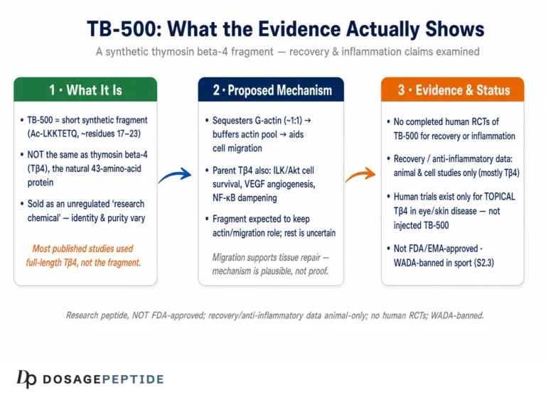
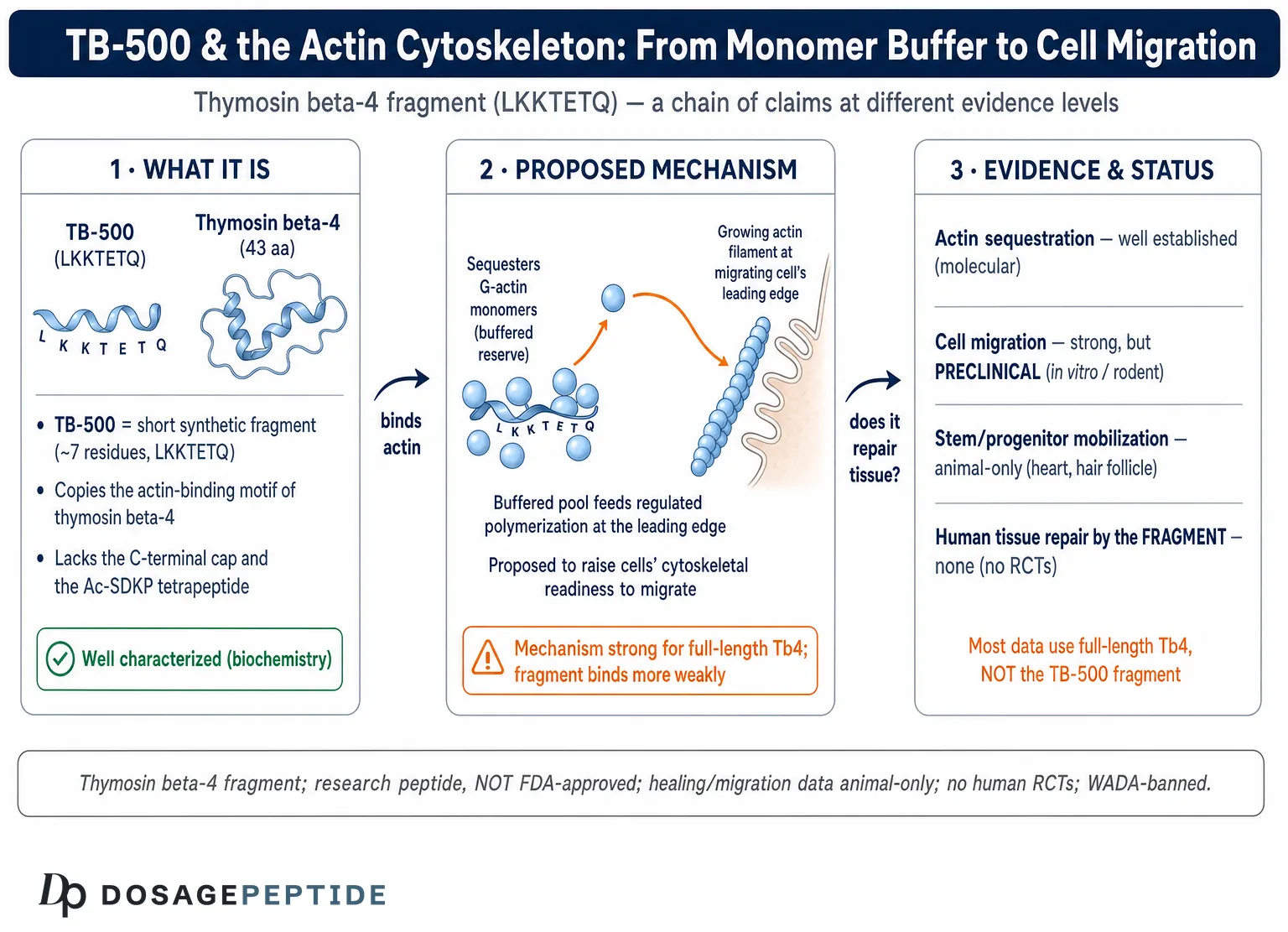
A further honesty note belongs up front. TB-500 is a research peptide. It is not approved by the U.S. Food and Drug Administration, the European Medicines Agency, or any comparable regulator for the treatment, cure, or prevention of any disease. Its healing, migration, and anti-inflammatory data are overwhelmingly animal-based; there are no published randomized controlled trials of the TB-500 fragment in humans for tissue repair. And it is prohibited in sport by the World Anti-Doping Agency at all times. With that framing fixed, we can look closely at the biology — which is legitimately interesting — without overstating what it means.
What TB-500 Actually Is — and Why the Name Matters
Thymosin β4 (Tβ4) is a small, 43-amino-acid, highly acidic peptide found in essentially every mammalian cell type and in high abundance in platelets and wound fluid. It belongs to the β-thymosin family and is, quantitatively, the major G-actin-sequestering molecule in most eukaryotic cells.1 Its job in normal cell physiology is not glamorous: it holds a reserve of unpolymerized actin monomers in a soluble, ready-to-use pool. That single biochemical function turns out to underpin a surprising range of downstream effects — cell movement, wound closure, angiogenesis — which is why Goldstein, Hannappel, and Kleinman once described Tβ4 as an actin-sequestering protein that “moonlights” to repair injured tissue.1
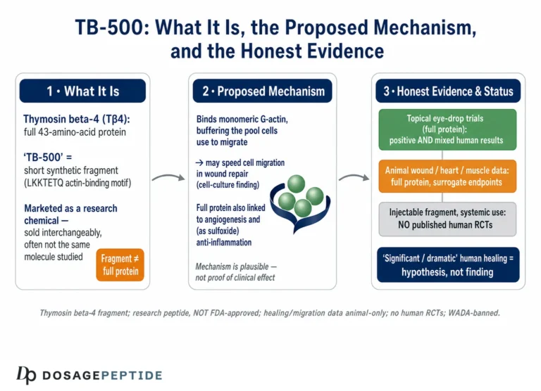
“TB-500,” strictly speaking, is not the same molecule. The name is a product label for a synthetic peptide corresponding to a short segment of Tβ4 — the actin-binding motif built around the sequence LKKTETQ, roughly residues 17–23 of the parent protein. This heptapeptide reproduces the central actin-binding function of Tβ4 but lacks the flanking regions of the full molecule.10 In practice, the research-chemical market uses “TB-500” loosely: some vials contain the short fragment, some contain sequences marketed as an acetylated or otherwise modified analog, and some vendors use “TB-500” and “thymosin β4” interchangeably even when the contents differ. This sloppiness is not trivial. It means that when someone cites a Tβ4 study to justify a claim about TB-500, they may be attributing to a seven-residue fragment the behavior of a 43-residue protein that carries additional functional domains.
Why does the distinction matter for cytoskeletal regulation specifically? Because the actin-binding motif is indeed the core of Tβ4’s monomer-sequestering activity, so the fragment can plausibly reproduce that one function. But the full molecule also harbors an N-terminal tetrapeptide, Ac-SDKP, that is enzymatically cleaved off and carries its own anti-inflammatory and anti-fibrotic activity,11 and other regions implicated in binding partners beyond actin. A fragment stripped down to LKKTETQ keeps the actin story but discards much of the rest. For an honest treatment of “how TB-500 affects cytoskeletal regulation,” the fair statement is: the fragment is designed to reproduce the actin-sequestering mechanism, and most experimental evidence for that mechanism comes from the parent peptide. We will return to this caveat repeatedly, because it is the single most important qualifier on the entire topic. Readers who want the broader repair context can compare this with the site’s discussion of TB-500 in tendon and ligament repair, which examines the same molecule through a musculoskeletal rather than a cell-biology lens.
The Cytoskeleton in Motion: Why Actin Dynamics Govern Cell Migration
To understand how a small actin-binding peptide could influence something as large-scale as tissue repair, it helps to start with why the actin cytoskeleton is the engine of cell movement in the first place. Actin exists in two interconverting forms: globular monomers (G-actin) floating in the cytosol, and filaments (F-actin), the long double-helical polymers that give cells shape and generate mechanical force. Cell migration is, at its core, a spatially organized cycle of building and dismantling these filaments.
A migrating cell — whether a keratinocyte crawling across a wound, an endothelial cell forming a new capillary, or a progenitor cell homing toward injured tissue — extends a flat, actin-rich protrusion called a lamellipodium at its leading edge. That protrusion is driven by rapid actin polymerization: monomers are added to the growing (barbed) ends of filaments pushing against the membrane, while older filament ends are depolymerized at the rear. The cell then forms adhesions to the substrate at the front, generates contractile force through actomyosin, and releases its rear adhesions to move forward. Every step of this cycle depends on the cell having, at the right place and the right time, a supply of polymerization-competent actin monomers.
This is where monomer-binding proteins become rate-limiting. If every actin monomer in the cytosol were free, it would polymerize spontaneously and uncontrollably, wasting the cell’s ability to build filaments where and when they are needed. The cell therefore maintains a large buffered pool of sequestered monomers — actin held in a form that cannot self-assemble but can be released on demand. Thymosin β4 is the principal buffer for that pool.1 Profilin, another monomer-binding protein, works alongside it but does the opposite job: it hands monomers off to growing filament ends. The balance between these two — Tβ4 holding monomers back, profilin feeding them forward — is a central control knob for how fast and where a cell can polymerize actin.12
The conceptual payoff is this: a molecule that changes the size or dynamics of the sequestered-monomer pool is, in principle, positioned to influence how readily cells can remodel their cytoskeleton and migrate. That is the mechanistic seed from which every tissue-repair claim about Tβ4 and TB-500 grows. It is a real and well-understood seed. The open questions are all about how far it germinates in a living organism, and whether the seven-residue fragment does the same job as the whole protein.
| Protein | Binds | Effect on actin dynamics | Role in migration |
|---|---|---|---|
| Thymosin β4 | G-actin (ATP-actin monomer) | Sequesters monomer; blocks polymerization and nucleotide exchange26 | Maintains ready reserve of unpolymerized actin |
| Profilin | G-actin (barbed-end) | Promotes hand-off of monomer to growing filament ends12 | Feeds leading-edge polymerization |
| Cofilin / ADF | F-actin and ADP-actin | Severs and depolymerizes older filaments | Recycles monomers at the trailing edge |
| Arp2/3 complex | Filament sides | Nucleates branched networks | Builds the lamellipodial mesh at the front |
The table makes the division of labor concrete. Tβ4 is not a motor and not a nucleator; it is a reservoir manager. Its influence on migration is indirect but foundational — it sets the availability of the raw material that profilin, cofilin, and the Arp2/3 complex then deploy.
How Thymosin β4 Sequesters G-Actin: the Molecular Mechanics
The mechanism by which Tβ4 holds an actin monomer is now understood at atomic resolution, which is unusual for a peptide of this kind and worth appreciating precisely because it distinguishes established structural biology from the looser claims that circulate downstream. A 2004 crystallographic study by Irobi and colleagues solved the structure of a Tβ4-actin complex (using a stabilized gelsolin-Tβ4 hybrid) at roughly 2 Å resolution and showed how the peptide wraps around the monomer.2
Tβ4 is intrinsically disordered in solution — a floppy chain with little fixed structure — but it folds upon binding actin. It engages the monomer through two contact regions connected by a central helix. An N-terminal segment (containing the LKKTETQ motif that defines the TB-500 fragment) clamps onto one face of the actin monomer near subdomains 1 and 2, while a C-terminal segment reaches across to contact the opposite end of the monomer, near subdomains 3 and 4. The net effect is that Tβ4 caps both ends of the actin monomer — the barbed end and the pointed end — simultaneously.2
Capping both ends is the structural basis for sequestration. A free actin monomer polymerizes by adding to filament ends through exactly these surfaces; if both are occluded, the monomer physically cannot join a filament. Beyond steric blockade, Tβ4 binding also strongly inhibits the exchange of the nucleotide (ATP/ADP) bound in the actin monomer’s cleft, locking the monomer in a stable, storage-competent state.1 The binding is 1:1 and transient rather than permanent — the complex has a modest affinity and a fast off-rate, which is precisely what a buffer needs. A buffer that bound too tightly would trap actin permanently; one that bound too weakly would not hold a reserve. Tβ4 sits in the useful middle.
The release side of the cycle was clarified by later structural work on the Tβ4/profilin exchange. Because the two proteins’ binding footprints on actin overlap only slightly, actin can be shuttled from Tβ4 to profilin and thence onto a growing filament, converting stored monomer into polymer without a large energetic penalty.12 This is the molecular hand-off that turns a static reserve into usable building material at the leading edge.
Two honest qualifications belong here. First, this beautifully resolved mechanism is a property of full-length Tβ4. The N-terminal LKKTETQ region present in TB-500 is necessary for actin binding but, on its own, the isolated heptapeptide binds actin far more weakly than the intact peptide, because the C-terminal contact that caps the second end is missing. In other words, the fragment reproduces part of the clamp, not the whole two-ended cap. How faithfully “TB-500” reproduces the sequestering function therefore depends heavily on exactly which sequence a given vial contains. Second, sequestration is a cell-intrinsic, cytosolic event. Much of the interesting tissue-repair biology attributed to Tβ4 involves the peptide acting extracellularly or being taken up by cells — contexts where the clean crystal-structure story is only part of the picture, and where additional, less well-defined receptor or signaling interactions may be at play.
From Monomer Buffer to Cell Migration
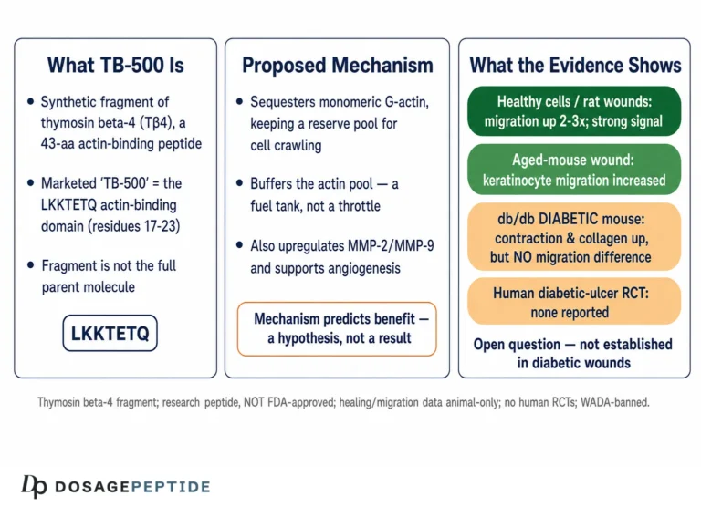
How does managing a monomer reservoir translate into a cell actually moving faster toward a wound? The link was first demonstrated cleanly in the context of dermal wound healing. In a foundational 1999 study, Malinda and colleagues showed that adding Tβ4 — topically or systemically — accelerated re-epithelialization of full-thickness wounds in rats and mice, increasing wound closure by roughly 42% at four days and up to 61% at seven days relative to saline controls.3 Critically, they tied the macroscopic effect to a microscopic one: in a Boyden-chamber assay, Tβ4 stimulated the directional migration of keratinocytes two- to three-fold, with activity detectable at picogram quantities.3
The interpretation offered was mechanistically coherent with the actin biology. By modulating the available monomer pool, Tβ4 helps keratinocytes and fibroblasts at the wound margin reorganize their cytoskeletons and crawl across the defect, and it stimulates endothelial-cell migration that supports new vessel growth.13 Later work extended the migration story to matrix remodeling: Tβ4 was shown to promote the expression of matrix metalloproteinases during wound repair, enzymes that clear a path through the extracellular matrix so that migrating cells can advance.1
There is an apparent paradox worth confronting, because a careful reader will notice it. If Tβ4 sequesters actin — that is, holds it back from polymerizing — why would adding more of it promote migration rather than freeze cells in place? The resolution is that migration is not about maximizing polymerization everywhere; it is about controlling it in space and time. A well-stocked, dynamically exchangeable monomer buffer lets a cell polymerize rapidly and repeatedly at the leading edge while recycling filaments at the rear. Too little buffered monomer, and the cell cannot sustain the cycles of protrusion; the reserve runs dry. Tβ4 supplied to the system appears to enhance the cell’s capacity for this regulated, cyclical remodeling rather than to gum it up. It is the difference between a warehouse that keeps an assembly line supplied and one that hoards inventory — the same stock, deployed differently, produces opposite outcomes. This is also why simple in-vitro dose-response can be non-monotonic and why extrapolating an optimal “dose” from a dish to an organism is fraught.
The migration effect is not limited to skin cells. Endothelial cells, corneal epithelial cells, and various progenitor populations have all shown enhanced motility in the presence of Tβ4 across preclinical assays.1 This breadth is what makes the molecule genuinely interesting as a research tool for studying migration — and also what makes it easy to over-sell, because “promotes cell migration in a dish” is a long way from “heals injuries in patients.”
It is also worth being precise about what “directional” migration means, because the distinction separates plausible biology from wishful thinking. In a Boyden chamber, cells move from an upper well toward a lower well containing a chemoattractant; a molecule that merely increases random cell speed (chemokinesis) is not the same as one that biases movement toward a target (chemotaxis). The wound-healing significance of Tβ4 depends on the latter — cells arriving at the defect, not just moving faster in place. The available assays suggest Tβ4 supports both increased motility and directional bias, which is the combination a repair signal would need.13 Even so, the readout in these experiments is cell arrival across a membrane over hours, not functional restoration of an organ over weeks, and the two should never be conflated. A dish full of well-migrated cells is a hypothesis about repair, not a demonstration of it.
Beyond Actin: the ILK–PINCH–Akt Migration-and-Survival Axis
If Tβ4 only sequestered actin, its effects would be confined to cytoskeletal mechanics. But one of the most cited findings in the field suggests it also plugs into intracellular signaling that couples migration to cell survival — a combination that is unusually well suited to tissue repair, where cells must both move into a hostile injured environment and stay alive once there.
In a 2004 Nature paper, Bock-Marquette and colleagues reported that Tβ4 promotes the migration and survival of cardiac cells and improves outcomes after experimental myocardial infarction in mice.4 The mechanistic core of the paper was that Tβ4 forms a functional complex with the adaptor protein PINCH and integrin-linked kinase (ILK), and that this complex activates the pro-survival kinase Akt (protein kinase B). After coronary artery ligation, Tβ4-treated hearts showed upregulated ILK and Akt activity, enhanced early cardiomyocyte survival, and improved cardiac function.4
This matters conceptually because ILK sits at focal adhesions — the very structures that link the actin cytoskeleton to the extracellular matrix and that a migrating cell must continually assemble and disassemble. A molecule that both manages the actin monomer supply and engages the adhesion-signaling machinery is touching two complementary halves of the migration apparatus, while the Akt arm simultaneously suppresses apoptosis. The elegance of that dual action is a large part of why the 2004 paper generated so much subsequent interest in Tβ4 as a cardiac-repair candidate.
Honesty requires two caveats, though. First, the cardiac-reprogramming and epicardial-regeneration claims that grew out of this line of work became contested; at least one later study reported that Tβ4 treatment after myocardial infarction did not reprogram epicardial cells into new cardiomyocytes, tempering some of the more ambitious regenerative narratives. The field’s enthusiasm outran its reproducibility in places, which is a normal and instructive part of how preclinical biology matures. Second — again — these are full-length Tβ4 experiments in animals. Whether the LKKTETQ fragment engages the ILK/PINCH complex at all is not something these studies establish. The migration-plus-survival mechanism is a Tβ4 property; its attribution to “TB-500” is an inference, not a demonstrated fact.
Stem and Progenitor Cell Migration: What the Animal Data Show
The title’s emphasis on “stem cell migration” points to the most compelling — and most preclinical — part of the story. Several influential animal studies suggest that Tβ4 can mobilize resident stem and progenitor populations from their niches and guide them toward sites of injury, where they contribute to repair through both direct differentiation and paracrine (signal-secreting) effects.
The clearest example comes from the heart. In a 2007 Nature study, Smart and colleagues showed that Tβ4 is required for coronary vessel development and, in the adult, can reactivate quiescent epicardial cells — the outer layer of the heart. Treated adult epicardial explants showed restored progenitor outgrowth and differentiation into endothelial cells, smooth muscle cells, and fibroblasts, with the peptide inducing epicardial progenitor mobilization and neovascularization in the injured adult heart.5 The picture that emerged was of a dormant reservoir of repair-competent cells that Tβ4 could partially reawaken — a striking result, and one that fits the migration mechanism, since mobilizing a cell out of a niche is fundamentally a motility problem.
A second, well-worked example is the hair follicle. Philp and colleagues reported that Tβ4 accelerates hair growth in rats and mice by activating hair-follicle stem cells in the bulge region, increasing their migration and differentiation.6 A follow-up specifically framed the effect as stem-cell migration and differentiation, showing that follicle clonogenic keratinocytes closely related to bulge stem cells migrated and differentiated in response to nanomolar Tβ4, with associated increases in protease production that would help cells move through their surroundings.67 The follicle work is valuable precisely because the bulge is one of the best-defined adult stem-cell niches, making the migration readout relatively clean.
Beyond these, Tβ4 has been reported to increase circulating endothelial progenitor cells and to recruit them to sites of vascular injury and ischemia in animal models, consistent with a role in mobilizing bone-marrow-derived repair cells. The logic connecting sequestration to niche mobilization is that leaving a niche is itself a migration event: a stem cell held in place by its adhesions and local architecture must remodel its cytoskeleton to detach, polarize, and crawl outward. A signal that improves a cell’s cytoskeletal readiness could, in principle, lower the threshold for that exit — which is exactly the behavior the epicardial and follicle studies observed.56 The caveat, once again, is that “mobilization” in an explant or a treated mouse is a controlled experimental readout; whether an injected fragment reproduces it in an intact human body, at a tolerable and specific dose, is entirely unestablished. This vascular-progenitor angle connects tissue repair to angiogenesis, a theme the site explores for other peptides in its overview of KLOW peptides in angiogenesis and tissue repair. The common thread across heart, follicle, and vasculature is that Tβ4’s pro-migratory action is not confined to differentiated cells; it appears to reach into stem and progenitor compartments, which is what makes the “regenerative” label scientifically defensible at the preclinical level.
| Study / model | Cell population | Reported finding | Evidence level |
|---|---|---|---|
| Malinda 1999, rodent dermal wounds3 | Keratinocytes, endothelial cells | 2–3× migration; faster re-epithelialization | Animal + in vitro |
| Bock-Marquette 2004, mouse MI4 | Cardiomyocytes, endothelial cells | ILK/Akt activation; migration + survival; better function | Animal + in vitro |
| Smart 2007, adult mouse epicardium5 | Epicardial progenitors | Progenitor mobilization; neovascularization | Animal + explant |
| Philp 2004/2007, rodent follicle67 | Hair-follicle bulge stem cells | Increased migration, differentiation, hair growth | Animal + in vitro |
| RGN-259 ocular trials89 | Corneal epithelium | Improved healing of epithelial defects (mixed later results) | Human (topical, full Tβ4) |
Reading the table honestly, the pattern is unmistakable: the stem-and-progenitor migration evidence is real, mechanistically coherent, and almost entirely preclinical. The one column with human data is ocular — and, as we will see, even that is mixed and uses full-length Tβ4 as a topical eye drop, not systemic TB-500.
Cytoskeletal Regulation in Tissue Repair: Assembling the Picture
Putting the mechanism together, a plausible and largely evidence-backed narrative of how cytoskeletal regulation feeds tissue repair looks like this. After injury, Tβ4 is released in abundance from platelets and damaged cells into the wound environment. Locally, it acts on resident and recruited cells to support the cytoskeletal remodeling that migration requires, drawing keratinocytes, fibroblasts, endothelial cells, and progenitor populations toward the defect.13 As those cells arrive, complementary activities attributed to the molecule — promotion of angiogenesis, upregulation of matrix-remodeling proteases, support of cell survival via the ILK/Akt axis, and modulation of the inflammatory response — help convert cell arrival into functional repair.14
The anti-inflammatory arm deserves a specific mention because it is mechanistically distinct from the actin story. The N-terminal tetrapeptide Ac-SDKP, cleaved from Tβ4 by enzymes such as prolyl oligopeptidase and meprin-α, has its own anti-inflammatory and anti-fibrotic activity and has been shown to reduce fibrosis in animal models of lung, kidney, and heart injury.11 This is biologically important for “repair” because unchecked fibrosis produces scar rather than functional tissue — but it is also a caution flag for the TB-500 fragment, since a fragment built around LKKTETQ does not contain the N-terminal Ac-SDKP sequence and therefore would not be expected to deliver this anti-fibrotic action at all. The site’s companion piece on TB-500 for chronic inflammatory conditions takes up this inflammation angle in more depth.
The cleanest human-facing test of the repair hypothesis has been in the eye, where a topical formulation of full-length Tβ4 (RGN-259) was developed for corneal wound healing, dry eye, and neurotrophic keratopathy — conditions where accelerated epithelial-cell migration is exactly what is wanted. Early and mid-stage trials were encouraging: a Phase III study in neurotrophic keratopathy reported complete healing of persistent epithelial defects in a majority of treated patients versus a minority on placebo.8 Preclinical dry-eye models likewise showed benefits comparable to prescription drugs.9 But the trajectory has been bumpy: at least one later Phase III dry-eye/keratitis program missed its primary endpoint, with the sponsor attributing the miss partly to an unexpectedly strong placebo response. This mixed record is the reality of translation — a strong mechanism and promising early trials do not guarantee reproducible pivotal results.
The TB-500 Fragment Versus Full Tβ4: a Critical Evidence Caveat
It is time to make explicit the caveat that has shadowed every section. Nearly all of the mechanistic and preclinical evidence discussed — the crystal structures, the wound-healing acceleration, the ILK/Akt signaling, the epicardial and follicle stem-cell mobilization, the ocular trials — was generated with full-length thymosin β4. The compound sold and injected as “TB-500” is generally a short fragment centered on the LKKTETQ actin-binding motif.10
What follows from that? A few things, stated carefully:
- The actin-binding motif is genuinely shared. LKKTETQ is the core of Tβ4’s actin contact, so a fragment containing it can plausibly participate in monomer binding and, by extension, in migration effects. This is the legitimate basis for expecting some overlap in activity.
- But the fragment lacks the full two-ended cap. The C-terminal contact that lets intact Tβ4 cap both ends of the actin monomer is not present in the heptapeptide, so the isolated fragment is a weaker sequesterer than the whole protein.2
- The fragment lacks Ac-SDKP. The N-terminal anti-fibrotic/anti-inflammatory tetrapeptide is simply absent, so the fragment cannot be assumed to reproduce that arm of Tβ4 biology.11
- Direct fragment data are sparse. There is comparatively little rigorous, published, controlled research on the LKKTETQ fragment itself — and essentially no randomized human trials of it for tissue repair. Studies exist characterizing TB-500 and its metabolites in vitro and screening wound-healing activity, but these do not amount to the depth of evidence available for the parent peptide.1013
The practical upshot for a reader trying to think clearly: statements of the form “TB-500 activates stem-cell migration and repairs the heart” are borrowing the reputation of full-length Tβ4 and applying it to a fragment that may reproduce only part of the biology. The honest formulation is that the fragment is designed to reproduce Tβ4’s actin-sequestering function, that this function is plausibly linked to migration, and that direct high-quality evidence for the fragment’s tissue-repair efficacy — especially in humans — is largely absent. This distinction, glossed over almost everywhere in the consumer-facing literature, is the difference between an interesting research peptide and a proven therapy. It is also worth defining these terms precisely; the site’s peptide glossary lays out the vocabulary of fragments, analogs, and sequences used throughout this discussion.
What the Human Evidence Does — and Does Not — Support
Consolidating the human picture is quick, because there is not much of it, and none of it is what the marketing implies.
Full-length Tβ4, topical, ocular: The RGN-259 program represents the most rigorous human testing of the molecule’s repair biology. It produced genuinely randomized, double-masked, placebo-controlled data in neurotrophic keratopathy and dry eye, with some positive results and some endpoint misses.89 This supports the idea that Tβ4 can promote epithelial repair on the ocular surface, but it is indication-specific, uses local delivery to a small avascular tissue, and does not validate systemic tissue repair anywhere else.
Full-length Tβ4, systemic: Early-phase safety and dermal-healing work in humans has been reported — for example, characterization of Tβ4 as a regenerative peptide that accelerated dermal healing in preclinical models and in patients with certain wound types.10 These are small, preliminary, and far short of the pivotal trials that support an approved indication.
The TB-500 fragment, any route: There are no published randomized controlled trials of the LKKTETQ fragment in humans for tendon, ligament, muscle, cardiac, neurological, or general tissue repair. The widespread use of TB-500 in athletic and research-chemical contexts is not backed by human efficacy trials of the fragment itself.1013
A recent scoping review of Tβ4 and TB-500 in tissue healing and musculoskeletal repair reached a conclusion consistent with all of the above: the preclinical signal is broad and encouraging, but the clinical evidence base — particularly for the injectable fragment — is limited and not yet sufficient to support therapeutic claims.13 That is the honest state of the field as of 2026. Anyone interested in how this compares with the recovery-and-inflammation narrative can weigh it against the site’s analysis of whether clinical studies show TB-500 speeds recovery, which reaches similarly measured conclusions.
| Claim | What supports it | Honest evidence level |
|---|---|---|
| Tβ4 sequesters G-actin | Crystal structures, decades of biochemistry12 | Well established (molecular) |
| Tβ4 promotes cell migration | In-vitro migration assays; rodent wounds3 | Strong (preclinical) |
| Tβ4 mobilizes stem/progenitor cells | Epicardial, follicle, EPC animal studies56 | Moderate (animal only) |
| Tβ4 repairs human tissue | Ocular trials (mixed); small dermal data810 | Limited, indication-specific |
| TB-500 fragment repairs human tissue | No randomized human trials of the fragment | Absent |
Research Models, Handling, and Methodology
Understanding how this biology is actually studied clarifies what the data can and cannot bear. The methodology spans three tiers, and each answers a different question.
In vitro biochemistry and structural biology. The actin-sequestration mechanism rests on solution biochemistry (binding stoichiometry, nucleotide-exchange assays) and X-ray crystallography of Tβ4-actin complexes.2 These methods are ideal for establishing how the peptide binds actin but say nothing about tissue-level outcomes. Migration itself is typically measured with Boyden-chamber (transwell) assays and scratch-wound assays, which quantify directional motility of cultured cells — the assays that first linked Tβ4 to keratinocyte movement.3
Animal models. The repair claims come from rodent dermal-wound models, myocardial-infarction models (coronary ligation), corneal-injury models, and follicle/hair-growth models, plus fibrosis models used to characterize Ac-SDKP.34511 These are the gold standard for preclinical proof-of-concept, but rodent healing differs from human healing in important ways (rodents heal partly by contraction; humans by re-epithelialization), and effect sizes rarely transfer intact across species.
Human trials. As covered, meaningful human data are essentially limited to topical full-length Tβ4 in ophthalmology, with mixed pivotal outcomes.89 No validated large-scale human trial framework has been applied to the systemic TB-500 fragment for tissue repair.
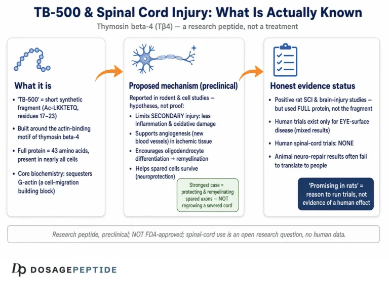
On handling, a brief and strictly educational note: TB-500 is typically supplied as a lyophilized (freeze-dried) powder in a sealed vial and, in research settings, reconstituted with sterile or bacteriostatic water. Standard research-peptide practice directs the diluent slowly against the vial wall and swirls rather than shakes, because vigorous agitation can shear and denature peptides; material is generally kept cold and dark and protected from repeated freeze-thaw cycles. The arithmetic of reconstitution — a fixed mass dissolved in a chosen volume sets the concentration — is the same for any peptide, and the site’s discussion of TB-500 in spinal-cord-injury research illustrates how these parameters are documented in a neurological-repair context. None of this handling detail, it must be said, creates efficacy where controlled evidence is absent; good technique preserves whatever activity the molecule has and nothing more.
Safety, Regulatory Status, and Anti-Doping
The safety and regulatory picture is where clarity matters most, because it is where the gap between research interest and sanctioned use is widest.
Safety. In the human ocular trials, topical full-length Tβ4 was generally well tolerated over the studied durations.8 But that reassurance does not transfer to systemic injection of a fragment sourced outside regulated channels. There are no long-term human safety data for injectable TB-500, no established dosing, and legitimate theoretical concerns worth naming: because the molecule promotes angiogenesis and cell migration, questions have been raised about whether it could, in principle, support the growth or spread of pre-existing malignancy — a concern that is biologically reasonable to flag even though it has not been resolved in humans. Product-quality risk is also real: research-chemical peptides vary in purity and may carry impurities or endotoxin unrelated to the molecule’s intrinsic properties.
Regulatory status. Neither full-length Tβ4 nor the TB-500 fragment is approved as a drug for tissue repair by the FDA, the EMA, or comparable regulators. RGN-259 remains investigational. TB-500 is sold only as a “research chemical,” explicitly not for human use, and marketing it for the treatment of any condition would be unlawful in most jurisdictions.
Anti-doping. This point is unambiguous and important for athletes. The World Anti-Doping Agency prohibits TB-500 at all times, in and out of competition, under the growth-factor provisions of its Prohibited List (Section S2), with related substances also captured under Section S0 (non-approved substances). It is a non-threshold substance: any detectable presence constitutes an anti-doping rule violation, exposing an athlete to multi-year sanctions.14 For anyone subject to WADA-compliant testing, use is a regulatory hazard regardless of the underlying pharmacology.
The synthesis is straightforward. TB-500 occupies the same ambiguous space as many research peptides: a legitimately interesting mechanism, a broad but preclinical evidence base concentrated on the parent molecule, essentially no human trial support for the fragment, no regulatory approval, and an outright ban in sport. For the biology of the actin cytoskeleton it is a valuable probe; for the treatment of injury in people it is, honestly, an open research question rather than an answer. Readers building broader context on healing peptides may find the foundational overview explaining BPC-157 a useful comparison point, since the two compounds are often discussed together and share the same evidentiary limitations.
Frequently Asked Questions
How does TB-500 affect the actin cytoskeleton?
TB-500 is a fragment built around the LKKTETQ actin-binding motif of thymosin β4, the body’s major G-actin-sequestering peptide. The parent molecule binds monomeric (globular) actin one-to-one, capping both ends of the monomer so it cannot polymerize, and holds it in a soluble reserve that cells draw on to build filaments where and when they need them.12 By managing this monomer pool, thymosin β4 influences how readily cells can remodel their cytoskeleton and migrate. The TB-500 fragment is designed to reproduce this actin-binding function, though the isolated heptapeptide binds actin more weakly than the intact protein because it lacks the second, C-terminal contact.
Does TB-500 really cause stem cells to migrate to injured tissue?
In animal models, thymosin β4 has been shown to mobilize resident progenitor populations — adult epicardial progenitors in the heart and bulge stem cells in hair follicles — and to recruit endothelial progenitor cells toward vascular injury.56 This is a genuine and mechanistically coherent preclinical finding. However, it was demonstrated with full-length thymosin β4 in animals, not with the injectable TB-500 fragment in humans, and no randomized human trials confirm a stem-cell-migration benefit for the fragment.
Is TB-500 the same thing as thymosin β4?
Not exactly. Thymosin β4 is the full 43-amino-acid natural peptide; TB-500 is generally a short synthetic fragment corresponding to its actin-binding region.10 The fragment reproduces the core actin-binding motif but lacks other functional parts of the parent molecule — including the N-terminal Ac-SDKP tetrapeptide responsible for much of Tβ4’s anti-fibrotic and anti-inflammatory activity.11 Much marketing conflates the two, which leads to the fragment being credited with the parent molecule’s full body of evidence.
Is there any human evidence that TB-500 repairs tissue?
For the TB-500 fragment specifically, no — there are no published randomized controlled trials in humans for tissue repair. The strongest human data involve topical full-length thymosin β4 (RGN-259) for corneal and ocular-surface healing, and even those pivotal trials produced mixed results.89 A 2026 scoping review concluded that the clinical evidence base, especially for the injectable form, remains limited.13
Why would a molecule that “sequesters” actin help cells move?
It seems paradoxical, but migration is about controlling actin polymerization in space and time, not maximizing it everywhere. A well-stocked, dynamically exchangeable pool of monomers lets a cell polymerize rapidly at its leading edge while recycling filaments at the rear. Thymosin β4 manages that reserve, and supplying it appears to enhance a cell’s capacity for the cyclical remodeling that migration requires rather than freezing the cytoskeleton in place.13
What is the ILK/Akt pathway and why is it relevant?
Beyond actin binding, thymosin β4 was reported to form a complex with PINCH and integrin-linked kinase (ILK) and to activate the survival kinase Akt, improving both migration and survival of cardiac cells after experimental infarction in mice.4 This links cytoskeletal remodeling to cell-survival signaling — a useful combination for repair — but it is an animal finding for full-length Tβ4, and some downstream regenerative claims from this line of work were later contested.
Is TB-500 approved or legal to use?
It is not approved as a drug for any condition by the FDA, EMA, or other major regulators. It is sold only as a research chemical, not for human use, and it is prohibited in sport at all times by the World Anti-Doping Agency under the growth-factor and non-approved-substance provisions of the Prohibited List, with no permitted threshold.14
Are there safety concerns with TB-500?
Topical full-length Tβ4 was generally well tolerated in ocular trials, but there are no long-term human safety data for injectable TB-500.8 Because the molecule promotes angiogenesis and cell migration, a theoretical concern has been raised about whether it could support growth of pre-existing tumors, and unregulated research-chemical material varies in purity. Absence of demonstrated harm in a narrow setting is not the same as established safety for systemic use.
Could TB-500 become an approved regenerative therapy someday?
It cannot be ruled out — the mechanism is real and the preclinical migration data are genuine — but it would require dedicated, controlled human trials of the actual fragment, with functional repair endpoints, run under regulatory oversight. Given how often mechanistically promising repair agents fail in pivotal trials (the mixed ocular results are a case in point), a fragment starting from essentially no human efficacy data has a long road ahead.13
References
- Goldstein AL, Hannappel E, Kleinman HK. Thymosin β4: actin-sequestering protein moonlights to repair injured tissues. Trends Mol Med. 2005;11(9):421-429. PMID: 16099219. https://pubmed.ncbi.nlm.nih.gov/16099219/
- Irobi E, Aguda AH, Larsson M, et al. Structural basis of actin sequestration by thymosin-β4: implications for WH2 proteins. EMBO J. 2004;23(18):3599-3608. PMID: 15329672. PMCID: PMC517612. https://pmc.ncbi.nlm.nih.gov/articles/PMC517612/
- Malinda KM, Sidhu GS, Mani H, et al. Thymosin beta4 accelerates wound healing. J Invest Dermatol. 1999;113(3):364-368. PMID: 10469335. https://pubmed.ncbi.nlm.nih.gov/10469335/
- Bock-Marquette I, Saxena A, White MD, et al. Thymosin β4 activates integrin-linked kinase and promotes cardiac cell migration, survival and cardiac repair. Nature. 2004;432(7016):466-472. PMID: 15565145. https://pubmed.ncbi.nlm.nih.gov/15565145/
- Smart N, Risebro CA, Melville AAD, et al. Thymosin β4 induces adult epicardial progenitor mobilization and neovascularization. Nature. 2007;445(7124):177-182. PMID: 17108969. https://pubmed.ncbi.nlm.nih.gov/17108969/
- Philp D, Nguyen M, Scheremeta B, et al. Thymosin β4 increases hair growth by activation of hair follicle stem cells. FASEB J. 2004;18(2):385-387. PMID: 14657002. https://pubmed.ncbi.nlm.nih.gov/14657002/
- Philp D, Kleinman HK. Animal studies with thymosin β4, a multifunctional tissue repair and regeneration peptide (thymosin β4 induces hair growth via stem cell migration and differentiation). Ann N Y Acad Sci. 2007;1112:95-103. PMID: 17947589. https://pubmed.ncbi.nlm.nih.gov/17947589/
- Sosne G, Dunn SP, Kim C. 0.1% RGN-259 (Thymosin β4) ophthalmic solution promotes healing and improves comfort in neurotrophic keratopathy patients in a randomized, placebo-controlled, double-masked Phase III clinical trial. Int J Mol Sci. 2022. PMCID: PMC9820614. https://pmc.ncbi.nlm.nih.gov/articles/PMC9820614/
- Kim CE, Kleinman HK, Sosne G, et al. RGN-259 (thymosin β4) improves clinically important dry eye efficacies in comparison with prescription drugs in a dry eye model. Sci Rep. 2018;8:10500. PMCID: PMC6043477. https://pmc.ncbi.nlm.nih.gov/articles/PMC6043477/
- Treadwell T, Kleinman HK, Crockford D, et al. The regenerative peptide thymosin β4 accelerates the rate of dermal healing in preclinical animal models and in patients. Ann N Y Acad Sci. 2012;1270:37-44. https://onlinelibrary.wiley.com/doi/10.1111/j.1749-6632.2012.06717.x
- Conte E, Fagone E, Gili E, et al. Preventive and therapeutic effects of thymosin β4 N-terminal fragment Ac-SDKP in the bleomycin model of pulmonary fibrosis. Oncotarget. 2016;7(23):33841-33854. PMCID: PMC5085123. https://pmc.ncbi.nlm.nih.gov/articles/PMC5085123/
- Xue B, Leyrat C, Grimes JM, Robinson RC. Structural basis of thymosin-β4/profilin exchange leading to actin filament polymerization. Proc Natl Acad Sci U S A. 2014;111(43):E4596-E4605. PMID: 25313062. https://www.pnas.org/doi/10.1073/pnas.1412271111
- Thymosin beta-4 and TB-500 in tissue healing, regeneration, and musculoskeletal repair: a scoping review. Appl Sci. 2026;16(12):6202. https://www.mdpi.com/2076-3417/16/12/6202
- Banned Substances Control Group. TB-500 — status, risks, and bans in sport and military (WADA Prohibited List S0/S2 classification). https://www.bscg.org/blogs/single/tb-500-status-risks-and-bans-in-sport-and-military
Educational and research-use disclaimer: This article is provided solely for scientific and educational purposes. TB-500 is a research peptide fragment of thymosin β4 and is not approved by the FDA, EMA, or any comparable regulator for the treatment, cure, or prevention of any disease, including any use in tissue repair, wound healing, or regeneration. The cytoskeletal, migration, stem-cell, and healing data described here are overwhelmingly from cell-culture and animal studies, generated largely with full-length thymosin β4 rather than the TB-500 fragment, and no randomized controlled human trials support the fragment’s efficacy for tissue repair. TB-500 is prohibited in sport at all times by the World Anti-Doping Agency. Nothing here is medical advice or a recommendation for human use. Any legitimate investigation of this compound should occur within properly authorized preclinical or clinical research under appropriate oversight, and readers should consult qualified professionals and applicable regulations before making any decisions.