The title of this article states a relationship as though it were settled fact: that MOTS-C regulates AMPK activity when a cell is under energy stress. Before building anything on that foundation, it is worth pausing on what “regulates” is doing in that sentence. In the peptide-research literature, MOTS-C and AMP-activated protein kinase (AMPK) are indeed tightly linked — arguably MOTS-C’s connection to AMPK is the single best-characterized part of its biology.1 But the strength of that link, the direction of causality, the exact molecular steps, and above all the level of evidence behind each claim vary enormously depending on which experiment you are looking at. Some of it rests on clean genetic and pharmacological work in cells and mice; some of it is inference; and the human data are thin and mostly observational. An honest treatment has to hold all of that in view at once.
So this piece does not simply assert that MOTS-C flips an AMPK switch. It asks, mechanistically and skeptically, how a 16-amino-acid peptide encoded inside the mitochondrial genome could plausibly change the activity of the cell’s master energy sensor, what the primary data actually demonstrate, and where the story shades from established biology into hypothesis. The reader I have in mind is a researcher or a scientifically literate person who wants the mechanism laid out in detail but is tired of vendor copy that treats every mouse result as a promise. MOTS-C is not an approved drug for any condition, it is prohibited in sport, and essentially all of the AMPK mechanism described below was worked out in cell culture and rodents.9 Keeping that frame in mind is not a disclaimer to skim past; it is the correct scientific posture toward a molecule whose therapeutic story is still almost entirely preclinical.
AMPK: What the Cell’s Energy Sensor Actually Does
To understand how MOTS-C might regulate AMPK, you first need a clear picture of what AMPK is and how it is normally switched on, because the peptide’s proposed mechanism only makes sense against that background. AMPK is a heterotrimeric enzyme: a catalytic α subunit that carries the kinase activity, plus regulatory β and γ subunits.2 The γ subunit contains binding sites for adenine nucleotides, and this is the heart of the sensing mechanism. When a cell is well fed and energy is abundant, those sites are largely occupied by ATP. When energy is consumed faster than it is regenerated — during exercise, nutrient withdrawal, hypoxia, or any metabolic insult that drains the ATP pool — ATP is progressively replaced by ADP and AMP. The rising AMP:ATP and ADP:ATP ratios are the signal.3
Binding of AMP (and, more weakly, ADP) to the γ subunit produces a coordinated, three-part activation. First, it allosterically stimulates the kinase directly. Second, it promotes phosphorylation of a critical threonine residue — Thr172 — in the activation loop of the α subunit. Third, it protects that phosphorylated Thr172 from being removed by protein phosphatases.2 The net result is that a small shift in energy charge is amplified into a large change in kinase activity. Phosphorylation of Thr172 is the molecular event most researchers actually measure when they say “AMPK was activated,” and it is the readout that recurs throughout the MOTS-C literature.
Thr172 is placed there by upstream kinases, principally the tumor-suppressor kinase LKB1 (in complex with STRAD and MO25) and, in a parallel pathway, the calcium/calmodulin-dependent kinase CaMKK2, which responds to rising intracellular calcium rather than to the AMP:ATP ratio.3 This detail matters for the MOTS-C question because it means “activating AMPK” can be achieved by more than one route: by genuinely lowering the energy charge (which is what AMP mimetics do), by raising calcium (the CaMKK2 route), or by any intervention that tilts the balance of Thr172 phosphorylation versus dephosphorylation. As we will see, MOTS-C is thought to act largely through the first of these — by generating a signal that mimics energy deficit — rather than by touching the AMPK enzyme itself.
Once activated, AMPK behaves as a master regulator that restores energy balance by switching off ATP-consuming (anabolic) processes and switching on ATP-generating (catabolic) ones.2 It phosphorylates acetyl-CoA carboxylase (ACC1 and ACC2) to relieve the block on fatty-acid oxidation; it promotes glucose uptake by driving translocation of the GLUT4 transporter; it inhibits fatty-acid and cholesterol synthesis; it restrains mTORC1 to slow protein synthesis and cell growth; and it stimulates mitochondrial biogenesis and mitophagy, in part through the transcriptional coactivator PGC-1α and through autophagy machinery such as ULK1.2 In other words, AMPK is not a niche enzyme but a hub that reprograms cellular metabolism toward survival under stress. Any molecule that can reliably nudge AMPK is, by extension, a molecule that can reach a very large downstream network — which is precisely why the MOTS-C–AMPK link attracts so much attention, and also why extraordinary claims about it deserve careful scrutiny.
What MOTS-C Is and Where It Comes From
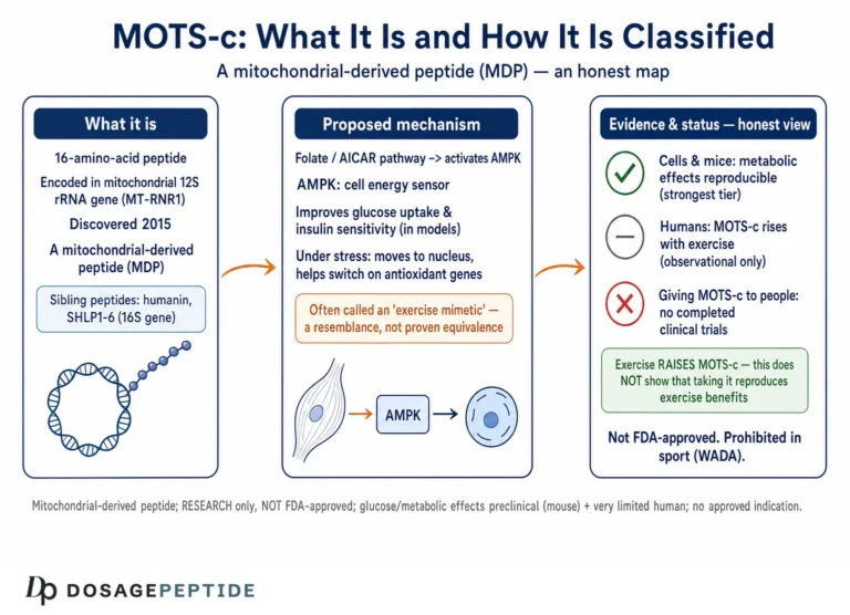
MOTS-C (Mitochondrial ORF of the Twelve-S rRNA type-c) is a peptide of 16 amino acids encoded not in the nuclear genome, as almost every other human peptide is, but within a short open reading frame in the mitochondrial 12S ribosomal RNA gene.1 It belongs to a small and unusual family called mitochondrial-derived peptides (MDPs), which also includes humanin and the SHLP series. The discovery that the mitochondrial genome — long regarded as encoding only a handful of respiratory-chain components and its own translation apparatus — also produces bioactive signaling peptides reframed the mitochondrion as an active endocrine and signaling organelle rather than a passive powerhouse. For readers who want the taxonomy of these molecules, the site’s overview of what MOTS-C is and how it is classified within mitochondrial-derived peptides lays out the family in more detail.
The functional logic of an MDP is elegant. Mitochondria are the cell’s primary sensors of energetic and oxidative state; they are where the AMP:ATP ratio is ultimately set by the rate of oxidative phosphorylation. A peptide encoded by the mitochondrial genome is, in principle, perfectly positioned to report on that state to the rest of the cell — a form of “retrograde” signaling from mitochondrion to nucleus and cytosol. MOTS-C appears to be exactly such a messenger: its expression and its subcellular location change in response to metabolic stress, and its best-documented effect is to engage the AMPK pathway that governs the cellular response to that stress.15
It is important to distinguish two versions of MOTS-C that are easily conflated. There is endogenous MOTS-C — the peptide your own mitochondria produce, present in tissues and circulating in plasma, whose levels rise with exercise and fall with age.7 And there is exogenous, synthetically manufactured MOTS-C, administered in the laboratory at pharmacological doses far above physiological concentrations. Much of the mechanistic work injects or applies synthetic peptide, which tells us what MOTS-C can do at high exposure but not necessarily what the endogenous peptide does at its natural levels. This distinction runs through the entire evidence base and is one of the main reasons for interpretive caution: demonstrating that a bolus of synthetic peptide activates AMPK in a mouse is not the same as demonstrating that endogenous MOTS-C is a physiologically decisive regulator of AMPK in humans.
The Core Mechanism: Folate Cycle, AICAR, and AMPK
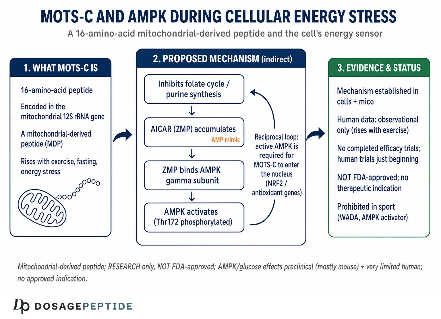
The central mechanistic claim — and the one with the most direct primary support — is that MOTS-C activates AMPK indirectly, by interfering with a specific branch of one-carbon (folate) metabolism. This is worth unpacking slowly, because the popular shorthand “MOTS-C activates AMPK” hides a genuinely interesting biochemical route.
In the original 2015 characterization, MOTS-C was shown to target the folate cycle and its tethered pathway of de novo purine biosynthesis.1 The folate cycle supplies one-carbon units for building purine rings. When MOTS-C inhibits this pathway, the consequence is not simply less purine synthesis; it is the accumulation of an intermediate. That intermediate is AICAR — 5-aminoimidazole-4-carboxamide ribonucleotide — whose monophosphorylated form (ZMP) is a well-known AMP mimetic. ZMP binds the AMPK γ subunit at the same nucleotide sites that AMP uses, and thereby triggers the same allosteric activation and Thr172-protecting effects that a genuine rise in AMP would.15 In effect, MOTS-C manufactures an internal AMP-mimicking signal by damming the folate pathway upstream of AICAR’s conversion, letting the intermediate pile up until it activates the energy sensor.
It is worth being precise about why AICAR is the pivotal molecule here. AICAR ribonucleotide (ZMP) is structurally similar enough to AMP that it occupies the same regulatory nucleotide sites on the AMPK γ subunit, yet it is metabolically “stuck” — it does not participate in the adenylate energy charge the way AMP does, so its accumulation reports a biosynthetic bottleneck rather than a true fall in ATP. In this sense MOTS-C exploits a quirk of purine metabolism: it converts a folate-pathway perturbation into a signal that the energy sensor reads as if ATP were running low, even when the adenylate pool itself is intact. This decoupling of the AMPK signal from the actual energy charge is what allows MOTS-C to act as a conditional amplifier of stress signaling rather than a mere reporter of ATP depletion. This “folate–AICAR–AMPK axis” is now the standard description of MOTS-C’s metabolic mechanism in the review literature.56 Several features make it satisfying as an explanation. It connects a mitochondrially encoded peptide to a cytosolic metabolic pathway to the canonical energy sensor in a chain of concrete biochemical steps, rather than invoking a vague “MOTS-C receptor.” It explains why MOTS-C’s effects overlap so heavily with those of AICAR and metformin, both of which converge on AMPK. And it fits the metabolic-stress framing: a peptide that mimics energy deficit is exactly what you would want a mitochondrial stress messenger to do.
But intellectual honesty requires flagging what remains unresolved. The precise enzymatic target of MOTS-C within the folate/purine pathway, the stoichiometry of the interaction, and whether the peptide acts by direct binding to an enzyme or through some intermediary have not been resolved with the rigor one would want. The AICAR-accumulation model is strongly supported by metabolomic and functional data, but MOTS-C almost certainly does more than this one thing — it also has direct nuclear actions (discussed below) that are AMPK-dependent rather than AMPK-causing, and the two roles can be hard to disentangle. The correct summary is that the folate–AICAR–AMPK route is the best-evidenced mechanism by which MOTS-C raises AMPK activity, not that it is the whole and final story.
There is also a conceptual subtlety worth naming. Because MOTS-C works by generating an AMP-mimetic signal rather than by lowering the true energy charge, it can activate AMPK without the cell actually being in an energy crisis. This is what makes it interesting as a candidate “exercise mimetic” — it engages the stress-response program in the absence of the stress. It is also a reason for caution: chronically forcing a survival program on cells that are not actually starving is not self-evidently benign, and the long-term consequences of pharmacological AMPK activation via this route in whole animals, let alone humans, are not well mapped.
The Reciprocal Loop: AMPK-Dependent Nuclear Translocation of MOTS-C
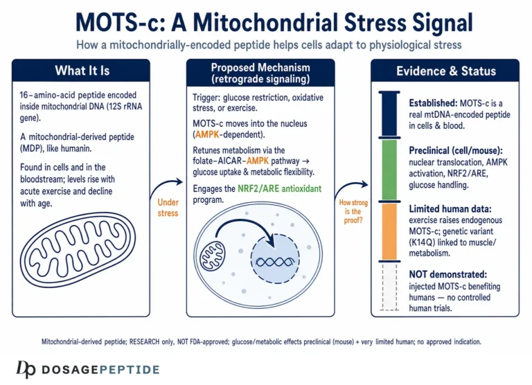
The relationship between MOTS-C and AMPK is not a one-way street, and this is one of the most important and most often oversimplified parts of the mechanism. A landmark 2018 study showed that MOTS-C, though normally cytosolic and mitochondrial, translocates to the nucleus in response to metabolic stress — and that this translocation itself requires AMPK activity.4
The experiments were clean. Different metabolic stressors — glucose restriction, serum deprivation, and oxidative-stress-inducing agents — transiently drove MOTS-C into the nucleus. When AMPK was inhibited pharmacologically or its α subunit was knocked down, stress-induced nuclear accumulation of MOTS-C was blocked.4 So AMPK sits both downstream and upstream of MOTS-C depending on the vantage point: MOTS-C helps activate AMPK through the folate–AICAR route, and activated AMPK is in turn required to route MOTS-C into the nucleus where it exerts a second class of effects. This is a feed-forward loop, not a linear pathway, and it is the reason simplistic “MOTS-C → AMPK” arrows on vendor infographics are misleading.
What does MOTS-C do once inside the nucleus? It regulates a broad set of stress-adaptive genes, and it does so in part by interacting with stress-responsive transcription factors — most notably NRF2 (NFE2L2), the master regulator of the antioxidant response.4 Under glucose restriction or oxidative challenge, MOTS-C bound to NRF2 and to the antioxidant response elements (AREs) of target genes, and its presence enhanced NRF2’s binding to those genes, boosting transcription of cytoprotective enzymes such as NQO1 and HO-1.4 Cells overexpressing wild-type MOTS-C were protected from glucose and serum deprivation, whereas mutants that could not enter the nucleus lost that protection — direct evidence that the nuclear, AMPK-dependent arm is functionally important, not incidental.
The upshot for the title’s question is nuanced. MOTS-C does not merely “turn on” AMPK and stop there. It participates in an integrated stress circuit in which AMPK activation, MOTS-C nuclear entry, and NRF2-driven antioxidant transcription reinforce one another during energy and oxidative stress. That is a richer and more defensible picture than the flat claim that MOTS-C is an AMPK agonist, and it is the picture the primary literature actually supports.45 Readers exploring how MOTS-C behaves under different physiological demands may find the site’s discussion of the function of MOTS-C in metabolic adaptation to physiological stress a useful complement to the molecular detail here.
Retrograde Signaling: Why a Mitochondrial Peptide Talks to AMPK at All
Step back from the biochemistry for a moment and ask the teleological question: why would the mitochondrial genome encode a peptide whose job is to engage AMPK? The answer illuminates the whole mechanism and explains why the MOTS-C–AMPK coupling is specifically a stress phenomenon rather than a housekeeping one.
Cells face a coordination problem. The vast majority of proteins that build and run mitochondria are encoded in the nucleus, yet the moment-to-moment energetic and redox state of the cell is set inside the mitochondrion, at the respiratory chain. For metabolism to adapt coherently, the nucleus needs continuous information about mitochondrial status — a flow of signals conventionally called retrograde (mitochondrion-to-nucleus) signaling, to distinguish it from the anterograde (nucleus-to-mitochondrion) control of mitochondrial gene expression. Classical retrograde signals include reactive oxygen species, calcium fluxes, and precisely the adenine-nucleotide ratios that AMPK reads.2 A mitochondrially encoded peptide that both promotes AMPK activation and, once AMPK fires, travels to the nucleus to shape transcription is a near-ideal retrograde messenger: it originates at the site of energy sensing and terminates at the site of adaptive gene expression.45
This framing does two things. It makes the MOTS-C–AMPK relationship intelligible as physiology rather than pharmacology — MOTS-C looks like a component of the cell’s built-in stress-response wiring, deployed when energy or redox balance is threatened. And it clarifies why the folate–AICAR route is such a fitting mechanism: by converting a metabolic-pathway perturbation into an AMP-mimetic signal, MOTS-C effectively translates “mitochondrial one-carbon metabolism is under strain” into the universal language of energy deficit that AMPK already understands. The peptide does not invent a new signal; it feeds into the oldest and most conserved energy-sensing system the cell has.3
The conserved nature of both partners reinforces the point. AMPK and its upstream activators are ancient, present from yeast to humans, precisely because energy sensing is non-negotiable for life. Mitochondrial-derived peptides, though discovered only recently, appear to be an evolutionarily old layer of this same regulatory fabric.11 Whether endogenous MOTS-C is a dominant retrograde regulator in humans or one modest input among many is exactly the question the current evidence cannot yet answer — but the architecture makes the hypothesis biologically reasonable rather than fanciful.
Downstream of AMPK: What Activation Actually Changes
Granting that MOTS-C can raise AMPK activity in cells and rodents, what follows metabolically? The reported downstream effects track closely with the canonical AMPK program, which is one reason the AMPK-centric interpretation is credible: MOTS-C’s metabolic phenotype looks like an AMPK phenotype.
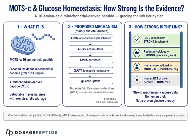
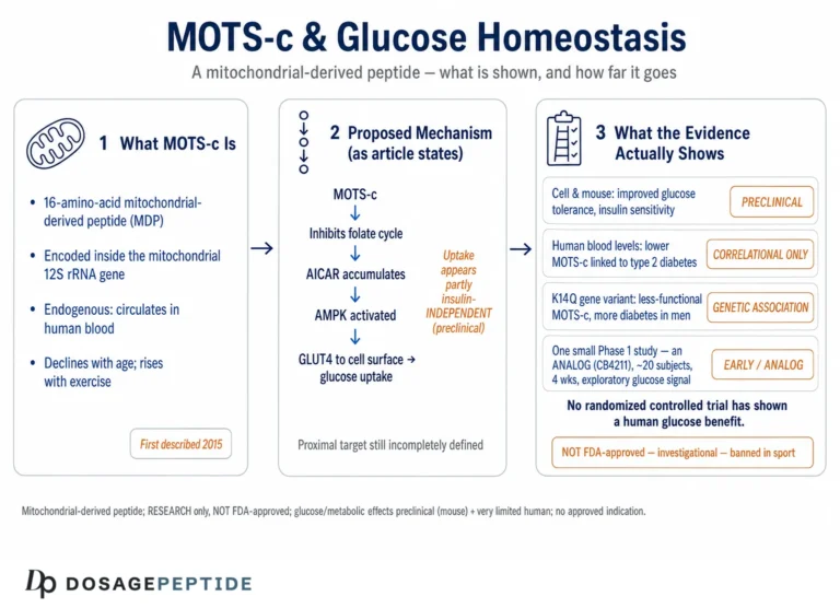
The most consistently reported effect is enhanced glucose disposal. In skeletal muscle — identified early as MOTS-C’s primary target tissue — the peptide promotes glucose uptake in a manner consistent with AMPK-driven GLUT4 mobilization, and it improves insulin sensitivity in models of diet-induced and age-related insulin resistance.16 A metabolomic study in obese mice found that just a few days of MOTS-C treatment lowered glucose and insulin and shifted plasma metabolites — sphingolipids, monoacylglycerols, dicarboxylates — in directions consistent with improved fatty-acid oxidation and reduced insulin resistance.8 These changes are exactly what AMPK activation, acting through ACC inhibition and increased fat oxidation, would be expected to produce.
Beyond glucose handling, AMPK activation by MOTS-C has been linked to increased fatty-acid oxidation, restrained lipogenesis, stimulation of mitochondrial biogenesis via PGC-1α, and enhanced cellular stress resistance through the NRF2 arm described above.45 In aggregate these effects sketch a molecule that pushes cells toward oxidative, catabolic, stress-resilient metabolism — the same direction exercise and caloric restriction push them, which is the basis for the “exercise-mimetic” framing. The site’s review of the evidence that MOTS-C modulates glucose homeostasis in various conditions collects the glucose-specific findings in one place.
Two cautions temper this tidy narrative. First, most of these downstream endpoints were measured in cells and mice, often with supraphysiological peptide doses; the leap to human physiology is unproven. Second, showing that MOTS-C changes a downstream marker (say, phospho-ACC or glucose uptake) and that the change is blocked by AMPK inhibition establishes AMPK-dependence, but it does not always establish that the folate–AICAR route is the sole upstream trigger; alternative or parallel mechanisms cannot be excluded from such experiments alone. The honest reading is that MOTS-C reproduces the AMPK downstream program robustly in preclinical models, and that the weight of evidence favors AMPK as the central node — while acknowledging that mechanistic completeness is still a work in progress. A further caveat is scale: the metabolic improvements reported in rodents were typically produced by repeated administration of synthetic peptide over days to weeks, and the effect sizes, while real within those experiments, were measured against the exaggerated metabolic dysfunction of diet-induced or aged mouse models rather than against a healthy human baseline. None of this establishes that a person taking MOTS-C would see comparable glucose or lipid changes, and it is precisely that unbridged gap — from a strong rodent signal to an unproven human effect — that the phrase “preclinical only” is meant to keep in view.
| Step | Molecular event | Evidence context |
|---|---|---|
| 1. Trigger | Metabolic/energy stress (exercise, fasting, glucose restriction, oxidative stress) raises MOTS-C expression and activity | Cell + mouse; human observational for exercise47 |
| 2. Folate-cycle inhibition | MOTS-C interferes with the folate cycle and de novo purine biosynthesis | Cell + mouse metabolomics1 |
| 3. AICAR/ZMP accumulation | Blocked pathway causes AICAR to build up; its monophosphate mimics AMP | Cell + mouse15 |
| 4. AMPK activation | ZMP binds AMPK γ subunit; allosteric activation + Thr172 phosphorylation/protection | Cell + mouse12 |
| 5. Nuclear translocation | Active AMPK is required for MOTS-C to enter the nucleus under stress | Cell4 |
| 6. Transcriptional response | Nuclear MOTS-C co-regulates NRF2/ARE genes (NQO1, HO-1); antioxidant + adaptive program | Cell4 |
| 7. Metabolic output | ↑ glucose uptake (GLUT4), ↑ fatty-acid oxidation, ↑ mitochondrial biogenesis, ↑ stress resistance | Mostly mouse168 |
Energy Stress in Context: Exercise, Fasting, and the Endogenous Signal
The title specifies “during cellular energy stress,” and this qualifier is doing real work, because MOTS-C’s coupling to AMPK is most evident precisely under those conditions. The peptide is not a constitutive metabolic switch that runs at full tilt all the time; its expression, its subcellular location, and its downstream engagement of AMPK are all stress-responsive.45
Exercise is the cleanest physiological example, and it is also where the only meaningful human data live. In a 2021 study, acute exercise induced endogenous MOTS-C expression in human skeletal muscle and raised circulating levels, positioning MOTS-C as an exercise-responsive peptide that rises exactly when muscle energy demand and AMPK activity spike.7 The same work showed, in mice, that MOTS-C is required for normal exercise capacity and that supplementing it in aged animals — beginning late in life, three times weekly — improved grip strength, gait, and treadmill endurance.7 The parsimonious interpretation is that endogenous MOTS-C is part of the muscle’s adaptive response to the energy stress of contraction, operating alongside the AMPK signaling that exercise classically activates.
Fasting, caloric restriction, and glucose withdrawal produce analogous effects at the cellular level: these are the very stressors that drive MOTS-C into the nucleus and engage the AMPK-dependent, NRF2-linked adaptive program.4 Oxidative stress does the same, consistent with MOTS-C’s role in inducing antioxidant genes. The recurring theme is that MOTS-C behaves like a stress-conditional amplifier of the energy-sensing response rather than a standalone effector — it shows up when the cell is being challenged and helps tune the AMPK-centered reaction to that challenge.
This context also explains a genuine tension in interpreting the literature. Endogenous MOTS-C rises with AMPK-activating stress, which makes it hard, from correlational human data alone, to say whether MOTS-C is driving AMPK, riding along with it, or both. The causal, MOTS-C-drives-AMPK claim rests on the interventional cell and mouse experiments — knockdowns, overexpression, folate-pathway metabolomics, AMPK-inhibitor blockade — not on the human exercise correlations.147 Conflating the two kinds of evidence is one of the most common errors in secondary write-ups, and it inflates the apparent strength of the human case.
How Strong Is the Evidence, Really?
Because the entire point of an honest treatment is to grade the evidence rather than assert conclusions, it is worth being explicit about what tier of proof supports each part of the MOTS-C–AMPK story. The mechanism is unusually well-developed for a research peptide, but “well-developed” is relative, and almost all of it is preclinical.
| Claim | Highest-quality evidence | Honest confidence |
|---|---|---|
| MOTS-C activates AMPK (Thr172) in cells | Cell culture, multiple labs14 | Reasonably strong (in vitro) |
| Mechanism is folate-cycle inhibition → AICAR → AMPK | Metabolomics + functional studies in cells/mice15 | Best-supported model; details incomplete |
| Nuclear translocation is AMPK-dependent | Mechanistic cell study with AMPK knockdown/inhibition4 | Strong within its model system |
| Improves insulin sensitivity / glucose handling | Multiple mouse studies168 | Consistent in rodents; not established in humans |
| Rises with exercise in humans | Human observational (muscle + plasma)7 | Real but correlational, small cohorts |
| Injected MOTS-C improves human metabolic outcomes | Registered/early-stage clinical work only10 | Not established; no approved use |
Several structural limitations cut across the whole field. Dosing is the first: mechanistic studies routinely use synthetic MOTS-C at concentrations that dwarf endogenous levels, so they define the peptide’s capacity to move AMPK, not its physiological contribution. Species is the second: the interventional causal chain lives in mouse and cell models, and metabolic peptides have a long history of impressive rodent results that shrink or vanish in humans. Measurement is the third: circulating MOTS-C assays vary in specificity, and comparing absolute levels across studies is fraught. And publication and commercial pressures are the fourth: MOTS-C is sold as a “research chemical,” and a great deal of confident online mechanism writing traces to vendors rather than to primary data.
None of this means the AMPK mechanism is wrong. On the contrary, the folate–AICAR–AMPK model is coherent, repeatedly reproduced in its core features, and mechanistically specific in a way that many peptide claims are not.145 It means the correct claim is bounded: in cells and in mice, MOTS-C activates AMPK during energy stress through a folate-cycle/AICAR route, and AMPK in turn gates MOTS-C’s nuclear, antioxidant actions. Extending that sentence to “MOTS-C regulates human energy metabolism” without the qualifiers is where honesty is lost. For a broader appraisal of how firmly the metabolic claims hold up, the site’s analysis of how strongly evidence links MOTS-C with glucose-homeostasis control applies the same grading discipline to the glucose endpoints.
MOTS-C Versus Direct AMPK Activators
One useful way to locate MOTS-C is to compare it with the pharmacological AMPK activators whose biology is far better understood. The comparison is instructive not because MOTS-C competes clinically with these agents — it does not, having no approved use — but because it clarifies what kind of AMPK activator MOTS-C is and how much less is known about it.
| Agent | How it engages AMPK | Evidence / status |
|---|---|---|
| Metformin | Indirect: inhibits mitochondrial complex I, raises AMP:ADP, plus AMPK-independent actions | Approved drug, decades of human data (type 2 diabetes) |
| AICAR (acadesine) | Direct AMP mimetic: its monophosphate (ZMP) binds the AMPK γ subunit | Long-used research tool; investigational; prohibited in sport |
| A-769662 / MK-8722 (direct activators) | Bind the AMPK β-subunit ADaM site, activate the enzyme directly | Research/preclinical pharmacology tools |
| Salicylate | Binds the ADaM site (aspirin metabolite) | Human exposure via aspirin; AMPK effect characterized |
| MOTS-C | Indirect: inhibits folate cycle → AICAR/ZMP accumulation → AMPK; plus AMPK-dependent nuclear signaling | Preclinical; no approved use; prohibited in sport19 |
The table exposes a telling point: MOTS-C’s proposed mechanism is mechanistically adjacent to AICAR, since both ultimately rely on ZMP-mediated occupancy of the AMPK γ site. In a sense MOTS-C is an endogenous way of generating an AICAR-like signal from within one-carbon metabolism, rather than supplying the mimetic directly.15 That is genuinely novel and biologically interesting. But note the right-hand column: the agents above MOTS-C either are approved medicines with extensive human data (metformin), or are decades-old characterized research tools (AICAR, A-769662, salicylate). MOTS-C sits at the bottom precisely because, despite an appealing mechanism, its human evidence is minimal and its regulatory status is that of an unapproved experimental substance that is banned in competitive sport under the metabolic-modulator/AMPK-activator category of the WADA list.9
A further contrast is worth drawing. Metformin’s AMPK engagement, for all its clinical importance, turned out to be only part of its mechanism, and pinning down which of its effects are truly AMPK-dependent took years of careful work. That history is a cautionary tale for MOTS-C: a molecule can genuinely activate AMPK and still have its overall physiology poorly captured by the AMPK story alone. Expect the MOTS-C picture to grow more complicated, not simpler, as it is studied.
Research Models and How the Mechanism Was Established
Understanding the methods behind the MOTS-C–AMPK claims is the best defense against over-reading them. The mechanism rests on a stack of complementary approaches, each with characteristic strengths and blind spots.
Cell-culture and biochemical work. The foundational evidence for AMPK activation, folate-cycle inhibition, and AICAR accumulation came from cultured cells combined with metabolomic profiling, which could show the buildup of pathway intermediates and the resulting phospho-AMPK signal.1 The nuclear-translocation and NRF2-interaction findings likewise came from cell systems, using AMPK knockdown and inhibitors to establish dependence, chromatin immunoprecipitation to show DNA binding, and translocation-deficient mutants to prove functional necessity.4 These are powerful for mechanism but say nothing about whole-organism physiology or dose realism.
Rodent models. Mouse studies extended the mechanism to physiology: diet-induced and age-related insulin-resistance models showed that MOTS-C administration improved glucose handling and body composition, and metabolomic analyses in obese mice tied those improvements to AMPK-consistent shifts in lipid and one-carbon metabolism.168 The exercise study added loss-of-function and aged-animal supplementation data.7 Mice are the workhorse here, and appropriately so, but rodent metabolism differs from human metabolism in ways that have repeatedly humbled translational expectations.
Human studies. Human data are the thinnest tier and are almost entirely observational: measurements of endogenous MOTS-C in muscle and plasma, associations with insulin sensitivity, age, and fitness, and the exercise-induction findings.7 Interventional human trials of administered MOTS-C are, at the time of writing, early-stage or registered rather than completed and peer-reviewed with robust efficacy readouts; a registered clinical study examining MOTS-C for insulin sensitivity in people with prediabetes and overweight/obesity illustrates that formal human evaluation is only now getting underway.10 Until such trials report, any statement that MOTS-C “regulates AMPK” in humans in a therapeutically meaningful way is an extrapolation from mice, not a demonstrated human fact.
The methodological bottom line is that the MOTS-C–AMPK mechanism is a well-built preclinical edifice with a thin human foundation. That is a normal and not disreputable state for an actively researched molecule — but it is very different from an established therapy, and it should be described as such.
Open Questions and What Would Settle Them
A mark of honest science writing is naming the questions that remain genuinely open, rather than papering over them. For the MOTS-C–AMPK relationship, several matter enough to change how one should read the whole field.
What is the exact molecular target in the folate/purine pathway? The AICAR-accumulation model is well supported at the level of metabolites and function, but the precise enzyme MOTS-C engages — and whether it does so by direct binding, allosteric modulation, or an indirect route — has not been pinned down with structural rigor.15 Resolving this would require binding and structural studies that identify a physical target, not just downstream metabolomic footprints. Until then, “MOTS-C inhibits the folate cycle” is a phenomenological description, not a molecular one.
How much does endogenous MOTS-C contribute at physiological concentrations? Nearly all causal experiments use synthetic peptide at high exposure. Loss-of-function approaches — genetic or antibody-based neutralization of endogenous MOTS-C, with AMPK activity as the readout under defined stress — would establish whether the peptide is a decisive physiological regulator or a modulator whose importance is dose-dependent and context-specific. The exercise study’s loss-of-function mouse data point in the “physiologically relevant” direction, but the question is far from closed.7
Does the mechanism translate to humans? This is the decisive unknown. Human data are observational; the causal chain is rodent and cellular. Adequately powered, well-controlled human trials with mechanistic readouts — tissue phospho-AMPK, glucose kinetics, target-gene expression — would be needed to convert the preclinical model into a human fact. Registered studies are a start, but results are not yet in hand.10
Is chronic AMPK activation via this route safe and beneficial? Activating a survival program in cells that are not actually energy-starved is not automatically good over the long term. AMPK sits at the crossroads of growth, autophagy, and metabolism, and sustained pharmacological engagement could have unintended consequences that short rodent studies miss. Long-duration safety data in relevant models, and eventually humans, are absent.
Naming these gaps is not a knock on the science; it is the science. The MOTS-C–AMPK mechanism is one of the more concrete and reproducible stories in the research-peptide space, and precisely because of that it deserves to be described with the qualifiers intact rather than flattened into marketing certainty.
Handling and Research-Use Considerations
Because MOTS-C is most often encountered as a lyophilized (freeze-dried) powder in a sealed vial, a brief, strictly educational note on laboratory handling is appropriate — with the emphasis that this describes standard research-peptide practice, not a usage recommendation, and that MOTS-C is not an approved therapeutic for any indication.
Lyophilized peptides are generally reconstituted with sterile or bacteriostatic water for laboratory purposes. The diluent is directed slowly against the inside wall of the vial rather than sprayed onto the powder, and the vial is swirled gently rather than shaken, because vigorous agitation can shear peptide bonds and denature the material. The chosen diluent volume simply sets the concentration — a fixed mass of peptide in a larger volume yields a lower concentration per unit volume, which is the arithmetic behind any reconstitution chart. General principles of this kind are covered in the site’s peptide reconstitution guide, and terminology such as AMPK, Thr172, and lyophilization is defined in the peptide glossary for readers new to the vocabulary.
Storage and stability practices that recur across the research-peptide literature include keeping lyophilized material cool and dark (with freezing favored for long-term storage), refrigerating reconstituted solutions and using them within a limited window, minimizing exposure to light and heat, avoiding repeated freeze–thaw cycles, and maintaining aseptic technique. It bears repeating that meticulous handling changes nothing about the evidence question: a perfectly reconstituted, high-purity vial of MOTS-C is still a compound whose AMPK mechanism is preclinical and whose human efficacy is unestablished. Good technique preserves whatever activity the molecule has; it does not create clinical proof where none exists. A particular hazard with research-grade material is variable purity and provenance — impurities, endotoxin, and mislabeling are real risks that have nothing to do with the molecule’s intrinsic biology and everything to do with sourcing outside regulated channels.
Regulatory and Anti-Doping Status
MOTS-C’s regulatory position is unambiguous and frequently misrepresented, so precision matters. It is not approved as a drug for any condition by the U.S. Food and Drug Administration, the European Medicines Agency, or any comparable major regulator. It remains an experimental peptide investigated for metabolic and aging-related applications, with no recognized therapeutic indication.9 In the United States, it has been treated as a substance not permitted for use in compounded medications, reflecting continued regulatory caution about peptide products in this class.
In sport, the status is explicit: MOTS-C is prohibited under the World Anti-Doping Agency Prohibited List, specifically within the section covering metabolic modulators and, more precisely, activators of AMP-activated protein kinase.9 That categorization is itself a revealing endorsement of the AMPK mechanism — anti-doping authorities classify MOTS-C alongside AICAR and related agents precisely because its recognized biological signature is AMPK activation. Athletes subject to WADA-compliant testing should treat use as an anti-doping rule violation for which no therapeutic-use exemption is available, given the absence of any approved medical indication.9
Interventional human research is beginning. Registered clinical studies are examining administered MOTS-C for metabolic endpoints such as insulin sensitivity in at-risk populations, which is the appropriate, oversight-governed path for testing whether the compelling preclinical AMPK biology translates.10 Until those studies report with rigorous, peer-reviewed efficacy and safety data, the honest regulatory synthesis is straightforward: MOTS-C is an unapproved, experimental peptide with an interesting and reasonably well-characterized preclinical AMPK mechanism, no established human therapeutic benefit, and a clear prohibition in competitive sport. Any legitimate exploration belongs within properly authorized research, not informal use.
AICAR is a small-molecule AMPK activator studied as an “exercise mimetic” alongside these mitochondrial peptides — see What Is AICAR? for the mechanism and the honest, preclinical-only evidence.
Frequently Asked Questions
Does MOTS-C directly bind and activate the AMPK enzyme?
No — the best-supported model is indirect. MOTS-C is thought to inhibit the folate cycle and de novo purine biosynthesis, causing the intermediate AICAR to accumulate. AICAR’s monophosphate (ZMP) then binds the AMPK γ subunit as an AMP mimetic, producing allosteric activation and promoting phosphorylation of Thr172.15 So MOTS-C generates an AMP-like signal upstream of AMPK rather than docking onto the kinase itself. The precise enzymatic target within the folate/purine pathway is not fully resolved.
Why does energy stress matter for the MOTS-C–AMPK relationship?
MOTS-C is a stress-conditional signal, not a constitutive switch. Its expression, its movement into the nucleus, and its engagement of AMPK are all enhanced by metabolic stress — glucose restriction, serum deprivation, oxidative stress, fasting, and exercise.47 Under those conditions AMPK is already being activated by the rising AMP:ATP ratio, and MOTS-C appears to act as part of the same adaptive circuit. Outside of stress, the coupling is far less prominent.
Is the MOTS-C–AMPK link established in humans?
Not therapeutically. The causal, interventional evidence — folate-pathway metabolomics, AMPK-inhibitor blockade, knockdown and overexpression — comes from cells and mice.14 Human data are mostly observational: endogenous MOTS-C rises with exercise in muscle and plasma and correlates with metabolic status.7 Because MOTS-C rises alongside AMPK-activating stress, correlational human data cannot by themselves prove MOTS-C drives human AMPK. Interventional human trials are only now underway.10
What is the “reciprocal” relationship between MOTS-C and AMPK?
It is a loop, not a one-way arrow. MOTS-C helps activate AMPK via the folate–AICAR route, and separately, activated AMPK is required for MOTS-C to translocate into the nucleus under stress, where it partners with NRF2 to switch on antioxidant genes.4 AMPK is therefore both downstream of MOTS-C (as a target) and upstream of it (as a gatekeeper for its nuclear actions).
What does MOTS-C do once it reaches the nucleus?
It co-regulates stress-adaptive genes. In an AMPK-dependent manner, nuclear MOTS-C binds NRF2 (NFE2L2) and the antioxidant response elements of target genes such as NQO1 and HO-1, enhancing NRF2’s binding and boosting transcription of cytoprotective enzymes.4 Cells that overexpress wild-type MOTS-C resist glucose and serum deprivation, whereas nuclear-entry-deficient mutants do not — showing the nuclear arm is functionally important.
How does MOTS-C compare to metformin or AICAR?
All three converge on AMPK but by different routes. Metformin inhibits mitochondrial complex I (plus AMPK-independent actions) and is an approved diabetes drug with extensive human data. AICAR is a direct AMP mimetic used as a research tool. MOTS-C is mechanistically closest to AICAR, since it works by causing AICAR/ZMP to accumulate endogenously, but unlike these characterized agents it has no approved use and only preclinical efficacy evidence.19
Is MOTS-C approved or legal to use?
It is not approved as a drug for any condition by the FDA, EMA, or other major regulators, and it is prohibited in sport under the WADA Prohibited List as an activator of AMPK within the metabolic-modulator category.9 It is an experimental research substance; material sold outside regulated channels varies in purity and provenance. There is no recognized therapeutic indication for which a sport therapeutic-use exemption could be granted.
Could pharmacological MOTS-C become a metabolic therapy?
It cannot be ruled out, but it is unproven. The preclinical AMPK mechanism is coherent and reproducible in its core features, and registered human trials are beginning to test metabolic endpoints.10 History urges caution: many metabolic peptides with strong rodent data have underperformed in humans, and even metformin’s AMPK story proved more complex than first thought. Judgment should wait for rigorous, peer-reviewed human efficacy and safety data.
References
- Lee C, Zeng J, Drew BG, et al. The mitochondrial-derived peptide MOTS-c promotes metabolic homeostasis and reduces obesity and insulin resistance. Cell Metab. 2015;21(3):443-454. PMID: 25738459. https://pubmed.ncbi.nlm.nih.gov/25738459/
- Herzig S, Shaw RJ. AMPK: guardian of metabolism and mitochondrial homeostasis. Nat Rev Mol Cell Biol. 2018;19(2):121-135. PMID: 28974774. https://pubmed.ncbi.nlm.nih.gov/28974774/
- Hardie DG, Ross FA, Hawley SA. AMPK: a nutrient and energy sensor that maintains energy homeostasis. Nat Rev Mol Cell Biol. 2012;13(4):251-262. PMID: 22436748. https://pubmed.ncbi.nlm.nih.gov/22436748/
- Kim KH, Son JM, Benayoun BA, Lee C. The mitochondrial-encoded peptide MOTS-c translocates to the nucleus to regulate nuclear gene expression in response to metabolic stress. Cell Metab. 2018;28(3):516-524.e7. PMID: 29983246. https://pubmed.ncbi.nlm.nih.gov/29983246/
- Wan W, Zhang L, Lin Y, et al. Mitochondria-derived peptide MOTS-c: effects and mechanisms related to stress, metabolism and aging. J Transl Med. 2023;21:36. PMID: 36670507. PMCID: PMC9854231. https://pmc.ncbi.nlm.nih.gov/articles/PMC9854231/
- Zheng Y, Wei Z, Wang T. MOTS-c: a promising mitochondrial-derived peptide for therapeutic exploitation. Front Endocrinol (Lausanne). 2023;14:1120533. https://www.frontiersin.org/journals/endocrinology/articles/10.3389/fendo.2023.1120533/full
- Reynolds JC, Lai RW, Woodhead JST, et al. MOTS-c is an exercise-induced mitochondrial-encoded regulator of age-dependent physical decline and muscle homeostasis. Nat Commun. 2021;12(1):470. PMID: 33473109. https://pubmed.ncbi.nlm.nih.gov/33473109/
- Kim S-J, Miller B, Mehta HH, et al. The mitochondrial-derived peptide MOTS-c is a regulator of plasma metabolites and enhances insulin sensitivity. Physiol Rep. 2019;7(13):e14171. PMID: 31293078. PMCID: PMC6640593. https://pmc.ncbi.nlm.nih.gov/articles/PMC6640593/
- U.S. Anti-Doping Agency (USADA). What is the MOTS-c peptide? (MOTS-c is prohibited under the WADA Prohibited List, Section S4.4.1, Activators of AMP-activated protein kinase; not FDA-approved). https://www.usada.org/spirit-of-sport/what-is-mots-c-peptide/
- ClinicalTrials.gov. MOTS-c for improving insulin sensitivity in adults with prediabetes and overweight/obesity (registered clinical study; identifier NCT07505745). https://clinicaltrials.gov/study/NCT07505745
- Miller B, Kim S-J, Kumagai H, Yen K, Cohen P. Mitochondria-derived peptides in aging and healthspan. J Clin Invest. 2022;132(9):e158449. PMCID: PMC9057581. https://pmc.ncbi.nlm.nih.gov/articles/PMC9057581/
Educational and research-use disclaimer: This article is provided solely for scientific and educational purposes. MOTS-C is a mitochondrial-derived research peptide that is not approved by the FDA, EMA, or any comparable regulator for the treatment, cure, or prevention of diabetes, obesity, aging, or any other disease. The AMPK-related mechanisms described here were established predominantly in cell-culture and mouse models; direct human therapeutic efficacy has not been established, and interventional human trials remain early. MOTS-C is prohibited in sport by the World Anti-Doping Agency. Nothing here is medical advice or a recommendation for human use. Any legitimate investigation of this compound should occur within properly authorized preclinical or clinical research under appropriate oversight. Readers should consult qualified professionals and applicable regulations before making any decisions.