No peptide is an approved treatment for a “leaky gut”, and almost everything published on peptides and the intestinal barrier is preclinical — cell cultures and rodent models, not human outcomes. What is real, and measurable, is intestinal permeability itself: the rate at which defined molecules cross the gut lining, which is genuinely elevated in conditions such as Crohn’s disease and active celiac disease.
This page keeps those two things apart. It explains how the barrier is built and how researchers actually measure it, then goes peptide by peptide — KPV, BPC-157, larazotide, teduglutide, LL-37 — and states for each one which model the data come from and how far it can honestly be pushed. The single peptide in this space with an approval attached, teduglutide, is approved for short bowel syndrome, not for sealing tight junctions. Nothing below is a treatment protocol.
Research Context: Why the Gut Barrier Became a Research Focus
The intestinal epithelium is the largest interface between the human body and the external environment. Estimates of its surface area vary, but even conservative measurements place it at roughly 30–40 square meters — a single layer of cells separating a nutrient-rich, microbe-dense lumen from the sterile internal milieu.[1] That this monolayer must simultaneously absorb water, electrolytes, and nutrients while excluding bacteria, bacterial products, and dietary antigens makes it one of the most demanding barriers in physiology.
Interest in this barrier accelerated as researchers linked its dysfunction to conditions ranging from inflammatory bowel disease (IBD) to celiac disease, and as mechanistic tools made it possible to quantify permeability with precision. Alessio Fasano’s identification of zonulin as an endogenous regulator of tight junctions in the early 2000s catalyzed a wave of interest by suggesting that barrier permeability might be a modifiable, upstream driver of disease rather than a passive downstream consequence.[2] That hypothesis remains scientifically contested, as discussed below, but it reframed the barrier as a potential target.
It is in that context that peptides entered the conversation. Because tight-junction assembly, mucus production, and epithelial repair are all regulated by signaling molecules — many of them peptidic — investigators began asking whether exogenous peptides could shift barrier function in controlled models. The honest starting point for any reader is this: the underlying gut-barrier biology is well characterized and reproducible, whereas the peptide-intervention literature is overwhelmingly preclinical, heterogeneous in quality, and in several cases dominated by a small number of research groups.
What Is the Intestinal Epithelial Barrier?
The barrier is not a single structure but a layered defense system. Understanding each layer is essential before evaluating any claim that a peptide “repairs” it.
Enterocytes and the epithelial monolayer
The physical barrier is a single sheet of columnar epithelial cells — predominantly absorptive enterocytes, interspersed with goblet cells (mucus), Paneth cells (antimicrobial peptides), enteroendocrine cells (hormones), and specialized microfold (M) cells over lymphoid tissue. This monolayer renews itself every three to five days from stem cells in the intestinal crypts, one of the fastest turnover rates in the body. That rapid renewal is itself a barrier defense: damaged or infected cells are shed and replaced quickly.
Tight junctions: claudins, occludin, and ZO-1
Between adjacent enterocytes sits the apical junctional complex, and at its most apical point the tight junction — the structure that determines paracellular (between-cell) permeability. Tight junctions are built from transmembrane proteins that physically bridge the gap between cells, anchored to intracellular scaffolding that links them to the actin cytoskeleton.[3] The key players are:
- Claudins — a family of more than 20 proteins that form the actual size- and charge-selective paracellular pores and seals. Some claudins (e.g., claudin-2) form pores that increase permeability to small ions and water, while others (e.g., claudin-1, claudin-3) tighten the barrier. The claudin expression profile of a given tissue largely sets its baseline “leakiness.”[4]
- Occludin — a transmembrane protein involved in junction regulation and signaling; its phosphorylation state and membrane localization change during barrier disruption.
- Zonula occludens proteins (ZO-1, ZO-2, ZO-3) — intracellular scaffolding proteins that connect claudins and occludin to the perijunctional actomyosin ring. ZO-1 is the most widely used immunostaining marker for intact tight junctions in experimental work.
Critically, tight junctions are dynamic, not static. The perijunctional actomyosin ring can contract — via myosin light-chain kinase (MLCK) activation — to transiently open the paracellular pathway, a physiological mechanism that also becomes pathological when chronically activated by inflammatory cytokines such as TNF-α and IFN-γ.[5] This MLCK pathway is a recurring target in mechanistic peptide studies.
The three-pathway model of paracellular flux
A useful conceptual framework, developed largely from the work of Turner and colleagues, divides paracellular permeability into distinct routes, and it clarifies why different assays and different insults produce different results. The pore pathway is a high-capacity, size- and charge-selective route governed by claudins; it admits small ions and molecules (roughly under 4–8 Å) and is what expression of a pore-forming claudin like claudin-2 increases. The leak pathway is a lower-capacity, less selective route that permits passage of larger molecules (up to several nanometers) and is regulated by occludin, ZO-1, and the MLCK-driven actomyosin ring — this is the route most engaged by inflammatory cytokines. Finally, the unrestricted pathway describes frank loss of barrier integrity at sites of epithelial damage or cell death, where the monolayer is physically breached rather than merely loosened.[3] This distinction matters because a probe like small-ion conductance mainly reports the pore pathway, whereas a 4-kDa FITC-dextran reports the leak or unrestricted pathways. A peptide that alters one pathway may leave the others untouched, so “improved permeability” in a study is meaningful only when the specific pathway and probe are named.
Transcellular versus paracellular transport
Permeability is not solely a between-cell phenomenon. The transcellular route — through the cell itself — handles the regulated absorption of nutrients via specific transporters and receptors, and it is also the route by which some antigens are sampled by specialized cells for immune surveillance. When researchers discuss “barrier integrity,” they usually mean the paracellular seal, but a complete picture recognizes that the epithelium is simultaneously a selective absorptive surface. This dual role is precisely why the intestine cannot simply be made “maximally tight”: nutrient uptake and immune sampling require controlled, regulated permeability, and pathology is a matter of dysregulation rather than any permeability at all.
The mucus layer and secretory IgA
Above the cells sits a biochemical barrier. Goblet cells secrete gel-forming mucins — principally MUC2 in the intestine — that organize into two layers in the colon: a dense inner layer that is essentially sterile and firmly attached to the epithelium, and a looser outer layer colonized by commensal bacteria.[6] The small intestine has a single, more penetrable mucus layer supplemented heavily by antimicrobial peptides.
Embedded in and above the mucus is secretory immunoglobulin A (sIgA), the dominant antibody isotype at mucosal surfaces — the body produces more IgA than all other antibody classes combined, most of it destined for mucosal secretions. sIgA performs “immune exclusion” — binding microbes and antigens to prevent their attachment to and translocation across the epithelium — without triggering the inflammatory cascade that other antibody classes provoke. It is transported across the epithelium by the polymeric immunoglobulin receptor and released into the lumen bound to a secretory component that protects it from proteolysis. Paneth cells, located at the base of small-intestinal crypts, contribute α-defensins (cryptdins in mice), lysozyme, and other antimicrobial peptides that shape the microbiota and keep the inner mucus layer relatively clear of bacteria. Together, mucus, sIgA, and antimicrobial peptides constitute a chemical and immunological barrier that operates before anything reaches the tight junctions — which means “barrier dysfunction” can originate at any of these layers, not only at the junctions themselves. This layered redundancy is why reducing the entire barrier to a single tight-junction measurement, as much popular discussion does, misrepresents how the system actually fails.
The immune barrier beneath
Below the epithelium, the lamina propria houses a dense population of immune cells — dendritic cells sampling luminal contents, regulatory and effector T cells, plasma cells producing IgA, innate lymphoid cells, and macrophages. This gut-associated lymphoid tissue does not just respond to breaches; it actively conditions barrier function through cytokine signaling. The result is a continuous feedback loop in which immune tone and epithelial integrity regulate each other — a loop that becomes self-amplifying in chronic inflammation.
What Does “Intestinal Permeability” Actually Mean — Science vs Popular Usage?
Few phrases have drifted further from their scientific origins than “leaky gut.” Precision here is not pedantry; it is the difference between a measurable physiological parameter and a marketing slogan.
The scientific definition
In physiology, intestinal permeability is a quantifiable property: the rate at which defined molecules cross the epithelium through paracellular (between-cell, tight-junction-dependent) or transcellular (through-cell) routes. It is measured, not diagnosed by symptom. Increased paracellular permeability — sometimes called “barrier dysfunction” — is a real, reproducible finding in several conditions, most robustly in Crohn’s disease (where increased permeability can precede clinical relapse and is seen in a subset of healthy first-degree relatives) and in celiac disease during active gluten exposure.[5]
The popular usage — and why it is problematic
In wellness culture, “leaky gut syndrome” has been promoted as a unifying explanation for a sprawling list of complaints — fatigue, brain fog, joint pain, mood disorders, food sensitivities, autoimmune disease — and as a target for supplements, elimination diets, and injectable peptides. The scientific reality is more restrained. Major gastroenterology bodies do not recognize “leaky gut syndrome” as a standalone diagnosis, because increased permeability has not been shown to be a sufficient or specific cause of these conditions; in many cases it appears to be a consequence of inflammation rather than its trigger, and its presence does not reliably predict who is symptomatic.[7]
This distinction matters for the peptide literature. A study showing that a peptide reduces FITC-dextran flux in a chemically injured mouse intestine is a legitimate mechanistic finding about permeability. It is not evidence that the peptide “cures leaky gut” in a person, because the human syndrome the phrase implies is not a defined, validated disease. Readers evaluating any peptide claim should watch for exactly this slippage from a measured laboratory endpoint to an unvalidated clinical promise.
Zonulin as a Tight-Junction Regulator: Promise and Controversy
No molecule better illustrates both the appeal and the fragility of barrier science than zonulin, so it deserves its own section.
The zonulin hypothesis
Zonulin was described by Fasano and colleagues as an endogenous protein that reversibly opens tight junctions — the human analogue of zonula occludens toxin from Vibrio cholerae. In this model, certain triggers (notably gliadin from gluten, and some enteric bacteria) stimulate zonulin release, which acts via the epidermal growth factor receptor and protease-activated receptor 2 to disassemble tight junctions and increase paracellular permeability.[2] The protein was later biochemically identified as pre-haptoglobin-2, a precursor of the common haptoglobin protein. The hypothesis was attractive because it offered a single, druggable, upstream switch — and it directly motivated the development of the peptide larazotide as a zonulin antagonist.
The scientific critique
The zonulin story carries important caveats that responsible coverage must state plainly. A central problem is measurement. The widely used commercial “zonulin” ELISA has been shown in independent work not to detect pre-haptoglobin-2 as claimed; instead, mass-spectrometry analyses indicate the assay cross-reacts with other proteins such as properdin and complement C3, meaning many published “serum zonulin” studies may not be measuring zonulin at all.[8] Beyond the assay, correlations between circulating “zonulin” and directly measured intestinal permeability have been inconsistent across studies, and some groups have struggled to reproduce key elements of the mechanism.
The honest summary is that zonulin identified a genuine and important idea — that tight junctions are actively, reversibly regulated — while the specific biomarker and some downstream claims remain scientifically unsettled. This is precisely why the clinical peptide built on the hypothesis (larazotide) is the most informative case study: it took the concept all the way into large human trials, and the outcome, discussed below, is instructive regardless of how the zonulin debate resolves.
How Do Researchers Measure Barrier Function?
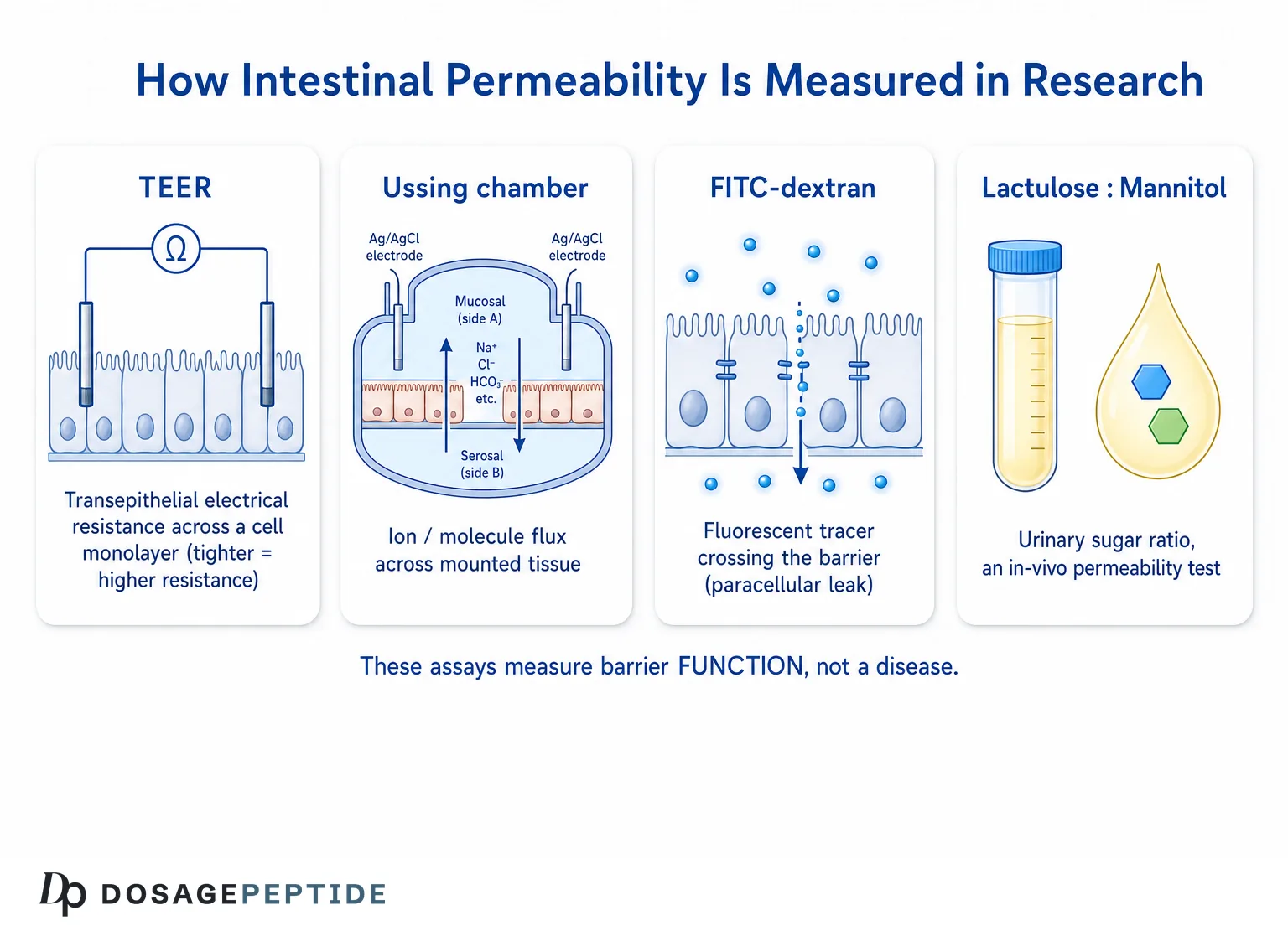
Because so many peptide claims rest on a single assay, understanding the measurement toolkit is essential to reading the literature critically. Each method answers a slightly different question, and each has limitations.
| Method | What it measures | Setting | Key limitation |
|---|---|---|---|
| Ussing chamber | Ion flux, tissue conductance, and tracer flux across mounted live tissue | Ex vivo (excised tissue or monolayers) | Tissue viability limited to hours; removed from in vivo blood flow and neural input |
| TEER (transepithelial electrical resistance) | Electrical resistance across a cell monolayer — a proxy for tight-junction integrity | In vitro (e.g., Caco-2 monolayers) | Reflects ionic/paracellular seal only; cell lines are not full intestine |
| FITC-dextran flux | Passage of fluorescent dextran (typically 4 kDa) from lumen to blood | In vitro and in vivo (rodents) | Probe size and gavage method affect results; largely a rodent readout |
| Lactulose/mannitol ratio | Urinary recovery ratio of two orally ingested sugars | In vivo (humans and animals) | Confounded by gastric emptying, renal function, bacterial degradation; probes mainly small-intestinal permeability |
| Immunostaining (ZO-1, occludin, claudins) | Localization and abundance of junction proteins | Ex vivo tissue/cells | Structural snapshot, not a functional permeability measure |
Why the model choice matters for peptide claims
Each assay also answers a question at a different level of biological organization, and conflating them is a common source of overstatement. The lactulose/mannitol test is the closest thing to a validated in vivo human permeability measure: two non-metabolized sugars are ingested, and their urinary recovery ratio reflects small-intestinal permeability, since the larger lactulose crosses mainly through a compromised paracellular route while the smaller mannitol crosses more freely and controls for non-barrier variables. Yet even this test is confounded by gastric emptying, intestinal transit time, renal clearance, and bacterial degradation of the sugars, which is why results vary between laboratories and why the test is used more in research than in routine clinical care. Newer probes using differently sized polyethylene glycols or multi-sugar panels attempt to distinguish small- from large-intestinal permeability, but no single assay is a definitive gold standard.
A recurring issue in the peptide literature is that a compound may “work” in one model and not translate to another. A peptide that raises TEER in a Caco-2 monolayer has shown an effect on a cancer-derived cell line under artificial culture conditions — a legitimate but preliminary signal. A peptide that lowers the urinary lactulose/mannitol ratio in a human trial has cleared a far higher bar. When evaluating any barrier peptide, the first question is always: in what model, and how far is that model from human physiology? The gap between an in-vitro monolayer, a preclinical/animal colitis model, and a randomized human trial is enormous, and it is the gap where most overstatement occurs. For readers who want to keep these tiers and terms straight, our peptide research glossary defines the measurement methods and evidence categories used throughout this article.
The Microbiome, Short-Chain Fatty Acids, and Barrier Maintenance
No account of the gut barrier is complete without the microbiota, because much of the barrier’s day-to-day maintenance is outsourced to bacterial metabolism — a point of well-established biology that provides essential context for any peptide claim.
Butyrate and colonocyte energetics
Commensal bacteria in the colon ferment dietary fiber into short-chain fatty acids (SCFAs) — principally acetate, propionate, and butyrate. Butyrate is the preferred energy substrate of colonocytes, supplying a large fraction of their metabolic needs, and its oxidation helps maintain the physiologically low-oxygen environment of the healthy colonic epithelium.[16] Beyond fueling the cells, butyrate has been reported in experimental systems to support tight-junction assembly and mucus production and to exert anti-inflammatory effects partly through histone-deacetylase inhibition and regulatory T-cell induction. When SCFA production falls — through fiber-poor diets or dysbiosis — colonocyte energetics can shift in ways associated with reduced barrier function in models.
Why microbiome context matters for peptide claims
This background reframes many “barrier repair” discussions. In a great many experimental settings, barrier integrity tracks closely with microbial composition, diet, and SCFA availability — variables that are powerful, well-documented, and entirely independent of any injectable peptide. A study that alters barrier function without controlling for diet and microbiota is difficult to interpret, and a peptide effect must be distinguished from the far larger and better-established influence of what the microbiota is producing. Responsible reading keeps this hierarchy in view: the microbiome–diet–SCFA axis is established barrier biology, whereas peptide interventions are a comparatively small and preliminary layer on top of it.
Mechanisms Studied: How Peptides Are Thought to Influence the Barrier
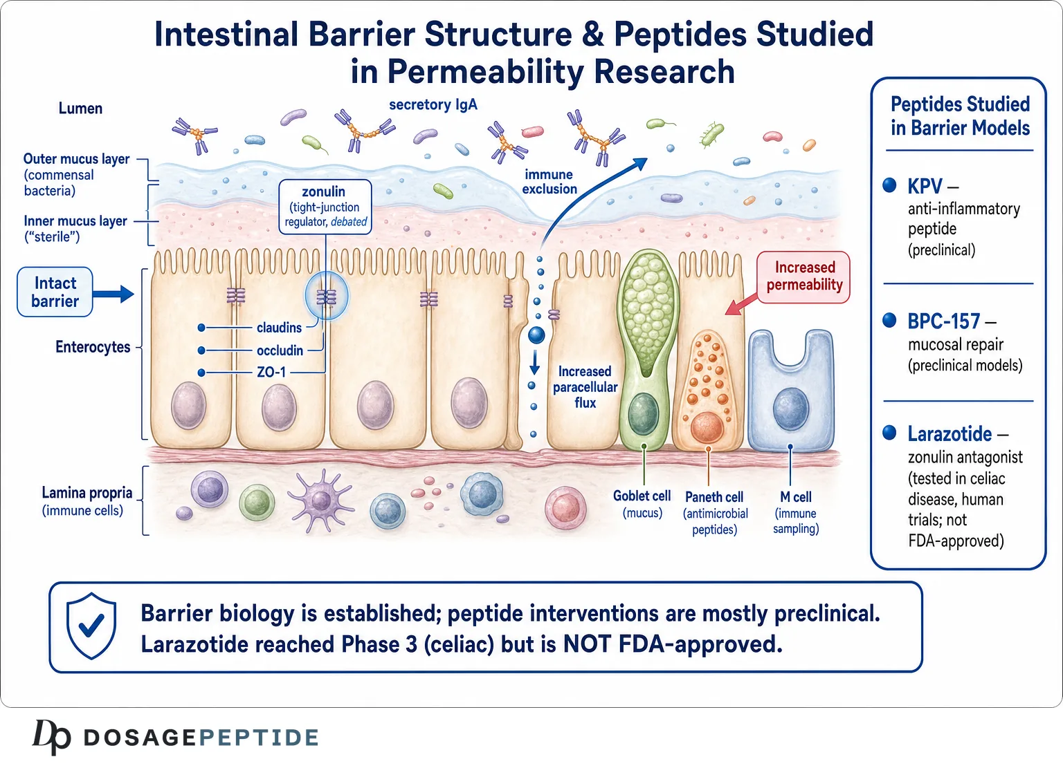
This section addresses the core mechanism question — how peptides are hypothesized to act on barrier function — before turning to specific molecules. It is important to read what follows as a map of proposed and studied mechanisms in experimental systems, not as established effects in humans.
Anti-inflammatory signaling that indirectly protects the junction
The most consistently studied route is indirect. Because inflammatory cytokines (TNF-α, IFN-γ, IL-1β) drive tight-junction disassembly via NF-κB and MLCK, any peptide that dampens this inflammatory signaling can preserve barrier integrity as a downstream consequence — without acting on claudins directly. Melanocortin-derived peptides such as KPV are the archetype here: their reported effect on the epithelium in colitis models is largely attributed to suppression of pro-inflammatory transcription rather than a direct structural action on the junction.[9]
Direct modulation of tight-junction assembly
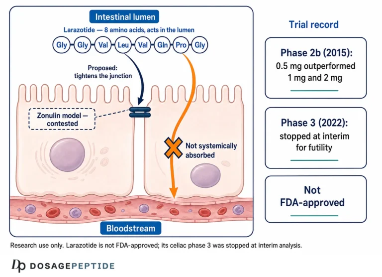
A second, more specific mechanism is direct interference with the signaling that opens junctions. This is the design logic of larazotide, an octapeptide intended to block the zonulin-triggered rearrangement of the actin cytoskeleton and thereby keep tight junctions assembled during a gluten challenge.[10] Mechanistically this is the cleanest “barrier peptide” concept because the intended target is the junction-regulating pathway itself.
Trophic and repair signaling
A third route is promotion of epithelial growth and wound healing. The clearest validated example is not an investigational peptide but an FDA-approved one: teduglutide (a glucagon-like peptide-2 analogue) is approved for short bowel syndrome and acts as an intestinotrophic factor, increasing villus height and crypt depth and improving fluid absorption.[11] It is included here as a mechanistic reference point — proof that a gut peptide can reach approval — while noting that its indication is intestinal failure, not “leaky gut,” and its mechanism is trophic growth rather than tight-junction sealing. Preclinical peptides such as BPC-157 are hypothesized to act partly through angiogenic and growth-factor-related repair pathways, a theme discussed in its own section.
Antimicrobial and mucus-supporting mechanisms
Finally, some peptides are studied for their effect on the chemical barrier rather than the physical one. Host antimicrobial peptides such as the cathelicidin LL-37 and defensins shape the microbiota and support mucus integrity, and their dysregulation is studied in IBD. LL-37 in particular sits at the intersection of antimicrobial defense and epithelial signaling; readers can explore this in our overview of LL-37 cathelicidin antimicrobial and immune research. The point for barrier biology is that “barrier support” can mean acting on the mucus/antimicrobial layer, the immune tone, the repair machinery, or the junctions directly — four distinct mechanisms too often collapsed into a single vague claim.
KPV: A Melanocortin Tripeptide in Intestinal-Inflammation Models
KPV (lysine–proline–valine) is the C-terminal tripeptide of α-melanocyte-stimulating hormone (α-MSH) and one of the more thoroughly characterized small peptides in intestinal-inflammation research. It is important to state the tier at the outset: KPV is a research compound studied in preclinical (cell and animal) models. It is not FDA-approved for any indication, and it is not an approved treatment for any gut condition in humans.
What the preclinical work shows
In cultured intestinal epithelial and immune cells, KPV has been reported to reduce pro-inflammatory signaling — decreasing NF-κB activation and downstream cytokine production — at low concentrations. A notable feature of its proposed pharmacology is uptake via the intestinal peptide transporter PepT1, which is expressed on enterocytes and upregulated during inflammation; this transporter-mediated entry has been proposed to deliver KPV directly to inflamed epithelium.[9] In mouse models of chemically induced colitis (DSS and TNBS), orally or rectally administered KPV has been reported to reduce disease activity scores, weight loss, and histological inflammation.[9]
Because barrier disruption in these colitis models is inflammation-driven, an anti-inflammatory effect can translate into preserved epithelial integrity — which is the indirect mechanism described earlier. This is the scientifically defensible way to describe KPV’s relationship to the gut barrier: it is studied primarily as a melanocortin-pathway anti-inflammatory that may secondarily protect the epithelium in inflamed tissue, not as a direct tight-junction sealant.
The melanocortin connection
KPV’s biology sits within the broader melanocortin system. The parent molecule, α-MSH, is a well-studied anti-inflammatory neuropeptide, and its C-terminal tripeptide (KPV) retains a portion of that activity while lacking the pigmentary (melanocortin-receptor) actions of the full sequence. Because KPV appears to act at least partly through receptor-independent, intracellular routes — interfering with NF-κB nuclear translocation — researchers have been interested in whether it can deliver anti-inflammatory effects without the systemic hormonal actions of α-MSH. This decoupling is scientifically appealing, but it remains a hypothesis explored in reductionist systems, and the precise molecular target of KPV inside cells is still not fully resolved. A related tripeptide amide, KdPT, has been studied in parallel for similar properties, underscoring that this is an active but still early research area rather than a settled therapeutic story.
PepT1-targeted delivery as a design concept
One of the more interesting translational ideas around KPV is exploitation of PepT1. Because PepT1 expression on the epithelium increases in inflamed intestine, a peptide that enters through this transporter could, in principle, concentrate itself where inflammation is greatest — a form of disease-targeted delivery. Investigators have gone further, studying nanoparticle and hydrogel formulations intended to deliver KPV to inflamed colon tissue in mouse models. These are legitimate and creative preclinical approaches, but they reinforce rather than diminish the central caveat: the science is at the stage of engineering delivery in animals, not demonstrating clinical benefit in people.
Context and honest limitations
The KPV literature is genuinely interesting but remains confined to cell culture and rodent models, with no controlled human efficacy trials for gut-barrier or IBD endpoints. Effects observed at defined concentrations in a dish or in gavaged mice do not establish what happens in a human gastrointestinal tract, and questions of oral bioavailability, dosing, and long-term safety in people are unresolved. Readers seeking the broader picture on this molecule can consult our explainer on what KPV is as an anti-inflammatory peptide for gut, skin, and beyond, and the reference-format handling and reconstitution details are compiled on the KPV 10 mg vial dosage protocol reference page — presented strictly as research-setting reference material, not as instructions for human use.
BPC-157: Preclinical Mucosal-Repair Research
BPC-157 (“body protection compound-157”) is a synthetic pentadecapeptide derived from a sequence in human gastric juice, and it is among the most widely discussed — and most cautiously interpreted — peptides in gut-repair research. The evidence tier must be stated bluntly: BPC-157 is preclinical/animal-only. It is not FDA-approved, has no completed randomized controlled human efficacy trials for gastrointestinal indications, and much of its literature originates from a limited number of affiliated research groups — a concentration of authorship that is itself a recognized limitation when weighing reproducibility.
What preclinical studies report
In rodent models, BPC-157 has been reported to accelerate healing across a range of gastrointestinal injuries — gastric and intestinal ulcers, anastomotic wounds, and chemically or NSAID-induced mucosal damage — and to influence colitis severity.[12] Proposed mechanisms in these studies include promotion of angiogenesis (partly via VEGF-related signaling), modulation of nitric oxide pathways, and interaction with growth-factor and cytoskeletal repair processes. In the barrier context specifically, the relevant claim is mucosal repair — restoring an injured epithelial surface — rather than acute tight-junction modulation in intact tissue.
Stability and the “stable gastric peptide” framing
Part of BPC-157’s appeal in the literature is a reported resistance to degradation in gastric juice, which is invoked to argue for oral activity — unusual for a peptide, since most are digested. This claimed stability is central to how the compound is positioned, but it is worth noting that pharmacokinetic characterization of BPC-157 in humans is essentially absent from the peer-reviewed record; there is little published data on absorption, distribution, half-life, or metabolism in people. A compound’s behavior in a rodent gut and its behavior in a human one can differ substantially, and claims of oral efficacy that rest on animal data plus a stability argument should be treated as provisional.
The reproducibility and translation gap
Two cautions are essential. First, the striking breadth of reported effects — spanning gut, tendon, brain, and vasculature — is scientifically unusual and warrants skepticism until confirmed by independent laboratories and, ultimately, human trials. Second, positive rodent healing data have historically failed to translate to humans in many therapeutic areas; a robust effect in a mouse ulcer model is a hypothesis-generating result, not clinical proof. For a more detailed, tier-aware treatment, see our coverage of what research says regarding BPC-157 healing effects on the gut and inflammation and the mechanistic discussion of how BPC-157 influences inflammation and mucosal repair in IBD models. The consistent message across both is that this is animal-model science, and any human application would be experimental and unproven.
Larazotide Acetate: The Clearest Clinical Example
If KPV and BPC-157 illustrate preclinical barrier research, larazotide acetate (formerly AT-1001) illustrates what happens when a barrier peptide is taken all the way into large human trials — and why the results are so instructive.
Rationale and early trials
Larazotide is an eight-amino-acid peptide designed as a zonulin antagonist and tight-junction regulator, intended to be taken orally and act locally in the gut lumen to reduce gliadin-triggered permeability in celiac disease. Early-phase studies explored whether it could blunt the increase in intestinal permeability and symptoms provoked by gluten challenge in patients with celiac disease.[10] A subsequent randomized, placebo-controlled Phase 2b trial in patients with persistent symptoms despite a gluten-free diet reported that a low dose of larazotide reduced symptoms compared with placebo — a genuinely encouraging signal, and the strongest human evidence for any tight-junction-targeting peptide to date.[13]
The Phase 3 outcome — and why it matters
The critical, and often omitted, part of the story is what happened next. Larazotide advanced to a large Phase 3 trial (the CeD-PRO program) for symptomatic celiac disease, and in 2022 the sponsor discontinued the program after a pre-planned interim analysis indicated the study was unlikely to meet its primary efficacy endpoint (a futility determination).[14] As a result, larazotide is not FDA-approved for celiac disease or any other indication.
How larazotide was engineered to stay in the gut
A design detail worth appreciating is that larazotide was intended to be minimally absorbed — to act locally at the luminal surface of the small intestine rather than systemically. This is the opposite of the systemic-injection framing that surrounds many research peptides, and it reflects a coherent pharmacological logic: if the target is the apical tight junction facing the lumen, a poorly absorbed oral peptide can act precisely where it is needed while limiting systemic exposure. That larazotide could be manufactured, dosed orally, and tested at scale in humans distinguishes it sharply from compounds that exist only as injectable research chemicals, and it is part of why its clinical result carries so much weight for the field.
This outcome is the single most important data point in the entire “barrier peptide” field, and it cuts in a sober direction. Larazotide had everything the others lack: a specific mechanism, a defined patient population, positive earlier-phase data, and a rigorous development program. It still failed to demonstrate efficacy at the confirmatory stage. That does not prove the barrier hypothesis is wrong — endpoints, dosing, and disease heterogeneity all complicate interpretation — but it is a powerful reminder of how often promising mechanisms and mid-stage signals fail to survive definitive testing. Any claim that a preclinical peptide reliably “heals the gut barrier” in humans should be read against the fact that the most advanced clinical candidate in this exact space did not clear its Phase 3 bar.
Other Peptides and Barrier-Adjacent Research
Beyond the three focal molecules, several other peptides appear in barrier-related literature at varying evidence tiers.
GLP-2 analogues (teduglutide)
As noted, teduglutide is an FDA-approved GLP-2 analogue for short bowel syndrome. Its relevance to barrier discussion is as a validated proof-of-concept that a peptide can therapeutically enhance intestinal structure and absorptive function — but its approved use is a specific malabsorptive condition, and its mechanism is intestinotrophic (promoting mucosal growth), not correction of a generalized “permeability” problem.[11]
Antimicrobial peptides: LL-37 and defensins
Host-defense peptides shape the chemical barrier. Reduced Paneth-cell α-defensin expression is associated with ileal Crohn’s disease, and cathelicidin LL-37 is studied for its dual antimicrobial and epithelial-signaling roles.[15] These are largely studied as endogenous components of barrier defense and as disease biomarkers rather than as injectable interventions with human efficacy data. Our LL-37 research overview covers this area in more depth.
Growth factors and other signals
Epidermal growth factor, keratinocyte growth factor, trefoil factor peptides, and glucagon-like peptide pathways all participate in mucosal maintenance and repair in experimental systems. Trefoil factors, for instance, are small secreted peptides that stabilize the mucus layer and promote epithelial restitution — the rapid migration of cells to reseal a wound — and they are studied as endogenous protectors of the surface. These molecules appear frequently in barrier reviews, but with the exception of the approved GLP-2 analogue they remain investigational or endogenous-physiology topics rather than validated barrier therapies, and none is an approved intervention for generalized intestinal permeability.
Current Evidence Level: A Tier-by-Tier Summary
Pulling the field together, the single most useful thing a reader can carry away is an accurate mental map of which claim sits at which evidence tier. The table below summarizes the peptides discussed. Note how sharply the tiers differ and how none of the investigational barrier peptides has a positive definitive human efficacy result.
| Peptide | Highest evidence tier for gut barrier | Regulatory status | Honest one-line summary |
|---|---|---|---|
| Teduglutide (GLP-2 analogue) | FDA-approved (short bowel syndrome) | Approved — specific indication | Proof a gut peptide can be approved; indication is intestinal failure, not “leaky gut” |
| Larazotide acetate | Reached Phase 3 (celiac disease) | Not approved; Phase 3 missed primary endpoint | Most advanced barrier peptide; failed confirmatory efficacy |
| KPV | Preclinical (cell + rodent colitis) | Not approved; research compound | Melanocortin anti-inflammatory; no human efficacy trials |
| BPC-157 | Preclinical (rodent mucosal repair) | Not approved; research compound | Broad animal claims; concentrated authorship; no RCTs |
| LL-37 / defensins | Endogenous biology + disease-association studies | Not therapeutics for barrier use | Studied as native barrier components and biomarkers |
How to read this table
The vertical distance in this table is the whole story. Well-established gut-barrier biology sits at the top of the evidence hierarchy; the peptide interventions marketed for “leaky gut” sit near the bottom. The one peptide that combined a specific mechanism with a full clinical program (larazotide) did not succeed at Phase 3, and the two most heavily promoted “barrier repair” peptides (KPV and BPC-157) have no controlled human efficacy data for these endpoints at all. That is not a reason to dismiss the research — preclinical work is how progress starts — but it is a decisive reason to reject any marketing that presents these as proven human gut treatments.
Limitations, Open Questions, and Research-Only Framing
Every section above carries caveats; this section consolidates them so no reader leaves with an inflated impression.
Limitations of the underlying models
Cell monolayers (Caco-2) are cancer-derived and lack the mucus layer, immune cells, microbiota, and blood supply of real intestine. Rodent colitis models (DSS, TNBS) are chemically induced and do not reproduce the multifactorial pathology of human IBD. Even the best human permeability assays — the lactulose/mannitol test — are confounded by gastric emptying, renal function, and bacterial sugar degradation. A positive result in any one system is a signal, not a conclusion.
Limitations of the peptide literature specifically
- Publication concentration. Some peptide bodies of work (notably BPC-157) are dominated by a small number of affiliated groups, raising reproducibility questions that only independent replication can resolve.
- Absent human efficacy data. For KPV and BPC-157, there are no completed randomized controlled human trials demonstrating gut-barrier or disease benefit.
- The larazotide precedent. The one rigorously tested barrier peptide failed its Phase 3 primary endpoint — a caution against assuming mechanism plus early data equals efficacy.
- Biomarker uncertainty. The zonulin assay controversy means a portion of the human “permeability” literature may rest on measurements that do not detect what they claim to.
- Manufacturing and identity. Research-chemical peptides are not pharmaceutical products; purity, identity, and sterility are not guaranteed outside validated manufacturing, an important consideration entirely separate from efficacy.
The “leaky gut” diagnosis question
To restate the central honesty point: increased intestinal permeability is a real, measurable phenomenon associated with defined conditions, but “leaky gut syndrome” as a standalone diagnosis explaining diffuse symptoms is not a validated clinical entity recognized by major gastroenterology organizations.[7] No peptide discussed here is an approved treatment for such a diagnosis, and none should be described as treating, curing, or preventing disease.
A practical checklist for evaluating a barrier-peptide claim
Readers encountering assertions about peptides and the gut barrier can apply a few disciplined questions that separate signal from marketing:
- What model produced the result? An in-vitro monolayer, an animal colitis model, and a human randomized trial are not interchangeable; the further from human physiology, the more provisional the claim.
- What exactly was measured? A named endpoint (TEER, FITC-dextran flux, lactulose/mannitol ratio, ZO-1 staining) is meaningful; a vague claim to “heal the gut lining” is not.
- Who did the work, and has it been replicated? Findings concentrated in a single research group await independent confirmation.
- Is a disease being implied? Any language that a peptide “treats,” “cures,” or “prevents” a condition — or targets an unvalidated “leaky gut” diagnosis — exceeds what the evidence supports.
- Is regulatory status stated honestly? With the narrow exception of teduglutide for short bowel syndrome, the peptides discussed here are not approved gut-barrier therapies.
Applied consistently, this checklist tends to collapse expansive marketing claims back down to their actual evidentiary footing, which for most barrier peptides is preclinical.
Where the research may go
Legitimate open questions remain genuinely interesting: whether transporter-targeted delivery (like PepT1 for KPV) can concentrate anti-inflammatory peptides at inflamed sites; whether repair peptides can be validated by independent groups; whether better permeability biomarkers than the current zonulin assay can be developed; and whether tight-junction modulation can succeed clinically with better-defined endpoints than those larazotide used. These are the productive frontiers — and they are frontiers precisely because the answers are not yet in.
Frequently Asked Questions
Are any peptides FDA-approved to fix a “leaky gut”?
No. No peptide is FDA-approved to treat “leaky gut,” and “leaky gut syndrome” is not a recognized standalone diagnosis. Teduglutide, a GLP-2 analogue, is approved — but for short bowel syndrome, a specific malabsorptive condition, and it works by promoting mucosal growth rather than sealing tight junctions. Every other peptide discussed in the barrier context is investigational or preclinical, and none is an approved treatment for intestinal permeability.
What is the difference between “intestinal permeability” and “leaky gut”?
Intestinal permeability is a measurable physiological property — the rate at which defined molecules cross the epithelium — that can be increased in conditions such as Crohn’s and active celiac disease. “Leaky gut,” in popular usage, is a broader wellness label linking that permeability to many diffuse symptoms. The measurable phenomenon is real and studied; the sweeping syndrome is not a validated clinical diagnosis, and the two should not be conflated.
Is the zonulin blood test reliable?
It is scientifically contested. Independent analyses have found that the widely used commercial “zonulin” ELISA may not detect pre-haptoglobin-2 as claimed and appears to cross-react with other proteins such as properdin and complement C3. This means some published serum-zonulin results may not be measuring zonulin at all, and clinicians and researchers should interpret such tests with considerable caution.
Does KPV repair the gut barrier?
KPV is studied in cell-culture and rodent colitis models as a melanocortin-derived anti-inflammatory peptide. In those models it can reduce inflammation and, indirectly, help preserve epithelial integrity in inflamed tissue. However, there are no controlled human trials demonstrating gut-barrier repair, and KPV is not FDA-approved for any use. It is best described as a preclinical research compound, not a proven human treatment.
Is BPC-157 proven to heal the gut in humans?
No. BPC-157’s gut-healing data come almost entirely from rodent models, and much of the literature originates from a small number of affiliated research groups. There are no completed randomized controlled human efficacy trials for gastrointestinal indications, and it is not FDA-approved. Positive animal results are hypothesis-generating, not clinical proof, and any human use would be experimental and unproven.
What happened with larazotide, the celiac peptide?
Larazotide acetate, a zonulin-antagonist peptide, showed encouraging results in earlier-phase celiac disease studies but did not succeed in its Phase 3 program, which was discontinued around 2022 after an interim futility analysis. It is not FDA-approved. Its trajectory is important because it was the most rigorously tested tight-junction peptide, and its confirmatory failure cautions against overinterpreting early-stage barrier-peptide data.
How do scientists actually measure the gut barrier?
Common tools include Ussing chambers (ion and tracer flux across live tissue), TEER (electrical resistance across cell monolayers), FITC-dextran flux (fluorescent tracer passage), the urinary lactulose/mannitol ratio in living subjects, and immunostaining of junction proteins like ZO-1 and claudins. Each measures a different aspect of the barrier and carries limitations, so results in one model do not automatically translate to human physiology.
Can these peptides be used safely at home for gut health?
This article is educational and does not provide human-use guidance. The peptides discussed are, in most cases, unapproved research compounds without established human safety or efficacy data for gut-barrier endpoints, and research-chemical products are not manufactured to pharmaceutical standards. Questions about personal health should be directed to a qualified, licensed medical professional rather than resolved from preclinical literature.
Is increased permeability a cause or a consequence of disease?
It can be either, and often it is a consequence. In Crohn’s disease, increased permeability can precede relapse and appears in some healthy relatives, suggesting an upstream role in that context. In many other situations, permeability rises as a result of existing inflammation rather than triggering it. Because it is not specific or uniformly causal, its mere presence does not establish a disease or predict who is symptomatic.
References
- Helander HF, Fändriks L. Surface area of the digestive tract — revisited. Scand J Gastroenterol. 2014.
- Fasano A. Zonulin and its regulation of intestinal barrier function: the biological door to inflammation, autoimmunity, and cancer. Physiol Rev. 2011.
- Turner JR. Intestinal mucosal barrier function in health and disease. Nat Rev Immunol. 2009.
- Günzel D, Yu ASL. Claudins and the modulation of tight junction permeability. Physiol Rev. 2013.
- Clayburgh DR, Shen L, Turner JR. A porous defense: the leaky epithelial barrier in intestinal disease. Lab Invest. 2004.
- Johansson MEV, Larsson JMH, Hansson GC. The two mucus layers of colon are organized by the MUC2 mucin. Proc Natl Acad Sci USA. 2011.
- Camilleri M. Leaky gut: mechanisms, measurement and clinical implications in humans. Gut. 2019.
- Scheffler L, et al. Widely used commercial ELISA does not detect precursor of haptoglobin2, but recognizes properdin as a potential second member of the zonulin family. Front Endocrinol (Lausanne). 2018.
- Kannengiesser K, et al. Melanocortin-derived tripeptide KPV has anti-inflammatory potential in murine models of inflammatory bowel disease. Inflamm Bowel Dis. 2008.
- Kelly CP, et al. Larazotide acetate in patients with coeliac disease undergoing a gluten challenge: a randomised placebo-controlled study. Aliment Pharmacol Ther. 2013.
- Jeppesen PB, et al. Teduglutide reduces need for parenteral support among patients with short bowel syndrome with intestinal failure. Gastroenterology. 2012.
- Sikiric P, et al. Stable gastric pentadecapeptide BPC 157: novel therapy in gastrointestinal tract. Curr Pharm Des. 2011.
- Leffler DA, et al. Larazotide acetate for persistent symptoms of celiac disease despite a gluten-free diet: a randomized controlled trial. Gastroenterology. 2015.
- A study of the efficacy and safety of larazotide acetate for the treatment of celiac disease (CeD-PRO). ClinicalTrials.gov NCT03569007.
- Ostaff MJ, Stange EF, Wehkamp J. Antimicrobial peptides and gut microbiota in homeostasis and pathology. EMBO Mol Med. 2013.
- Donohoe DR, et al. The microbiome and butyrate regulate energy metabolism and autophagy in the mammalian colon. Cell Metab. 2011.
Research-use disclaimer: This article is provided strictly for educational and informational purposes and describes preclinical and clinical research on gut-barrier biology and peptides. It is not medical advice, and nothing here constitutes a therapeutic recommendation, dosing instruction, or claim that any peptide diagnoses, treats, cures, or prevents any disease. The peptides discussed are, in most cases, unapproved research compounds. Consult a qualified, licensed healthcare professional for any health concern.