DES(1-3)IGF-1 and IGF-1 LR3 are both engineered analogs of human IGF-1 built to do the same job — escape capture by the IGF binding proteins (IGFBPs) — but they are built differently, and the published animal data point in a direction most readers do not expect. The short answer: in rodent studies, both analogs are cleared from plasma faster than native IGF-1, not slower, and LR3 is the faster-clearing of the two analogs. The widely repeated claim that LR3 has a half-life of roughly a day while DES lasts twenty minutes is not supported by any peer-reviewed pharmacokinetic study we could locate, and the direct animal comparison that does exist reports the opposite ordering.
That single fact is why these two compounds are not interchangeable, and why a figure derived from one is structurally wrong for the other. Below is what the literature actually establishes, what it does not, and where the popular numbers come from. Neither compound is approved for human use anywhere in the world.
DES(1-3)IGF-1 vs IGF-1 LR3: Comparison Table
| Property | DES(1-3)IGF-1 | IGF-1 LR3 | Native IGF-1 (reference) |
|---|---|---|---|
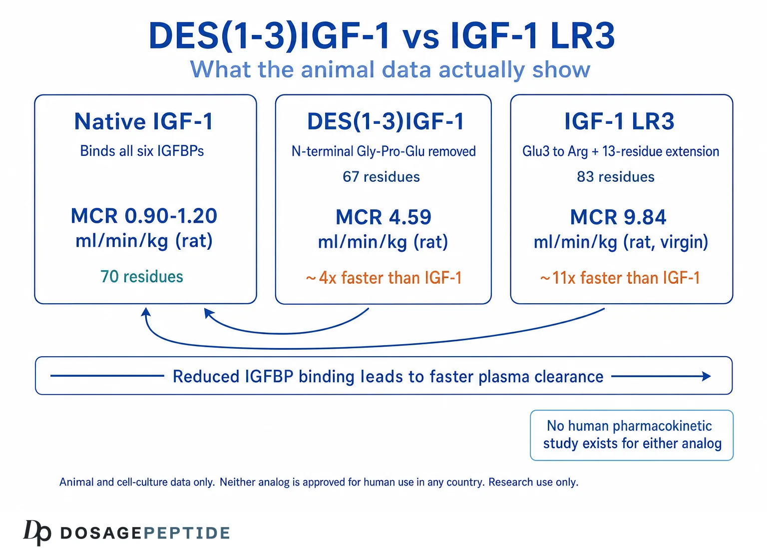
| Structure | Human IGF-1 with the N-terminal tripeptide Gly-Pro-Glu removed (67 residues) | Human IGF-1 with Glu3 replaced by Arg, plus a 13-residue N-terminal extension (the first 11 amino acids of methionyl porcine growth hormone followed by Val-Asn)[4] | 70 residues |
| How it evades IGFBPs | Loss of the glutamate at position 3[7] | Substitution of arginine for the glutamate at position 3[5] | Does not — binds IGFBPs with high affinity |
| IGFBP binding | Greatly reduced; native IGF-1 competes at least 60-fold better for binding protein[1] | Binds “very poorly” to the IGF-binding protein tested (bovine IGFBP-2)[5] | High affinity for all six IGFBPs |
| Type 1 IGF receptor binding | Essentially unchanged versus IGF-1[1] | Roughly 3-fold weaker than IGF-1[6] | Reference |
| Potency in IGFBP-secreting cell cultures | Generally about 10-fold greater than IGF-1[7] | Comparable to DES(1-3); both exceed IGF-1[4] | Reference |
| Potency in cells that secrete no IGFBPs | Advantage disappears | Less potent than IGF-1[4] | Reference |
| Plasma clearance, rat IV bolus | MCR 4.59 ml/min/kg vs IGF-1 at 1.20 in the same study[2] | MCR 9.84 (virgin rats) and 9.19 (pregnant rats) ml/min/kg, vs IGF-1 at 0.90 and 2.88 in the same study[3] | 0.90–2.88 ml/min/kg depending on model |
| Direction versus IGF-1 | Roughly 4× faster clearance | Roughly 11× faster in virgin rats; roughly 3× faster in pregnant rats, where IGFBP-3 is already low | — |
| Detection window after IM dose in rats | Unchanged parent detected up to 24 h[10] | Disappeared rapidly after 4 h[10] | — |
| Published human half-life | None located | None located | Characterised for mecasermin in its FDA labelling |
| Regulatory status | Research chemical; not approved for human use anywhere; prohibited in sport[10] | Research chemical and cell-culture reagent; not approved for human use anywhere; prohibited in sport[10] | Mecasermin (Increlex) is FDA-approved, but only for growth failure in pediatric patients 2 years and older with severe primary IGF-1 deficiency[13] |
Reference material for each compound is catalogued separately in our IGF-1 DES 1 mg vial reference page and our IGF-1 LR3 1 mg vial reference page, which document how these compounds are handled in laboratory settings rather than prescribing any human use.
Why IGFBPs Are the Whole Story
In circulation, native IGF-1 is almost entirely bound. It travels predominantly in a large ternary complex of roughly 150 kDa, and this complex is what protects it from filtration and degradation. In rat plasma, labelled IGF-1 is found mainly in that 150 kDa complex, whereas labelled LR3 is detected largely as free peptide[3].
This produces a causal chain that governs everything else about these analogs:
- Lower IGFBP affinity means a larger free fraction of peptide.
- A larger free fraction means more peptide available to engage the type 1 IGF receptor — hence higher measured potency in any cell culture that secretes IGFBPs into the medium.
- But the same free fraction is also exposed to filtration and proteolysis, so it is removed from plasma faster.
Potency and persistence are therefore not independent dials. In these molecules they are the same dial, turned in opposite directions. An analog that is more potent because it dodges the binding proteins is, for exactly that reason, shorter-lived in plasma. The literature makes this explicit: Ballard and colleagues concluded that plasma binding proteins inhibit transfer of the growth factors to tissue sites of action, and proposed that IGF analogs cleared rapidly from blood may have greater biological potencies in vivo[2].
The cleanest demonstration that potency is entirely context-dependent comes from a cell-line comparison: in chicken embryo fibroblasts, which secrete no detectable IGFBPs into the medium, Long[Arg3]-IGF-1 was less potent than plain IGF-1[4]. Remove the binding proteins and the analog’s entire advantage evaporates, leaving only its slightly weaker receptor affinity. Terminology used throughout this section is defined in our peptide research glossary.
What DES(1-3)IGF-1 Actually Is
DES(1-3)IGF-1 is human IGF-1 with the first three residues — glycine, proline, glutamate — removed from the N-terminus. It is not purely synthetic in origin: it has been isolated from bovine colostrum, human brain and porcine uterus, and is thought to arise from post-translational cleavage of IGF-1[7].
A systematic truncation series established which residue matters. Removing one or two residues changed almost nothing. Removing the third — the glutamate — produced roughly a ten-fold jump in potency in an L6 myoblast protein-synthesis assay, while receptor competition was essentially unchanged. Removing five residues collapsed potency by nearly two orders of magnitude. Critically, native IGF-1 and the des(1) and des(1-2) forms competed for binding protein at least 60-fold better than the shorter peptides[1]. The potency gain is a binding-protein effect, not a receptor effect.
What DES has been studied in
The in vivo work is animal-only and dates largely from the early 1990s. In research models, DES(1-3)IGF-1 increased body length in GH-deficient lit/lit mice at a tenth the dose required for equivalent IGF-1 effects[8], and improved nitrogen balance in rats made catabolic by dexamethasone[6]. These are preclinical rodent findings. No controlled human efficacy trials of DES(1-3)IGF-1 were located.
What IGF-1 LR3 Actually Is
IGF-1 LR3 is a larger and more heavily modified molecule than most readers assume. It carries two changes: the glutamate at position 3 is replaced by arginine, and a 13-residue extension is fused to the N-terminus — specifically the first 11 amino acids of methionyl porcine growth hormone followed by the dipeptide Val-Asn[4]. The extension was not designed to extend duration of action. It was an expression-system feature: the hydrophobic N-terminal extension facilitated correct folding of the analog in E. coli and gave very high yields of biologically active product[4]. The name “Long” refers to the peptide chain, not to how long it lasts — a coincidence of nomenclature that has probably done more to spread misinformation than any single vendor claim.
Substituting arginine for Glu3 reduced IGFBP binding sharply while leaving receptor binding slightly reduced relative to IGF-1, and the authors concluded that reduced IGFBP binding rather than increased receptor binding explains the greater biological potency[5].
LR3 as a cell culture reagent
LR3-IGF-1 is a mainstream biotechnology reagent. In serum-free Chinese hamster ovary cell cultures expressing recombinant proteins, LongR3 was compared directly against insulin and was better able to sustain viability of both tested cell lines under production conditions[12]. Its resistance to IGFBPs is precisely what makes it useful in a culture flask, where binding proteins secreted by the cells would otherwise sequester added IGF-1.
That industrial provenance has a practical consequence. Anti-doping analysts identified a black-market injection vial whose contents were His-tagged Long-R3-IGF-1 — a form carrying a hexahistidine purification tag normally used for biochemical studies, and which the authors judged was likely a by-product of laboratory work rather than material made for injection[11]. Further mechanistic background is collected in our overview of what IGF-1 LR3 is and how it is studied, and the comparison with a different class of IGF-1 variant is covered in MGF versus IGF-1 LR3 in muscle research.
What the Pharmacokinetic Evidence Does and Does Not Establish
The commonly circulated half-life figures for these analogs — roughly 20 to 30 minutes for DES, roughly 20 to 30 hours for LR3, and a resulting “60-fold difference” — do not appear in the peer-reviewed pharmacokinetic literature. We could not locate a published human pharmacokinetic study of either compound. These numbers circulate in forum posts and vendor product descriptions. They should be treated as community-derived folklore, not as data.
What the published literature does contain is animal clearance work, and it points the other way.
| Study | Model | Analog MCR (ml/min/kg) | IGF-1 control in same study | Ratio |
|---|---|---|---|---|
| Ballard et al. 1991[2] | Rats, IV bolus, labelled tracer | DES(1-3)IGF-1: 4.59 | 1.20 | ~3.8× faster |
| Bastian et al. 1993[3] | Virgin rats, IV bolus, labelled tracer | LR3IGF-1: 9.84 | 0.90 | ~11× faster |
| Bastian et al. 1993[3] | Pregnant rats (day 18), IV bolus | LR3IGF-1: 9.19 | 2.88 | ~3.2× faster |
Two points about how to read that table honestly. First, the DES and LR3 figures come from two different studies, so comparing them to each other is an indirect cross-study inference — though both were performed by the same Adelaide research group using the same tracer methodology, and each contains its own internal IGF-1 control, which makes the normalised ratios reasonably comparable. A later study from the same programme did examine IGF-1, des-(1-3)IGF-1 and LR3IGF-1 together in a single rat model of chronic renal failure, and reported that total clearance of LR3IGF-1 was significantly increased in that setting[9]. Second, and more importantly, both analogs are unambiguously cleared faster than native IGF-1 within their own studies. That within-study comparison is the robust finding.
The pregnancy arm of the Bastian study is a natural experiment that confirms the mechanism. On day 18 of gestation, when circulating IGFBP-3 is dramatically reduced, native IGF-1 clearance more than tripled — moving toward the value seen with LR3 — while LR3 clearance was essentially unchanged between pregnant and virgin animals[3]. Strip away the binding proteins and native IGF-1 starts behaving like the analogs. The binding proteins were the reservoir all along.
The one direct head-to-head
The most relevant modern study administered IGF-1 analogs intramuscularly to rats at 100 mcg/kg and tracked them by immunopurification and high-resolution mass spectrometry. Unchanged DES(1-3)-IGF-1 was detectable up to 24 hours after administration, whereas LongR-IGF-1 disappeared rapidly after 4 hours[10]. The same study characterised N-terminal degradation products of LongR-IGF-1, one of which remained detectable up to 16 hours.
Two caveats matter. This is a detection-window measurement of the intact parent molecule by mass spectrometry, not a formally derived elimination half-life. And it is a rodent study — although the authors noted that incubating the analogs in human whole blood in vitro produced the same degradation products, which they took to suggest the rat observations may extrapolate to humans. With those caveats stated, the direction is unambiguous and it is the reverse of the popular claim.
What this means for transferring a figure between them
A number derived for one of these analogs is not merely imprecise when applied to the other — it is structurally wrong, for three independent reasons:
- Different molecular weights. DES(1-3) is 67 residues; LR3 is 83, because it carries the 13-residue extension on top of the full 70-residue sequence. LR3 is therefore the substantially heavier molecule, and an equal mass of it contains meaningfully fewer molecules — so a mass-based figure and a molar figure do not translate between the two.
- Different receptor affinity. DES(1-3) binds the type 1 IGF receptor essentially like IGF-1[1]; LR3 binds it about three-fold more weakly[6]. Equal free concentrations do not produce equal receptor occupancy.
- Different clearance, in the direction opposite to the folklore. Per the animal data above, LR3 is the faster-clearing of the two, not the slower.
Anyone reasoning from the circulating “LR3 lasts a day” figure is reasoning from a premise that the available evidence contradicts. General laboratory handling considerations are covered in our peptide reconstitution guide.
Regulatory Status: Stated Precisely
Neither DES(1-3)IGF-1 nor IGF-1 LR3 is approved for human use in any jurisdiction. Both are research chemicals and laboratory reagents. Anti-doping researchers describe them plainly as substances that “were never approved for use in humans” yet are “readily available as black market products”[10].
Mecasermin is a different molecule. Mecasermin (brand name Increlex) is recombinant human IGF-1 — the native 70-residue sequence, not DES and not LR3. It is FDA-approved, and the labelled indication is narrow: treatment of growth failure in pediatric patients 2 years of age and older with severe primary IGF-1 deficiency, or with growth hormone gene deletion who have developed neutralizing antibodies to GH[13]. Readers routinely conflate the approval of mecasermin with approval of IGF-1 analogs generally. It is not one. No analog shares it.
Prohibited in sport. IGF-1 and its analogs are prohibited substances under the World Anti-Doping Agency’s Prohibited List, within the peptide hormones and growth factors class, at all times — in and out of competition[14]. LongR-IGF-1 and Des(1-3)-IGF-1 are named specifically in the anti-doping analytical literature as targets for detection[10].
Why proliferation signalling is the central safety question
IGF-1 signalling through the type 1 IGF receptor plays a documented role in cell proliferation and resistance to apoptosis. In renal cell carcinoma specifically, a published review reports that patients whose tumours overexpress the type 1 IGF receptor have a markedly higher risk of death than patients whose tumours do not — an association observed in tumour biology, not evidence that these analogs cause cancer[15]. The concern is not purely theoretical: the FDA-approved native IGF-1 product carries postmarketing reports of malignant neoplasms in pediatric patients, and its labelling directs prescribers to monitor all patients for the development of neoplasms[13].
Analogs engineered specifically to increase the free, receptor-available fraction of IGF-1 act on the same receptor without the physiological buffering that IGFBPs normally provide — and without any of the medical supervision, dose titration, or IGF-1 monitoring that accompanies the approved product. No published study establishes that either analog is safe in humans, at any dose, for any purpose. Related context is available in our discussion of what is and is not known about peptide safety.
Frequently Asked Questions
Is IGF-1 DES stronger than IGF-1 LR3?
In cell-culture systems that secrete IGF binding proteins, both analogs substantially outperform native IGF-1, and published comparisons place them in a similar range. DES(1-3)IGF-1 is generally reported as about ten-fold more potent than IGF-1, and one direct comparison ranked Long[Arg3]-IGF-1 and DES(1-3)IGF-1 together at the top of the potency order. Neither has been shown superior to the other in any human study, because no such study exists.
What is the half-life of IGF-1 DES?
No peer-reviewed human pharmacokinetic study establishing a half-life for DES(1-3)IGF-1 could be located. In rats given an intravenous bolus, its metabolic clearance rate was 4.59 ml/min/kg against 1.20 for native IGF-1 in the same experiment — roughly four times faster. In a separate rat study using intramuscular dosing, the unchanged parent compound remained detectable by mass spectrometry for up to 24 hours. The frequently quoted “20 to 30 minutes” figure is community-derived and unverified.
Does IGF-1 LR3 really have a 20 to 30 hour half-life?
That figure has no peer-reviewed basis that we could locate, and the available animal evidence contradicts it. In rats, LR3IGF-1 was cleared roughly ten times faster than native IGF-1, and after intramuscular administration the intact molecule disappeared rapidly after about four hours. The mechanism explains why: LR3 cannot bind the IGFBP-3 complex that normally protects IGF-1 in circulation, so it is exposed to clearance rather than sheltered from it.
Why is it called “Long” R3 if it does not last longer?
The “Long” prefix refers to the physical length of the peptide chain, not its duration of action. LR3 carries a 13-residue N-terminal extension derived from methionyl porcine growth hormone, which was introduced because the hydrophobic extension improved correct folding and yield during bacterial expression. The naming has been widely misread as a claim about half-life.
Can I use an IGF-1 LR3 figure for IGF-1 DES?
No. The two molecules differ in size (83 versus 67 residues), in type 1 IGF receptor affinity (LR3 binds roughly three-fold more weakly), and in plasma clearance. A figure derived for one carries none of those properties across to the other. This is a research reference site and does not provide human dosing guidance for either compound.
Is IGF-1 LR3 the same thing as Increlex?
No. Increlex is mecasermin, which is recombinant native human IGF-1 — the unmodified 70-residue sequence. IGF-1 LR3 is a structurally different engineered analog. Mecasermin’s FDA approval covers growth failure in pediatric patients with severe primary IGF-1 deficiency and does not extend to LR3, DES, or any other analog.
Why is LR3 sold as a cell culture reagent?
Because its resistance to IGF binding proteins is genuinely useful in a culture flask, where binding proteins secreted by the cultured cells would otherwise sequester added growth factor. Published bioprocess work compared LongR3 against insulin in serum-free CHO cell culture and found it better sustained cell viability under production conditions. Its status as an industrial reagent is one reason material of laboratory rather than pharmaceutical provenance circulates — including at least one documented case of a His-tagged research preparation appearing in an injection vial.
Are IGF-1 analogs banned in sport?
Yes. IGF-1 and its analogs sit within the peptide hormones and growth factors class of the WADA Prohibited List and are prohibited at all times, both in and out of competition. Validated mass spectrometry methods for detecting LongR-IGF-1, Des(1-3)-IGF-1 and R-IGF-I in serum have been published for anti-doping purposes.
What human data exist on these compounds?
Essentially none for efficacy or safety. The substantive published work on both analogs is preclinical: cell culture, rodent studies, and biotechnology reagent applications, most of it produced between 1989 and the late 1990s by a small number of research groups. Where this article states a numerical finding, it comes from an animal or in-vitro study and is described as such.
References
- Bagley CJ, May BL, Szabo L, et al. A key functional role for the insulin-like growth factor 1 N-terminal pentapeptide. Biochem J. 1989;259(3):665-71. https://pubmed.ncbi.nlm.nih.gov/2730580/
- Ballard FJ, Knowles SE, Walton PE, et al. Plasma clearance and tissue distribution of labelled insulin-like growth factor-I (IGF-I), IGF-II and des(1-3)IGF-I in rats. J Endocrinol. 1991;128(2):197-204. https://pubmed.ncbi.nlm.nih.gov/2005410/
- Bastian SE, Walton PE, Wallace JC, Ballard FJ. Plasma clearance and tissue distribution of labelled insulin-like growth factor-I (IGF-I) and an analogue LR3IGF-I in pregnant rats. J Endocrinol. 1993;138(2):327-36. https://pubmed.ncbi.nlm.nih.gov/7693845/
- Francis GL, Ross M, Ballard FJ, et al. Novel recombinant fusion protein analogues of insulin-like growth factor (IGF)-I indicate the relative importance of IGF-binding protein and receptor binding for enhanced biological potency. J Mol Endocrinol. 1992;8(3):213-23. https://pubmed.ncbi.nlm.nih.gov/1378742/
- King R, Wells JR, Krieg P, et al. Production and characterization of recombinant insulin-like growth factor-I (IGF-I) and potent analogues of IGF-I, with Gly or Arg substituted for Glu3, following their expression in Escherichia coli as fusion proteins. J Mol Endocrinol. 1992;8(1):29-41. https://pubmed.ncbi.nlm.nih.gov/1311930/
- Tomas FM, Knowles SE, Owens PC, et al. Insulin-like growth factor-I (IGF-I) and especially IGF-I variants are anabolic in dexamethasone-treated rats. Biochem J. 1992;282(Pt 1):91-7. https://pubmed.ncbi.nlm.nih.gov/1371669/
- Ballard FJ, Wallace JC, Francis GL, Read LC, Tomas FM. Des(1-3)IGF-I: a truncated form of insulin-like growth factor-I. Int J Biochem Cell Biol. 1996;28(10):1085-7. https://pubmed.ncbi.nlm.nih.gov/8930132/
- Gillespie C, Read LC, Bagley CJ, Ballard FJ. Enhanced potency of truncated insulin-like growth factor-I (des(1-3)IGF-I) relative to IGF-I in lit/lit mice. J Endocrinol. 1990;127(3):401-5. https://pubmed.ncbi.nlm.nih.gov/2280209/
- Gillespie CM, Hazel SJ, Walton PE, Martin AA. Effects of chronic renal failure on plasma clearance of insulin-like growth factor I, des-(1-3)IGF-I, and LR3IGF-I. Am J Physiol. 1996;271(4 Pt 1):E649-57. https://pubmed.ncbi.nlm.nih.gov/8897852/
- Mongongu C, Coudoré F, Domergue V, Ericsson M, Buisson C, Marchand A. Detection of LongR-IGF-I, Des(1-3)-IGF-I, and R-IGF-I using immunopurification and high resolution mass spectrometry for antidoping purposes. Drug Test Anal. 2021;13(7):1256-69. https://pubmed.ncbi.nlm.nih.gov/33587816/
- Kohler M, Thomas A, Walpurgis K, Terlouw K, Schänzer W, Thevis M. Detection of His-tagged Long-R³-IGF-I in a black market product. Growth Horm IGF Res. 2010;20(5):386-90. https://pubmed.ncbi.nlm.nih.gov/20675162/
- Morris AE, Schmid J. Effects of insulin and LongR(3) on serum-free Chinese hamster ovary cell cultures expressing two recombinant proteins. Biotechnol Prog. 2000;16(5):693-7. https://pubmed.ncbi.nlm.nih.gov/11027158/
- INCRELEX (mecasermin) injection, for subcutaneous use — US prescribing information. DailyMed, US National Library of Medicine. https://dailymed.nlm.nih.gov/dailymed/lookup.cfm?setid=a8b27a1b-a611-4f91-ad22-76d4b390c3ae
- World Anti-Doping Agency. The Prohibited List. https://www.wada-ama.org/en/prohibited-list
- Tracz AF, Szczylik C, Porta C, Czarnecka AM. Insulin-like growth factor-1 signaling in renal cell carcinoma. BMC Cancer. 2016;16:453. https://pubmed.ncbi.nlm.nih.gov/27405474/
Research use only. DosagePeptide is an independent research reference library. It does not sell peptides and does not provide medical advice. DES(1-3)IGF-1 and IGF-1 LR3 are research chemicals and laboratory reagents; neither is approved for human use in any jurisdiction, and both are prohibited in sport. Every quantitative finding described above comes from in-vitro or animal research and is reported as an observation in research models, not as guidance for any person. Nothing on this page is a recommendation to administer any substance to a human being, and no content here is intended to diagnose, treat, cure or prevent any disease.