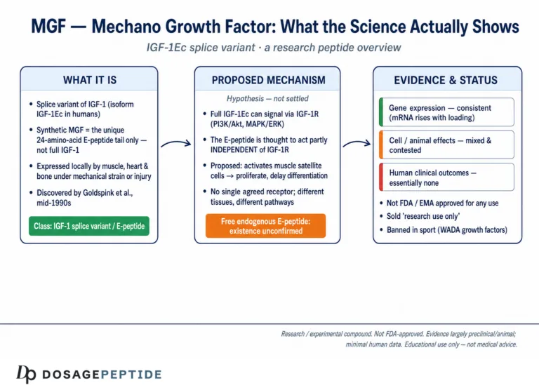
Mechano Growth Factor (MGF) and IGF-1 Long R3 (IGF-1 LR3) are frequently grouped together in the peptide-research literature because both trace back to a single gene — insulin-like growth factor 1 — yet they are strikingly different molecules with different origins, different pharmacokinetics, and, in preclinical models, different biological behavior. The central research question this article examines is a mechanistic one: if MGF is a naturally occurring splice product of the IGF-1 gene that acts locally and transiently in mechanically loaded muscle, while IGF-1 LR3 is an engineered analog built to evade the body’s clearance system and act systemically for hours, how do these two “IGF-1 variants” actually differ at the level of receptor engagement, binding-protein interaction, and satellite-cell biology? This is a comparison of laboratory research reagents, not a comparison of approved therapeutics, and the honest answer requires separating what has been demonstrated in cells and animals from what remains hypothesis.
MGF vs IGF-1 LR3: the short version
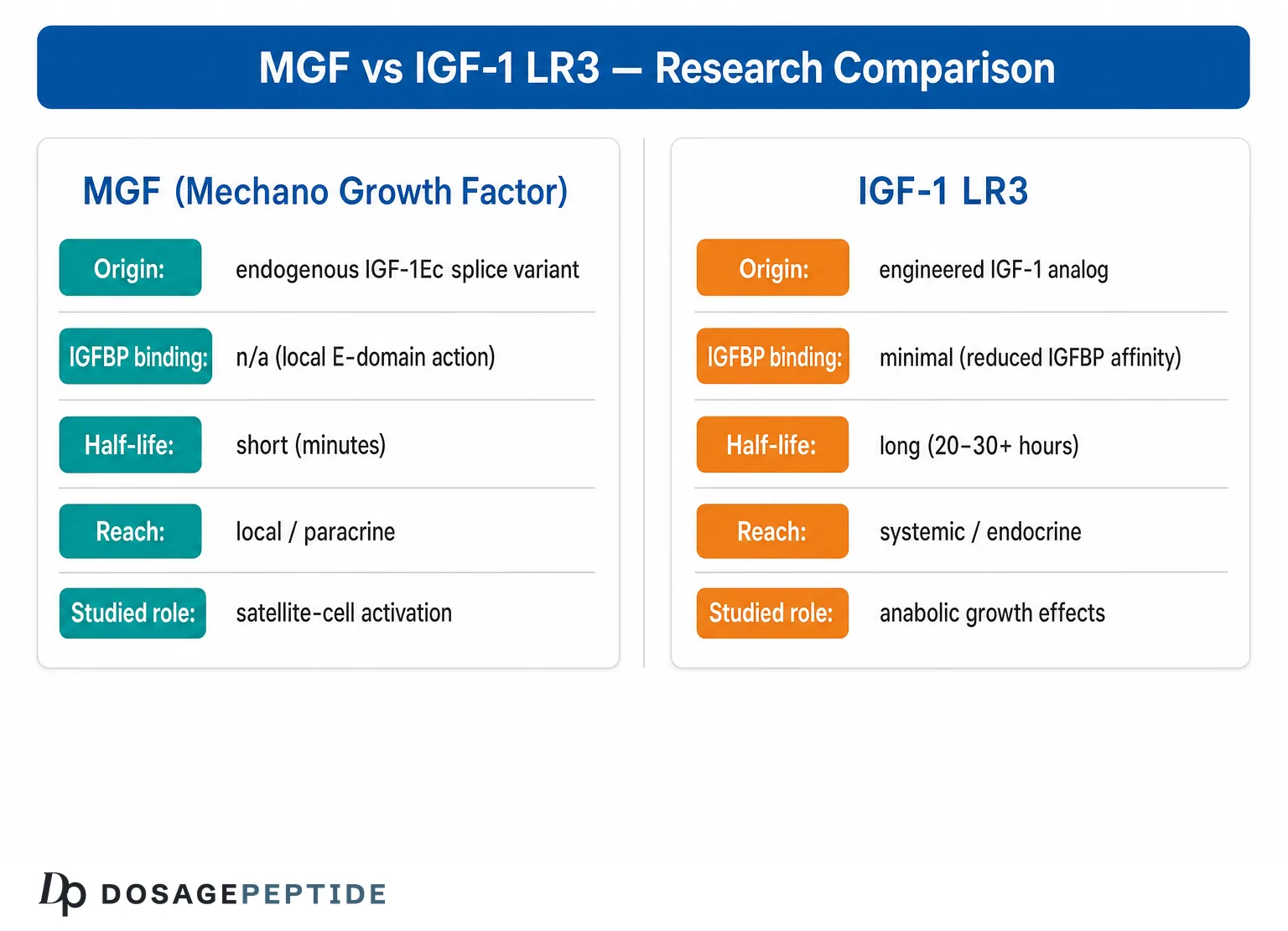
| MGF (IGF-1Ec) | IGF-1 LR3 | |
|---|---|---|
| Origin | Natural splice variant of the IGF-1 gene, produced in muscle after mechanical loading | Engineered analog, manufactured as a laboratory reagent |
| Where it acts | Locally, in the tissue that produced it (autocrine/paracrine) | Systemically, wherever the circulation carries it |
| Persistence | Configured for transience: short-lived and confined to the tissue that made it | Engineered for persistence in vitro. In the body it is more complicated — escaping the binding proteins also forfeits the reservoir that keeps IGF-1 in circulation |
| Receptor | The E-peptide’s proposed action does not depend on the IGF-1 receptor, which is precisely what is disputed | The type 1 IGF receptor, unambiguously |
| Evidence quality | Contested. Key satellite-cell findings have proved difficult to replicate. | Well characterised as a cell-culture reagent. No human outcome data. |
| Muscle growth in humans | Not demonstrated | Not demonstrated |
And PEG-MGF? Attaching polyethylene glycol to the MGF peptide is an attempt to fix its defining weakness: it is built to be transient. Pegylation is a well-established way of slowing clearance, but it changes how long a molecule circulates — not what that molecule does. It does not turn a locally acting E-peptide into a systemic anabolic agent, and it does nothing to settle the underlying dispute about whether the E-peptide has an independent biological action at all. No controlled human pharmacokinetic study defines a half-life for PEG-MGF either. A longer-lasting version of a contested molecule is still a contested molecule.
So which one “builds muscle”? Neither has been shown to, in a human being, in a controlled trial. Both are research reagents; the hypertrophy story comes from cell culture, rodents and gene-transfer experiments, none of which transfers to injecting a peptide into a person. The sections below explain exactly where each line of evidence comes from and where it breaks down. Handling references: MGF 5 mg vial, PEG-MGF 2 mg vial, IGF-1 LR3 1 mg vial.
Research Context: Why Compare Two Molecules From the Same Gene?
The IGF-1 gene is not a simple one-gene-one-protein arrangement. Through alternative splicing and alternate promoter usage, a single primary transcript can generate several distinct messenger RNAs that share a common mature IGF-1 core but differ in their C-terminal “E-domain” extensions.[1] Two of the products that emerge from this system have attracted disproportionate attention in muscle and sports-science circles: the locally expressed splice variant known as IGF-1Ec, or Mechano Growth Factor, and a wholly synthetic laboratory analog called Long R3 IGF-1. Confusingly, both are marketed to researchers under the umbrella of “IGF-1 variants,” and the naming overlap has fostered a persistent misconception that they are interchangeable or that they work by the same route.
They are not the same. MGF is an endogenous splice product — something the body itself produces in response to mechanical strain and tissue damage — whose distinguishing feature is a unique E-domain peptide.[2] IGF-1 LR3, by contrast, is an entirely artificial construct designed in a laboratory to solve a specific pharmacological problem: the rapid inactivation of IGF-1 by its binding proteins.[3] Understanding the contrast between a naturally spliced local signal and an engineered long-acting analog is the backbone of this article, and it is the foundation for reading the primary literature honestly rather than through the lens of marketing copy. For readers new to the terminology used throughout, the site’s peptide research glossary defines terms like splice variant, autocrine signaling, and binding protein that recur below.
A note on what “research reagent” means here
Neither MGF nor IGF-1 LR3 is an FDA-approved drug for any indication in humans. Both are sold and studied as research chemicals and laboratory reagents. IGF-1 LR3 in particular has a well-documented, legitimate industrial use as a cell-culture growth supplement, where it substitutes for insulin or native IGF-1 in serum-free media.[4] Everything discussed below should be read as an account of findings in cell lines, isolated tissues, and animal models — not as evidence of safety or efficacy in people, and not as a recommendation for human use of any kind.
What Is the IGF-1 Gene and How Does Alternative Splicing Create Variants?
The human IGF-1 gene contains six exons whose transcription is controlled by two promoters. The mature 70-amino-acid IGF-1 peptide — the molecule that circulates in blood and mediates most of growth hormone’s downstream anabolic effects — is encoded by exons 3 and 4. Exons 5 and 6 are alternatively spliced and code for three different C-terminal E-peptides: Ea (from exon 6), Eb (from exon 5), and Ec (from a combination of fragments of exons 5 and 6).[1] The result is that the same gene can yield transcripts commonly labeled IGF-1Ea, IGF-1Eb, and IGF-1Ec, each carrying the identical mature IGF-1 core but a different tail. Because the mature core is identical across isoforms, whatever isoform-specific biology exists must reside in the E-domain or in how the E-domain changes the way the mature molecule is processed, transported, or presented to its receptor.
In skeletal muscle, this splicing is dynamic and appears to be regulated by physical activity. When rodent muscle is subjected to mechanical damage or stretch combined with electrical stimulation, the IGF-1Ec/Eb splice variant is rapidly and transiently upregulated, then declines within a few days, after which the systemic IGF-1Ea variant rises more slowly.[5] This temporal sequence — a fast, local pulse followed by a slower, sustained systemic response — is the origin of the entire “local versus systemic IGF-1” framework that dominates this research area. It is also the biological rationale for why researchers came to treat the E-domain of the mechanically induced variant as a signal in its own right, distinct from the mature IGF-1 attached to it.
The E-domain reading-frame shift
The feature that makes MGF distinctive is a small insertion in the E-domain. In humans there is a 49-base-pair insert (a slightly longer insert in rodents) that produces a reading-frame shift during splicing, generating a mature isoform with a unique C-terminal peptide sequence that does not exist in the other IGF-1 variants.[6] It is this unique C-terminal stretch — often synthesized in the lab as a standalone 24-amino-acid peptide — that has been the object of most MGF research. Whether that peptide is ever actually cleaved and released as an independent signaling molecule in living tissue is one of the field’s genuinely unresolved questions, discussed in the limitations section below.
What Is MGF (Mechano Growth Factor / IGF-1Ec)?
Mechano Growth Factor is the name given by Geoffrey Goldspink’s group to the IGF-1 splice variant that is preferentially expressed in muscle following mechanical loading. The cDNA of this variant was cloned from active muscle and characterized as an autocrine factor — that is, one that acts on or near the cell that produces it — distinguished from the liver-derived, endocrine IGF-1 that has a systemic mode of action.[2] The reasoning behind the “local” designation was structural as much as functional: because the E-domain reading-frame shift alters the C-terminal sequence, the variant was predicted to interact differently with the binding proteins present in muscle interstitial spaces, keeping its action localized and its half-life short.
In practice, most MGF research does not use the full-length IGF-1Ec protein at all. It uses a synthetic peptide corresponding to the final 24 C-terminal residues of the E-domain — variously written as MGF-Ct24E, MGF-24aa-E, or simply “the MGF peptide.” This distinction is critical and frequently blurred: when the literature reports that “MGF” activates satellite cells or promotes myoblast proliferation, it is almost always referring to this isolated E-domain peptide, not to the intact spliced protein that carries a functional IGF-1 core.[7] The intact IGF-1Ec molecule would be expected to engage the IGF-1 receptor through its mature core; the isolated E-peptide, lacking that core, appears in several studies to act through a separate, still-uncharacterized mechanism. As the in-vivo evidence discussed later shows, however, this “isolated synthetic peptide” versus “E-domain expressed in context” distinction turns out to be one of the most important and least appreciated fault lines in the entire field.
The satellite-cell hypothesis
The central hypothesis around MGF is that its transient early expression after muscle injury serves to activate quiescent satellite cells — the resident muscle stem cells — pushing them into proliferation while delaying their terminal differentiation, thereby expanding the pool of myogenic precursors available for repair before the later, IGF-1Ea-driven phase completes the regeneration.[5] To understand why that division of labor is biologically appealing, it helps to recall how satellite cells behave: in resting muscle they sit quiescent beneath the basal lamina, marked by the transcription factor Pax7; on activation they upregulate the myogenic regulatory factor MyoD, proliferate as myoblasts, and only later express myogenin as they commit to differentiation and fusion. A signal that expands the proliferating MyoD-positive pool while restraining premature myogenin-driven differentiation would, in principle, enlarge the precursor reservoir before repair is finalized. This is exactly the behavior ascribed to the MGF E-domain, and the hypothesis is supported by the correlation between MGF upregulation and markers of satellite-cell activation such as M-cadherin and MyoD in the days immediately after damage.[5] Readers who want a dedicated overview of this molecule can consult the site’s explainer on what MGF is and how it is studied.
What Is IGF-1 LR3 (Long R3 IGF-1)?
IGF-1 LR3 is a bioengineered analog of human IGF-1, not a splice product. It is an 83-amino-acid protein created by two deliberate modifications to the native 70-amino-acid IGF-1 sequence. First, the glutamate at position 3 is replaced with an arginine — this is the “R3” substitution (arginine, single-letter code R, at position 3). Second, a 13-amino-acid extension is added to the N-terminus — the “Long” part of the name. The precise extension sequence has been documented as Met-Phe-Pro-Ala-Met-Pro-Leu-Ser-Ser-Leu-Phe-Val-Asn attached ahead of the modified IGF-1 chain.[8] Together, these two changes have one dominant consequence: they dramatically reduce the molecule’s affinity for the insulin-like growth factor binding proteins (IGFBPs).
Why does that matter? In the body, the overwhelming majority of circulating IGF-1 is not free — it is bound to a family of six IGFBPs that regulate its availability, transport, and clearance, and that generally restrain its biological activity. By engineering an analog that binds these proteins poorly, researchers created a form of IGF-1 that remains largely free and therefore available to engage the IGF-1 receptor over a much longer window.[9] The original N-terminally modified variants of this type, including LR3IGF-I and the related des(1-3)IGF-I, were shown to be consistently several-fold more potent than native IGF-1 in animal tissue-growth assays, a difference attributed directly to their reduced interaction with binding proteins.[3]
It is worth pausing on the logic of these modifications, because it reveals how deliberately IGF-1 LR3 was designed against a specific structural target. The N-terminal region of native IGF-1 is directly involved in IGFBP binding: the related analog des(1-3)IGF-I simply deletes the first three residues (Gly-Pro-Glu), and this alone sharply lowers IGFBP affinity while leaving receptor binding largely intact.[9] The Long R3 design pushes the same principle further: the Arg-for-Glu swap at position 3 removes a residue important for binding-protein contact, and the 13-residue N-terminal extension further perturbs the IGFBP interface. The net effect is an analog that is still recognized well by the IGF-1 receptor but poorly by the binding proteins — precisely the profile that makes it valuable as a persistent, free-ligand research tool.[3] Crucially, none of this engineering was aimed at muscle; it was aimed at defeating the binding-protein system, and the muscle-performance reputation was grafted on afterward.
The industrial reagent behind the marketing
It is worth emphasizing where IGF-1 LR3 actually earns its keep in legitimate science. Its long half-life and binding-protein resistance make it an excellent supplement for mammalian cell culture, and it is widely used to sustain the growth, viability, and productivity of Chinese hamster ovary (CHO) cells in serum-free media for biopharmaceutical manufacturing.[4] Transcriptome studies of CHO cells cultured with LongR3 as an IGF-1 substitute show it enhances proliferation and delays cell death.[10] It has also been evaluated as an IGF-1 replacement in in-vitro oocyte maturation media, where LongR3-IGF-1 supported bovine oocyte maturation comparably to native IGF-1.[11] These are the real, verifiable applications of the molecule — cell-culture and reproductive-biology reagents — and they underscore that its characterized biology is a story about cultured cells and animals, not human muscle. The site’s IGF-1 LR3 mechanism and research overview expands on this profile.
How Do the Two Molecules Work? Mechanisms Studied
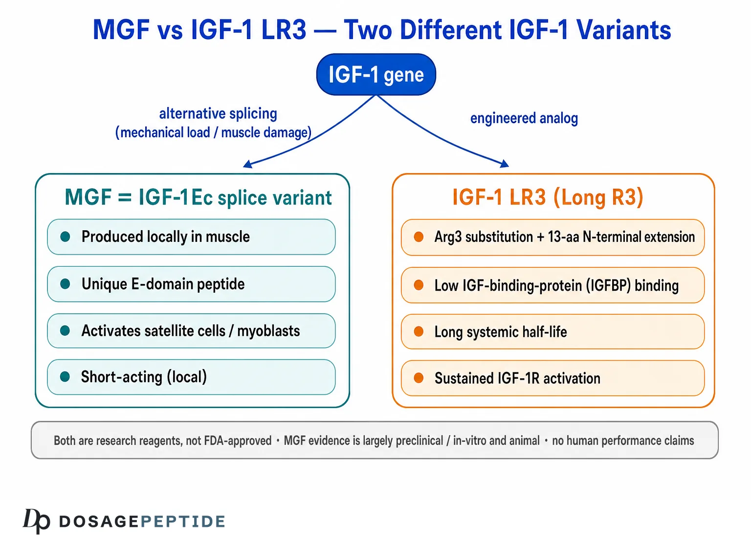
The mechanistic contrast between MGF and IGF-1 LR3 is best understood by asking three questions of each molecule: which receptor does it engage, how does it interact with binding proteins, and over what spatial and temporal scale does it act?
Receptor engagement
IGF-1 LR3 is, at its core, still IGF-1. Its mature domain binds and activates the type 1 IGF receptor (IGF-1R), triggering the canonical downstream cascades — principally the PI3K/Akt/mTOR axis that governs protein synthesis and the Ras/ERK axis associated with proliferation.[9] The engineering did not change the receptor it targets; it changed how long the molecule survives to keep targeting it. In muscle-cell models, sustained IGF-1R signaling through these pathways is well established as a driver of hypertrophy and myoblast proliferation, which is why a long-acting IGF-1R agonist is of interest to muscle biologists.[12] The Akt/PKB node sits at the center of this control: once activated downstream of IGF-1R, Akt simultaneously promotes protein synthesis through mTOR and glycogen synthase kinase-3β, and suppresses protein breakdown by phosphorylating and excluding the FoxO transcription factors that switch on the muscle-wasting ubiquitin-ligase program. This dual push-pull is why the IGF-1–Akt axis is considered a master regulator of muscle mass, although its exact role in the adult muscle response to mechanical load is, by the authors’ own account, less clearly defined than its role in development and regeneration.[20]
The synthetic MGF E-peptide is the more mechanistically puzzling of the two. Multiple studies report that its effects on myoblast proliferation and migration are not abolished by blocking the IGF-1 receptor, implying that the isolated E-domain peptide acts through a different, non-IGF-1R route.[13] In experiments on human myogenic precursor cells, the MGF-Ct24E peptide enhanced proliferation and cell migration through a mechanism explicitly shown to differ from IGF-1 receptor binding, up-regulating components of the fibrinolytic and metalloproteinase systems involved in cell movement.[14] The identity of the receptor or receptors that mediate these E-peptide effects remains, as of the current literature, unresolved — and, as the in-vivo work discussed below shows, the claim of full IGF-1R independence is itself contested by studies in which E-peptide-driven hypertrophy did require the receptor.
Binding-protein interaction and half-life
This is where the two molecules diverge most sharply, and it is the single most important axis of the comparison. Native IGF-1 and, by extension, the mature core of any IGF-1 variant are avidly captured by IGFBPs, which limits free concentration and shortens the effective signaling window. IGF-1 LR3 was purpose-built to escape this capture, and its reduced IGFBP affinity is precisely what confers its extended activity.[3] The intact MGF splice variant, by contrast, was proposed to bind a different binding protein present in muscle interstitial tissue — an interaction predicted to keep it unstable in the unbound form and thus to localize and time-limit its action.[2] In other words, one molecule was engineered for maximal persistence and the other is naturally configured for maximal transience.
The ternary complex and why binding proteins act as a reservoir
To understand the half-life paradox that follows, it helps to know what the binding proteins actually do to circulating IGF-1. In blood, most IGF-1 is not merely bound to a single IGFBP; the majority circulates in a large, roughly 150-kDa ternary complex composed of IGF-1, IGFBP-3 (or IGFBP-5), and a third protein called the acid-labile subunit (ALS).[24] This complex is too large to cross the capillary endothelium freely, so it functions as a slowly releasing circulating reservoir: it prolongs the systemic presence of IGF-1 while simultaneously keeping the bulk of it biologically restrained until it is handed off to tissues. A striking human illustration of how the system is organized comes from ALS deficiency, in which loss of the ternary complex profoundly lowers circulating IGF-1 yet produces only a mild growth phenotype — a discrepancy attributed to the continued action of locally produced, autocrine/paracrine IGF-1 in the tissues themselves.[24] That observation is quietly profound for this article’s theme: even in intact human physiology, local IGF-1 signaling can carry much of the growth load while circulating, protein-bound IGF-1 is dramatically reduced, which is exactly the “local matters” logic that made the MGF splice variant interesting in the first place.
There is an important caveat about half-life that the marketing rarely acknowledges. While analogs with reduced IGFBP affinity show increased potency in vitro, careful pharmacokinetic work has shown that such analogs are actually cleared more rapidly than native IGF-1 after parenteral administration in vivo, because the binding proteins that inhibit activity also serve as a circulating reservoir that prolongs systemic presence.[9] The relationship between IGFBP binding and functional half-life is therefore more nuanced than “LR3 lasts longer,” and this subtlety is frequently lost in popular summaries. A molecule that binds IGFBPs poorly gains free-ligand availability at any given moment but forfeits the reservoir that would otherwise keep it in circulation — so “more active per molecule” and “present in the body longer” are not the same property, and the second does not automatically follow from the first.
Local (autocrine/paracrine) versus systemic action
The conceptual heart of the comparison is spatial scale. MGF, as a splice product induced within loaded muscle fibers, is framed as an autocrine/paracrine signal — produced where it is needed, acting on neighboring cells, and disappearing quickly.[5] IGF-1 LR3, delivered exogenously and resistant to the binding proteins that would normally sequester it, is by design a systemic agent that circulates and can act on IGF-1 receptors throughout the body. This distinction has real biological consequences: a localized, self-limiting signal and a persistent, body-wide one are not merely different doses of the same thing, and the animal literature on localized IGF-1 expression illustrates why. A muscle-restricted, locally acting IGF-1 transgene sustained hypertrophy and preserved regenerative capacity in aging mouse muscle without producing the systemic pathology seen in whole-body IGF-1 overexpression models.[15] Locality, in short, appears to be a feature rather than a limitation.
MGF vs IGF-1 LR3: A Side-by-Side Comparison
The table below summarizes the mechanistic and pharmacological contrasts drawn from the preclinical literature. Every entry describes findings in cell or animal models or in vitro characterization; none should be read as a claim about effects, safety, or use in humans.
| Attribute | MGF (Mechano Growth Factor / IGF-1Ec) | IGF-1 LR3 (Long R3 IGF-1) |
|---|---|---|
| Origin | Endogenous alternative splice variant of the IGF-1 gene, induced by mechanical load/damage | Wholly synthetic engineered analog of IGF-1 |
| Structure | Mature IGF-1 core plus a unique E-domain from a reading-frame shift; research typically uses only the isolated 24-aa C-terminal E-peptide | 83-aa protein: full IGF-1 with Arg substituted at position 3 plus a 13-aa N-terminal extension |
| Primary receptor studied | Isolated E-peptide effects reported as largely IGF-1R-independent (receptor unidentified); in-vivo E-domain-driven hypertrophy has been reported as IGF-1R-dependent | Type 1 IGF receptor (IGF-1R), same as native IGF-1 |
| IGFBP interaction | Intact variant proposed to bind a distinct muscle-interstitial binding protein; kept unstable/local | Deliberately reduced IGFBP affinity to evade sequestration |
| Duration of action | Short and transient; expression peaks early then declines within days | Extended free-ligand availability in vitro; systemic in vivo pharmacokinetics are nuanced |
| Spatial scale | Local — autocrine/paracrine | Systemic when administered exogenously |
| Best-characterized legitimate use | Research tool for satellite-cell and myoblast biology | Cell-culture growth supplement (e.g., CHO cells); in-vitro reproductive biology reagent |
| Strength of muscle evidence | Preclinical/in vitro and animal; includes a notable failed independent replication and conflicting in-vivo E-peptide data | Cell and animal models; little muscle-specific in-vivo characterization published |
| Regulatory status | Not FDA-approved; research reagent | Not FDA-approved as a drug; research/manufacturing reagent |
For readers comparing dosing and reconstitution conventions used in laboratory handling of these reagents, the site maintains structured reference pages: the MGF vial dosage protocol reference and the IGF-1 LR3 vial dosage protocol reference. These are research-handling references, not usage recommendations.
What Does the Current Evidence Show for MGF in Muscle Research?
The MGF evidence base is genuinely interesting but must be read with care, because it contains both supportive findings and a prominent, well-powered contradiction. On the supportive side, the foundational cell-culture work reported that the distinct E-domain of MGF inhibits terminal differentiation of myoblasts while increasing their proliferation — a behavior opposite to that of mature IGF-1, and one that would fit the proposed role of expanding the precursor pool before differentiation.[13] Subsequent work on primary human muscle cell cultures found that the MGF-24aa-E peptide increased the proliferative lifespan and delayed senescence of satellite cells isolated from neonatal and young-adult muscle, though notably not from old-adult muscle.[6]
In transplantation-focused research, a synthetic MGF-Ct24E peptide was reported to improve the engraftment of human myogenic precursor cells in immunodeficient mice by modulating their proliferation and differentiation, with the authors again noting that the effect operated through a mechanism distinct from IGF-1 receptor binding.[16] A companion study identified a pro-migratory activity of the same peptide on human myogenic precursor cells, linking it to up-regulation of urokinase plasminogen activator and matrix metalloproteinase-7.[14] Beyond muscle repair, MGF expression has been examined in the context of inflammation; overexpression in a cardiotoxin muscle-injury model modulated inflammatory cytokine expression and delayed macrophage resolution, suggesting a role in the inflammatory phase of regeneration rather than a purely anabolic one.[17]
The replication problem
Any honest account of MGF must foreground a 2013 study from investigators at two pharmaceutical companies who set out specifically to reproduce the claimed effects of the MGF peptide — and largely could not. Concentrations of the peptide up to 500 ng/mL failed to increase proliferation of mouse C2C12 cells or primary human skeletal muscle myoblasts, failed to inhibit myoblast differentiation, and failed to activate the signaling responses previously attributed to it, even as mature IGF-1 and full-length IGF-1Eb produced robust responses in the same assays.[18] The authors went so far as to question whether the MGF peptide has any physiological role at all. This is not a fringe dissent; it is a serious, well-controlled challenge to the core in-vitro claims, and it means the MGF literature should be described as contested rather than settled. A balanced reading is that the satellite-cell activation hypothesis remains plausible and biologically motivated but is not robustly reproducible across laboratories.
Reconciling the Contradiction: What the In-Vivo E-Peptide Studies Add
The apparent standoff — positive early cell-culture reports on one side, a rigorous failed replication on the other — is not the whole story, and a fair reading requires bringing in a separate body of in-vivo work that studied the E-peptides differently. Rather than dosing cells with a chemically synthesized 24-amino-acid fragment, this line of research used viral vectors to make muscle fibers themselves express the rodent IGF-I isoforms (IGF-IA and IGF-IB, whose E-peptides are the rodent counterparts of the human Ea and Eb/Ec extensions). The distinction matters enormously: it is the difference between exposing cells to a free synthetic peptide of uncertain stability and having living tissue produce the E-domain in its natural molecular and cellular context.
The results of that approach are more supportive of an E-peptide role — but with heavy caveats that cut against the hype. Local viral delivery of the IGF-I isoforms promoted muscle hypertrophy in young mice, and the effect was isoform- and age-dependent: in growing animals both IGF-IA and IGF-IB enlarged muscle, whereas in older (six-month) animals only IGF-IA produced significant hypertrophy, suggesting the bioavailability or receptor affinity of the IGF-IB isoform diminishes with age.[21] This age dependence is a notable echo of the cell-culture finding that the MGF peptide extended proliferative lifespan in young but not old human satellite cells,[6] and it is a sobering signal that whatever the E-domain does, it may do it best precisely in the young, growing tissue that is least representative of the adult humans who are the target of marketing claims.
A follow-up study sharpened the picture by testing whether the E-peptides are actually required for local IGF-I to build muscle. When mature IGF-I lacking any E-peptide extension was expressed virally, it failed to increase muscle mass, whereas the E-peptide-bearing IGF-IA and IGF-IB isoforms drove hypertrophy — evidence that, in this system, the E-domain is necessary for the local anabolic effect rather than being an inert appendage.[22] The same work parsed the downstream genes carefully and found a split: some responses (such as induction of the survival gene Bcl-XL) were abolished in mice engineered to lack functional IGF-1 receptors on their muscle fibers, while others (such as induction of matrix metalloproteinase-13) persisted even without the receptor.[22] That result is the most honest way to characterize the receptor question: E-peptide biology appears to be partly IGF-1R-dependent and partly IGF-1R-independent, rather than cleanly one or the other — which is why blanket claims that “MGF works through its own receptor” overstate what the data support.
Perhaps the most important corrective for anyone imagining a performance benefit comes from a study that expressed the E-peptides alone, using an inactivating mutation to strip out mature IGF-I so that only the E-domain’s contribution remained. The isolated E-peptides did modulate ERK1/2 and Akt phosphorylation and increased satellite-cell proliferation, and the Eb-type peptide drove significant muscle hypertrophy — but that hypertrophy was IGF-1 receptor-dependent, and, critically, the added muscle mass came at the expense of muscle strength.[23] A bigger muscle that is proportionally weaker is a striking illustration of why “more mass” is not automatically “better function,” and it is exactly the kind of physiological consequence that popular summaries never mention. Taken together, the in-vivo E-peptide literature partially rehabilitates the idea that the E-domain does something, while simultaneously demolishing the simplistic version of the story: the effects are context-dependent, age-dependent, only partly receptor-independent, and not unambiguously beneficial even in mice.
What Does the Current Evidence Show for IGF-1 LR3?
The IGF-1 LR3 evidence base is, in a sense, the mirror image of MGF’s: its characterized biology is solid and reproducible, but it is almost entirely outside the muscle-performance context that popular interest assumes. The molecule’s defining property — reduced IGFBP affinity yielding greater in-vitro potency — is well documented, and the class of N-terminally modified IGF-1 analogs to which it belongs was rigorously characterized in the 1990s for its receptor and binding-protein interactions.[9] In dexamethasone-treated (catabolic) rats, LR3IGF-I and the related des(1-3)IGF-I induced marked gut growth and were consistently several-fold more potent than native IGF-1, directly demonstrating that reduced binding-protein interaction translates into greater in-vivo tissue-growth potency.[3] It is worth noting that the tissue in that landmark demonstration was gut, not skeletal muscle — a reminder that the analog’s proven potency is a general growth-factor effect on a sensitive splanchnic tissue, not a muscle-specific result.
Where the molecule truly shines is as a manufacturing and research reagent. As a serum-free cell-culture supplement, LongR3 outperformed insulin at sustaining the viability and productivity of recombinant-protein-expressing CHO cell lines.[4] Transcriptome analysis confirmed that supplementation shifts cells toward enhanced growth and delayed death,[10] and in reproductive biology it has served as a functional IGF-1 substitute during in-vitro oocyte maturation.[11] What is conspicuously thin is published, muscle-specific, in-vivo characterization of IGF-1 LR3 as a hypertrophy agent. The inference that a long-acting IGF-1R agonist should promote muscle growth is mechanistically reasonable — given the established role of IGF-1R/PI3K/Akt/mTOR signaling in muscle anabolism[12] and the central place of the Akt node in that control[20] — but it is largely an extrapolation from IGF-1 biology rather than a direct body of LR3-specific muscle trials.
Local vs Systemic IGF-1: Why the Distinction Matters
The most consequential lesson from putting these two molecules side by side is that how and where IGF-1 signaling is delivered may matter as much as how much of it there is. The animal literature is instructive here. When IGF-1 is expressed in a muscle-restricted, locally acting form, it can sustain myocyte hypertrophy and preserve age-independent regenerative capacity without the systemic pathology associated with whole-body IGF-1 elevation.[15] This is exactly the biological logic that made the local MGF splice variant interesting in the first place: nature appears to use a transient, compartmentalized IGF-1 signal for repair, precisely to avoid the metabolic disturbances that broad, sustained IGF-1 receptor activation would cause — including effects on glucose homeostasis.[2] The human ALS-deficiency picture reinforces the same point from the opposite direction: profoundly reduced circulating IGF-1 is compatible with near-normal growth when local tissue IGF-1 is intact, underscoring that the circulating pool and the local pool are not simply interchangeable.[24]
IGF-1 LR3 sits at the opposite pole of this spectrum. By design, it maximizes systemic, prolonged IGF-1 receptor engagement. From a research standpoint this makes it a powerful and convenient tool for driving IGF-1R signaling in a dish. But the same property that makes it useful in culture is what makes any extrapolation to human physiology fraught: broad, persistent activation of a receptor that regulates growth, metabolism, and cell survival across virtually every tissue is a fundamentally different proposition from a self-limiting local pulse. The IGF-1 axis is also mechanistically entangled with cell proliferation in ways that intersect with cancer biology, which is one reason the muscle-specific splice approach was originally pursued as a way to gain local anabolic signaling “without the oncogenic side effects observed for IGF-1.”[6] None of this constitutes a safety assessment for humans; it simply explains why local-versus-systemic is the axis researchers care about most.
How this connects to growth-hormone secretagogue research
Both molecules sit downstream of, or parallel to, the growth-hormone/IGF-1 axis that also motivates interest in growth-hormone secretagogues. Peptides such as the ghrelin-mimetic secretagogues are studied for their capacity to stimulate the body’s own growth-hormone pulse, which in turn drives hepatic IGF-1 — a fundamentally different strategy from administering an IGF-1 analog directly. Readers exploring how endogenous secretagogue signaling compares to direct IGF-1R agonism may find the overview of GHRP-2 growth-hormone-releasing peptide research a useful contrast, since it highlights the difference between amplifying a physiological hormone axis and bypassing it with an exogenous ligand.
What Are the Limitations of This Research?
The limitations here are substantial and deserve to be stated plainly, because they define the boundary between what is known and what is merely assumed.
The E-peptide identity problem
A foundational uncertainty hangs over the entire MGF field: it is not established that the isolated E-domain peptide actually exists as an independent, cleaved product in living tissue. A widely cited minireview noted that no analogous peptide product of the IGF-1 gene has been identified in or isolated from cultured cells, their conditioned medium, or in-vivo animal tissues or biological fluids — meaning the synthetic MGF peptide studied in the lab may not correspond to any naturally occurring signaling molecule.[7] Much of the MGF literature therefore studies the behavior of a synthetic reagent whose physiological counterpart has not been demonstrated to be released as a free peptide. This is precisely why the in-vivo studies that express the E-domain as part of an IGF-I isoform, rather than as a free fragment, are so valuable: they sidestep the assumption that a cleaved free peptide is the physiologically relevant species.[22]
Reproducibility and model dependence
As detailed above, the central in-vitro claims for the MGF peptide failed to replicate in a rigorous multi-company study.[18] Discrepancies of this kind often stem from differences in peptide synthesis quality, cell-line provenance, passage number, serum conditions, and assay endpoints, and they mean that positive findings should be treated as provisional. The most defensible synthesis is that the effect of a free synthetic MGF peptide on cultured myoblasts is weak or absent, while E-domain sequences expressed endogenously in vivo do produce measurable, though context- and age-limited, effects — two statements that are only superficially contradictory once the difference in experimental design is appreciated. For IGF-1 LR3, the limitation is different: the reproducible evidence is real but confined to cell culture and non-muscle animal models, so its muscle-hypertrophy reputation rests largely on inference from general IGF-1 biology rather than on direct, controlled muscle studies of the analog itself.
Pharmacokinetic oversimplification
Popular descriptions frequently reduce these molecules to a single number — “MGF lasts minutes, LR3 lasts a day” — but the underlying pharmacology is more complicated. Reduced IGFBP affinity increases in-vitro potency yet can actually accelerate in-vivo clearance because binding proteins serve as a stabilizing reservoir.[9] Half-life values circulated in non-scientific sources are rarely traceable to controlled human pharmacokinetic studies, because such studies for these specific reagents are essentially absent. Any single number offered for the “half-life” of either compound in humans should be treated as unsourced until a controlled human pharmacokinetic study is produced, and none currently is.
The species and translation gap
Nearly all of the mechanistic work discussed here comes from rodent tissue, immunodeficient mouse xenografts, isolated human cell cultures, or non-human animal models. Muscle regeneration timing, satellite-cell dynamics, splicing regulation, and IGFBP profiles all differ across species and across ages,[19] and effects observed in a young rodent or a cultured cell line do not transfer automatically to human physiology. The repeated finding that E-domain effects are strongest in young, growing tissue and blunted with age — seen both in cultured human satellite cells[6] and in virally treated mouse muscle[21] — is a particularly pointed reminder that the models most likely to show a positive result are the least representative of the adult humans who feature in performance narratives. There is no adequately controlled human efficacy or safety literature for either molecule as a muscle agent, and their use as unapproved substances carries risks that fall entirely outside the scope of what the research can address.
Regulatory and Safety Status
To be unambiguous: neither Mechano Growth Factor nor IGF-1 LR3 is approved by the FDA or comparable regulators as a drug for building muscle, treating injury, or any other human indication. IGF-1 itself, in its recombinant mecasermin form, is an approved therapeutic only for narrow endocrine indications such as severe primary IGF-1 deficiency — a status that does not extend to these variants and does not endorse their use for performance or body composition. MGF and IGF-1 LR3 are sold and handled as research chemicals and laboratory reagents; IGF-1 LR3 additionally has established legitimate use as a cell-culture supplement.[4] Both fall under anti-doping prohibitions on IGF-1 and its analogs/mimetics in competitive sport, where growth factors affecting muscle are banned at all times. The IGF-1 axis’s deep involvement in cell proliferation is a recognized theoretical concern that the field itself has repeatedly flagged.[6] Anyone encountering these compounds should treat them strictly as objects of laboratory study.
Related research: Follistatin-344 and myostatin inhibition.
Frequently Asked Questions
Are MGF and IGF-1 LR3 the same thing because they both come from IGF-1?
No. MGF (IGF-1Ec) is a naturally occurring splice variant of the IGF-1 gene produced locally in mechanically stressed muscle, defined by a unique E-domain peptide. IGF-1 LR3 is a synthetic 83-amino-acid analog engineered with an Arg3 substitution and a 13-residue N-terminal extension to reduce binding-protein affinity. They share the IGF-1 lineage but differ in origin, structure, receptor engagement, and duration of action, and they are not interchangeable in research.
Which one acts locally and which acts systemically?
MGF is framed as a local, autocrine/paracrine signal: it is produced within loaded muscle, acts on nearby cells, and is transient, with expression peaking early after injury and declining within days. IGF-1 LR3 is designed to act systemically, resisting the binding proteins that would normally sequester it so that it remains available to engage IGF-1 receptors over an extended window when administered exogenously in experimental settings.
Does the MGF peptide work through the IGF-1 receptor?
The honest answer is that it is contested. In several cell-culture studies the isolated MGF E-domain peptide’s effects on myoblast proliferation and migration were not blocked by inhibiting the IGF-1 receptor, implying a separate, still-unidentified mechanism. Yet in-vivo work in which the E-domain was expressed as part of an IGF-I isoform found that E-peptide-driven hypertrophy did depend on the IGF-1 receptor, while some individual gene responses did not. The receptor question is therefore best described as partly dependent and partly independent, not cleanly resolved.
Why is IGF-1 LR3 described as long-acting?
The “Long R3” modifications sharply reduce the analog’s affinity for insulin-like growth factor binding proteins, so more of it remains free and available to activate IGF-1 receptors, increasing in-vitro potency. However, pharmacokinetic research shows reduced binding-protein affinity can also speed clearance in vivo, since binding proteins act as a circulating reservoir. “Long-acting” is therefore an oversimplification of a more nuanced picture, and no controlled human pharmacokinetic study defines a specific human half-life for this reagent.
Is there strong evidence that either builds muscle in humans?
No. There is no adequately controlled human efficacy evidence for either molecule as a muscle agent. MGF’s muscle data are preclinical and in-vitro, a rigorous replication study failed to reproduce its core cell-culture effects, and even the supportive in-vivo E-peptide studies were done in mice, were age-dependent, and in one case produced larger but weaker muscle. IGF-1 LR3’s reproducible biology is in cell culture and non-muscle animal models. Muscle-growth reputations rest on extrapolation from general IGF-1 biology, not direct human trials.
What is IGF-1 LR3 actually used for legitimately?
Its best-documented legitimate application is as a cell-culture growth supplement. It substitutes for insulin or native IGF-1 in serum-free media, where it has been shown to sustain the growth, viability, and productivity of recombinant-protein-producing CHO cells and to support in-vitro oocyte maturation. These industrial and reproductive-biology uses, in cultured cells, are where its characterized value lies — not in human administration.
Why do researchers care about the local-versus-systemic distinction?
Because animal work suggests that a localized, transient IGF-1 signal can support muscle hypertrophy and regeneration while avoiding the metabolic disturbances and broad-tissue effects of sustained systemic IGF-1 receptor activation. A muscle-restricted IGF-1 transgene preserved regenerative capacity in aging mouse muscle without the pathology of whole-body overexpression, and humans lacking the circulating ternary complex still grow nearly normally on local tissue IGF-1. Delivery mode and location may matter as much as the total amount of signaling.
Are these substances legal or approved?
Neither is FDA-approved as a drug for muscle growth or any related human use. Both are handled as research chemicals and laboratory reagents, and both fall under anti-doping bans on IGF-1 and its analogs and mimetics in competitive sport. Recombinant IGF-1 (mecasermin) is approved only for narrow endocrine deficiency indications, which does not extend to these variants. They should be treated strictly as research materials.
Does exercise naturally produce MGF?
Research in rodents indicates that mechanical loading — stretch, resistance-type stimulation, or injury — rapidly and transiently upregulates the IGF-1Ec/Eb splice variant in muscle, followed by a slower rise in the systemic IGF-1Ea variant during repair. This exercise- and damage-responsive splicing is the biological observation that gave MGF its name. Whether a free E-peptide is actually released as a signaling molecule in tissue remains unresolved.
Research-use disclaimer: This article is provided for educational and informational purposes only and describes findings from laboratory, cell-culture, and animal research. It is not medical advice. MGF and IGF-1 LR3 are research reagents that are not approved by the FDA or other regulators for human use, and nothing here should be interpreted as a recommendation, endorsement, or protocol for self-administration. Compounds discussed are intended for use only by qualified researchers in appropriate laboratory settings. Consult a licensed healthcare professional for any medical question.
References
- Kasprzak A, Szaflarski W. Role of Alternatively Spliced mRNA Isoforms of IGF1 in Selected Human Tumors. Int J Mol Sci. 2020;21(19):6995.
- Goldspink G. Changes in muscle mass and phenotype and the expression of autocrine and systemic growth factors by muscle in response to stretch and overload. J Anat. 1999;194(Pt 3):323–334.
- Read LC, Tomas FM, Howarth GS, et al. Insulin-like growth factor-I and its N-terminal modified analogues induce marked gut growth in dexamethasone-treated rats. J Endocrinol. 1992;133(3):421–431.
- Morris AE, Schmid J. Effects of insulin and LongR3 on serum-free Chinese hamster ovary cell cultures expressing two recombinant proteins. Biotechnol Prog. 2000;16(5):693–697.
- Hill M, Goldspink G. Expression and splicing of the insulin-like growth factor gene in rodent muscle is associated with muscle satellite (stem) cell activation following local tissue damage. J Physiol. 2003;549(Pt 2):409–418.
- Kandalla PK, Goldspink G, Butler-Browne G, Mouly V. Mechano Growth Factor E peptide (MGF-E), derived from an isoform of IGF-1, activates human muscle progenitor cells and induces an increase in their fusion potential at different ages. Mech Ageing Dev. 2011;132(4):154–162.
- Matheny RW Jr, Nindl BC, Adamo ML. Minireview: Mechano-growth factor: a putative product of IGF-I gene expression involved in tissue repair and regeneration. Endocrinology. 2010;151(3):865–875.
- Zhao X, McBride BW, Trouten-Radford LM, Burton JH. Effects of insulin-like growth factor-I and its analogues on bovine hydrogen peroxide release by neutrophils and blastogenesis by mononuclear cells. J Endocrinol. 1993;139(2):259–265.
- Cascieri MA, Bayne ML. Analysis of the interaction of insulin-like growth factor I (IGF-I) analogs with the IGF-I receptor and IGF-binding proteins. Horm Res. 1994;41(Suppl 2):80–85.
- Becker J, Timmermann C, Rupp O, et al. Transcriptome analyses of CHO cells with the next-generation microarray CHO41K: development and validation by analysing the influence of the growth-stimulating IGF-1 substitute LongR3. J Biotechnol. 2014;178:23–31.
- Araujo MS, Guastali MD, Paulini F, et al. Molecular and cellular effects of IGF-1 and LongR3-IGF-1 on in vitro maturation of bovine oocytes: comparative study. Growth Horm IGF Res. 2020;55:101357.
- Zanou N, Gailly P. Skeletal muscle hypertrophy and regeneration: interplay between the myogenic regulatory factors (MRFs) and insulin-like growth factors (IGFs) pathways. Cell Mol Life Sci. 2013;70(21):4117–4130.
- Yang SY, Goldspink G. Different roles of the IGF-I Ec peptide (MGF) and mature IGF-I in myoblast proliferation and differentiation. FEBS Lett. 2002;522(1–3):156–160.
- Mills P, Lafrenière JF, Benabdallah BF, El Fahime EM, Tremblay JP. A synthetic mechano growth factor E peptide promotes myoblast proliferation with a modulation of the fibrinolytic system. Exp Cell Res. 2007;313(3):527–537.
- Musarò A, McCullagh K, Paul A, et al. Localized Igf-1 transgene expression sustains hypertrophy and regeneration in senescent skeletal muscle. Nat Genet. 2001;27(2):195–200.
- Mills P, Dominique JC, Lafrenière JF, Bouchentouf M, Tremblay JP. A synthetic mechano growth factor E peptide enhances myogenic precursor cell transplantation success. Am J Transplant. 2007;7(10):2247–2259.
- Sun KT, Cheung KK, Au SWN, Yeung SS, Yeung EW. Overexpression of Mechano-Growth Factor Modulates Inflammatory Cytokine Expression and Macrophage Resolution in Skeletal Muscle Injury. Front Physiol. 2018;9:999.
- Fornaro M, Hinken AC, Needle S, et al. Mechano-growth factor peptide, the COOH terminus of unprocessed insulin-like growth factor 1, has no apparent effect on myoblasts or primary muscle stem cells. Am J Physiol Endocrinol Metab. 2014;306(2):E150–E156.
- Moore DR, McKay BR, Tarnopolsky MA, Parise G. Blunted satellite cell response is associated with dysregulated IGF-1 expression after exercise with age. Eur J Appl Physiol. 2018;118(10):2225–2231.
- Schiaffino S, Mammucari C. Regulation of skeletal muscle growth by the IGF1-Akt/PKB pathway: insights from genetic models. Skelet Muscle. 2011;1(1):4.
- Barton ER. Viral expression of insulin-like growth factor-I isoforms promotes different responses in skeletal muscle. J Appl Physiol (1985). 2006;100(6):1778–1784.
- Barton ER, DeMeo J, Lei H. The insulin-like growth factor (IGF)-I E-peptides are required for isoform-specific gene expression and muscle hypertrophy after local IGF-I production. J Appl Physiol (1985). 2010;108(5):1069–1076.
- Brisson BK, Spinazzola J, Park S, Barton ER. Viral expression of insulin-like growth factor-I E-peptides increases skeletal muscle mass but at the expense of strength. Am J Physiol Endocrinol Metab. 2014;306(8):E965–E974.
- Domené S, Domené HM. The role of acid-labile subunit (ALS) in the modulation of GH-IGF-I action. Mol Cell Endocrinol. 2020;518:111006.