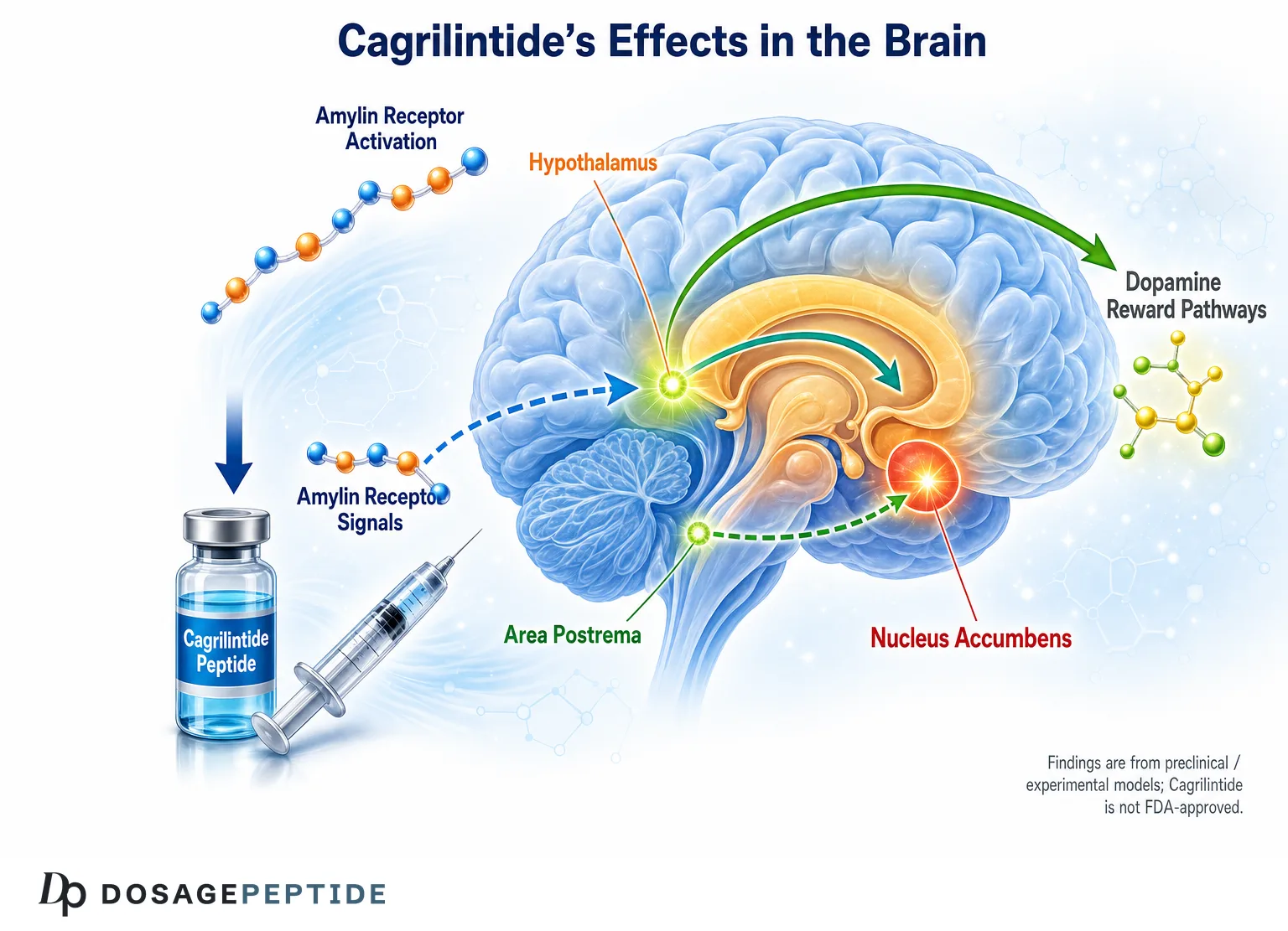
Cagrilintide is an investigational, long-acting amylin analog being studied primarily for weight management, and a central research question is how it engages the brain: cagrilintide brain satiety circuits appetite control is thought to run through the same amylinergic pathways that native amylin uses to signal fullness after a meal. This article reviews, at the correct evidence level, what is known about how cagrilintide and its parent hormone amylin act on the area postrema, nucleus tractus solitarius, hypothalamus, and mesolimbic dopamine system — and where the strongest claims (reward-circuit modulation) rest on rodent data rather than human brain imaging. Cagrilintide is not FDA-approved; it remains an experimental compound used in clinical trials and preclinical research.
Throughout, we separate what has been demonstrated in receptor and cell studies, what comes from rodent models, and what human trials have actually measured (largely appetite and body-weight endpoints, not direct neural readouts). If you are looking for the practical research-handling side rather than the neuroscience, see the companion pages on the Cagrilintide 5 mg dosage protocol and the Cagrilintide 10 mg dosage protocol.
What Is Cagrilintide and Why Does Its Brain Activity Matter?
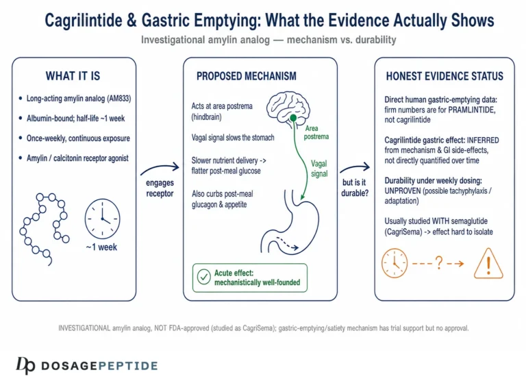
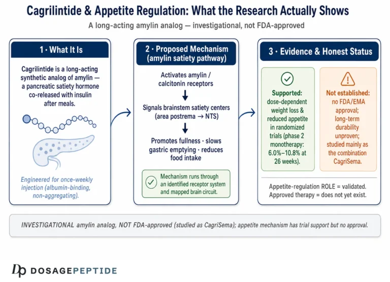
Cagrilintide is a synthetic, acylated analog of human amylin (islet amyloid polypeptide, IAPP), engineered for once-weekly dosing. Amylin is a 37–amino-acid peptide co-secreted with insulin from pancreatic β-cells in response to nutrient intake. In physiological terms, amylin is one of the body’s principal meal-termination signals: it slows gastric emptying, blunts postprandial glucagon, and — most relevant here — acts on the brain to promote satiation and reduce meal size. Cagrilintide was designed to reproduce and extend that satiety signal pharmacologically.
The reason cagrilintide’s brain activity matters is that appetite is fundamentally a central-nervous-system output. Peripheral effects such as delayed gastric emptying contribute to fullness, but the durable reduction in food intake seen with amylin agonists depends on receptors expressed in specific brainstem, hypothalamic, and midbrain nuclei. Understanding those circuits helps explain why amylin analogs reduce not only how much is eaten in a single sitting (homeostatic satiation) but, in preclinical models, how strongly animals will work for palatable, calorie-dense food (hedonic or reward-driven eating). For the peripheral half of the picture, see the discussion of how cagrilintide affects gastric emptying and postprandial metabolism.
An important honesty caveat frames everything below: the detailed reward-circuit findings come almost entirely from studies of amylin, salmon calcitonin, and amylin-receptor agonists in rats and mice. Cagrilintide itself has been characterized mechanistically in receptor pharmacology and in whole-animal weight studies, and its efficacy is established in human trials on appetite and weight, but there is no human neuroimaging dataset dissecting cagrilintide’s effects on dopaminergic reward pathways. Where the text says “amylin,” the finding is a class or mechanism observation; where it says “cagrilintide,” the finding is specific to the compound.
How Does Amylin Signaling Reach the Brain at the Molecular Level?
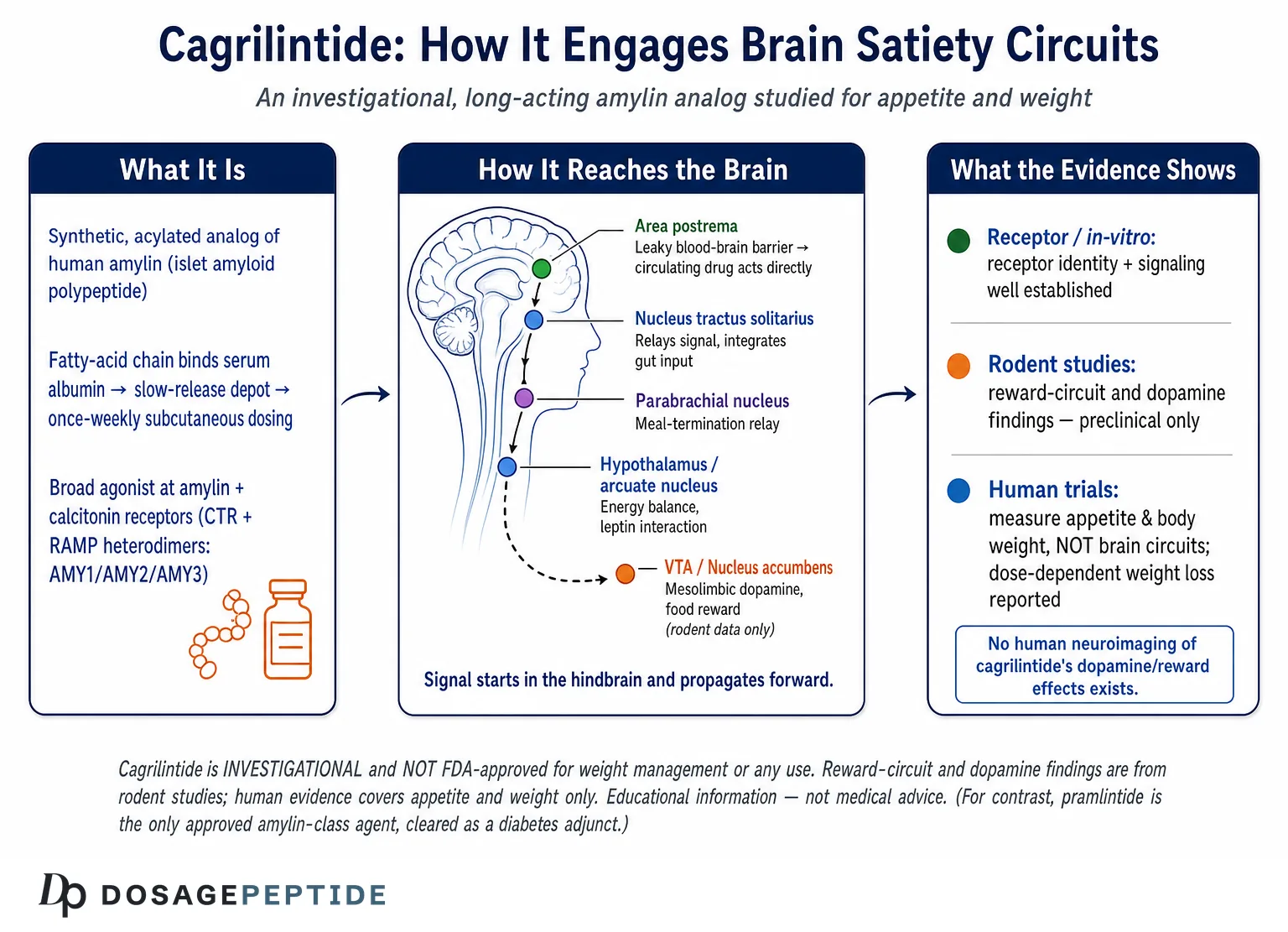
Amylin does not have a single dedicated receptor. Instead, the amylin receptor is a heterodimer: the core calcitonin receptor (CTR, a class B G-protein-coupled receptor) associates with one of three receptor activity-modifying proteins (RAMP1, RAMP2, or RAMP3). This pairing converts the calcitonin receptor into a high-affinity amylin receptor and defines three principal subtypes — AMY1, AMY2, and AMY3 — each with distinct pharmacology.[4] The presence of a RAMP is what gives amylin its selectivity over the closely related hormone calcitonin, and it explains why brain distribution of the response depends on where CTR and each RAMP are co-expressed.
When amylin (or cagrilintide) binds an AMY receptor, the CTR couples primarily to Gs, raising intracellular cyclic AMP and activating protein kinase A; downstream signaling also recruits ERK1/2 and, in some neurons, alters intracellular calcium handling. In the area postrema, amylin-responsive neurons increase their firing, and this activity depends on cGMP/PKG signaling in classic electrophysiology work. The net cellular effect in satiety-relevant neurons is depolarization and increased excitability, which is then relayed forward into the appetite-controlling network.
| Receptor subtype | Molecular composition | Relative amylin sensitivity | Notes relevant to appetite |
|---|---|---|---|
| AMY1 | CTR + RAMP1 | High | Implicated in central food-intake suppression; relevant in hindbrain and midbrain circuits |
| AMY2 | CTR + RAMP2 | Moderate | Less well characterized functionally in feeding behavior |
| AMY3 | CTR + RAMP3 | High | Together with AMY1, central to amylin’s energy-balance effects in rodent knockout work |
| CTR (no RAMP) | Calcitonin receptor alone | Lower for amylin | Prefers calcitonin; still activated by high amylin/analog concentrations |
The functional importance of RAMP1 and RAMP3 specifically has been reinforced by cagrilintide-focused mechanistic work. In a 2025 study, cagrilintide’s ability to lower body weight was shown to depend on brain amylin receptors assembled with RAMP1 and RAMP3, linking the compound’s in-vivo effect directly to defined receptor subtypes rather than to a nonspecific action.[2] This is one of the clearer examples of molecular-level evidence tying cagrilintide to a specific central mechanism.
Where Are Amylin Receptors Expressed in the Brain?
Amylin and calcitonin receptors, together with RAMPs, are distributed across the central nervous system. Autoradiographic and molecular studies place amylin-binding sites densely in the circumventricular organs — particularly the area postrema — where the blood-brain barrier is incomplete, allowing circulating amylin (and, by extension, a systemically dosed analog) to reach neurons directly.[11] Beyond the area postrema, receptor components and amylin-responsive neurons have been mapped in the nucleus tractus solitarius, lateral parabrachial nucleus, ventral tegmental area, and several hypothalamic nuclei including the arcuate nucleus.[5] This anatomically distributed expression is why amylin action is best understood as a network effect rather than a single-node switch.
Which Brain Regions Does Cagrilintide Act On to Control Satiety?
The canonical entry point for amylin’s satiety signal is the hindbrain, and from there the signal propagates rostrally into circuits that shape both how much and how rewarding food feels. The table below summarizes the principal nodes, and the subsections explain the evidence behind each.
| Region | Location | Proposed role in amylin action | Evidence level |
|---|---|---|---|
| Area postrema (AP) | Caudal hindbrain, circumventricular | Primary sensor; direct amylin binding, neuronal activation | Rodent lesion, electrophysiology, receptor mapping |
| Nucleus tractus solitarius (NTS) | Dorsal medulla, adjacent to AP | Relays AP signal; integrates vagal/gut input | Rodent c-Fos, tracing |
| Lateral parabrachial nucleus (LPBN) | Dorsolateral pons | Relay to forebrain; meal-termination signaling | Rodent |
| Arcuate nucleus (ARC) | Mediobasal hypothalamus | Interacts with leptin/melanocortin tone; long-term energy balance | Rodent |
| Ventral tegmental area (VTA) | Midbrain | Dopamine source; modulates food reward and motivation | Rodent |
| Nucleus accumbens (NAc) | Ventral striatum | Dopamine target; hedonic valuation of food | Rodent |
The Area Postrema and Nucleus Tractus Solitarius
The area postrema is widely regarded as amylin’s primary central target. Because it lies outside the blood-brain barrier, circulating amylin reaches AP neurons directly, and lesioning the AP in rodents abolishes much of amylin’s anorectic effect — a classic demonstration that this node is necessary, not incidental. Single-neuron studies have shown that amylin-receptor components and the leptin receptor are co-expressed in individual AP neurons, providing a cellular substrate for cross-talk between the short-term satiation signal (amylin) and the longer-term adiposity signal (leptin).[12] This co-expression is one mechanistic reason amylin agonists are of interest as leptin-sensitizing partners.
Activated AP neurons project to the adjacent nucleus tractus solitarius, which also receives vagal afferents carrying gut-distension and nutrient signals. The NTS integrates these inputs and forwards them to the parabrachial nucleus and hypothalamus. In whole-brain activity mapping, systemic amylin drives neuronal activation in a reproducible constellation of nuclei, and this pattern is markedly attenuated in mice lacking RAMP1 and RAMP3, again anchoring the response to defined amylin-receptor subtypes.[10] Intriguingly, chronic amylin exposure has also been reported to enhance neurogenesis in the adult rat area postrema, suggesting the signal may remodel the very circuit that senses it — though the behavioral significance of this remains an open preclinical question.[13]
The Parabrachial Nucleus and the Meal-Termination Relay
The lateral parabrachial nucleus is a critical relay for meal termination and malaise-related signals. Amylin-responsive projections from the hindbrain converge here before ascending to the forebrain. Distinguishing amylin’s physiological satiation from aversive “stop eating because you feel ill” signaling has been a recurring theme in the literature, because the parabrachial nucleus participates in both. An important nuance from rodent work is that amylin and salmon calcitonin appear to target genetically distinct neuronal populations across the hindbrain and midbrain, which may help separate the desirable satiety effect from nausea-associated pathways — a distinction with obvious relevance to tolerability.[5]
The Hypothalamus and Integration With Leptin
Within the hypothalamus, the arcuate nucleus is a hub where amylin signaling intersects with the melanocortin system and with leptin. Preclinical studies distributed amylin’s central actions across hypothalamic and hindbrain sites and showed that amylin can enhance the anorectic response to leptin, an effect that underlies the long-standing hypothesis that amylin restores leptin responsiveness in diet-induced obesity.[7] For cagrilintide specifically, the hypothalamic contribution is inferred from shared receptor biology rather than from compound-specific hypothalamic mapping, so it should be read as mechanistically plausible and class-supported rather than directly proven for the analog.
How Does Cagrilintide Influence Reward-Driven Eating and Dopamine?
The most compelling — and most frequently over-stated — part of the amylin story concerns reward. Eating is driven not only by homeostatic need but by the hedonic value of palatable food, encoded largely by the mesolimbic dopamine system: dopamine neurons in the ventral tegmental area projecting to the nucleus accumbens. A substantial rodent literature indicates that amylin receptors sit directly within this circuit and can dial down the motivational pull of calorie-dense food. This is the mechanistic basis for the idea that cagrilintide might reduce reward-driven eating, but it is essential to state plainly: the direct reward-circuit evidence is preclinical, and no human study has imaged cagrilintide’s effect on dopamine signaling.
Amylin Receptors on VTA Dopamine Neurons
Calcitonin/amylin receptors are expressed on VTA dopamine neurons, and activating amylin receptors in the VTA reduces food intake in rats — including intake of palatable, high-fat diet — without simply making the animal sick. Foundational work showed that amylin modulates the mesolimbic dopamine system to control energy balance, with VTA amylin-receptor activation suppressing feeding and reducing the motivation to obtain palatable food in operant tasks.[6] Because these effects are produced by delivering agonist directly into the VTA, they demonstrate that the reward node is sufficient on its own to change feeding, not merely a downstream bystander.
Nucleus Accumbens Dopamine and Hedonic Valuation
Downstream of the VTA, amylin signaling reduces phasic dopamine release in the nucleus accumbens core, and this suppression appears to mediate the hypophagia produced by VTA amylin-receptor activation. In other words, the behavioral output (eating less palatable food) tracks a measurable reduction in the dopamine signal that normally encodes food reward. This provides a neurochemical mechanism — not just a behavioral correlation — for how an amylin agonist could blunt the reinforcing value of highly palatable food.
Fat Versus Sugar, Impulsivity, and the VTA-to-Cortex Circuit
More recent rodent work has refined this picture in two directions. First, VTA amylin-receptor activation appears to modulate mesolimbic dopamine differently depending on whether the reward is fat or sugar, suggesting the circuit is not a uniform “reward off switch” but a nutrient-sensitive modulator.[8] Second, amylin has been shown to act on a ventral-tegmental-area-to-medial-prefrontal-cortex circuit to suppress food intake and, notably, to reduce impulsive food-directed behavior — connecting amylin signaling to cognitive control over eating, not just to raw consumption.[9] These findings are why amylin analogs are discussed in the context of reward-driven and even impulsive eating, but again they are rodent mechanistic studies; extrapolating them to human eating-behavior claims is not warranted, and cagrilintide must never be framed as a treatment for eating disorders.
What Makes Cagrilintide Long-Acting Compared With Native Amylin?
Native human amylin is pharmacologically inconvenient: it has a very short half-life (on the order of minutes) and an intrinsic tendency to aggregate into amyloid fibrils, which is both a stability problem and a biological liability. The first clinically used amylin analog, pramlintide, solved the aggregation problem with amino-acid substitutions borrowed partly from the non-amyloidogenic rat sequence, but it still required dosing at each meal because of its short duration of action.
Cagrilintide extends the concept. It is an acylated amylin analog: a fatty-acid (lipid) moiety is attached so that the peptide binds reversibly to serum albumin in the circulation. Albumin binding creates a slow-release depot effect and dramatically prolongs residence time, supporting once-weekly subcutaneous administration. Structural modifications also improve solubility and reduce the aggregation tendency of the parent hormone. The result is a compound that maintains sustained agonism at amylin (and calcitonin) receptors across a full week rather than producing a short satiety pulse. This pharmacokinetic profile is the practical reason cagrilintide is studied as a weekly agent alongside weekly GLP-1 receptor agonists such as semaglutide.
| Property | Native human amylin | Pramlintide | Cagrilintide |
|---|---|---|---|
| Dosing frequency (research use) | Not used therapeutically (unstable) | With each main meal | Once weekly |
| Approximate half-life | Minutes | Roughly 20–45 minutes | Days (supports weekly dosing) |
| Key structural feature | Amyloidogenic native sequence | Proline substitutions reduce aggregation | Lipidation + substitutions; albumin binding |
| Receptor targets | AMY1/2/3 (CTR+RAMP) | Amylin receptors | Amylin and calcitonin receptors (broad agonism) |
| Regulatory status | N/A | Approved (adjunct in diabetes) | Investigational, not FDA-approved |
One pharmacologically meaningful difference is that cagrilintide is often described as a broader agonist that engages both amylin and calcitonin receptors, whereas selective amylin agonists engage the AMY subtypes more narrowly. This broader receptor coverage may contribute to durable efficacy, but it also means the compound’s central footprint could include calcitonin-receptor-bearing neurons, a consideration that is still being characterized. Half-life values in the table are approximate and drawn from general pharmacology of these agents; exact human parameters for cagrilintide are reported in its clinical trial literature.
What Does the Evidence Actually Show, Organized by Study Type?
Because YMYL content demands precision about evidence level, this section separates the literature into receptor/in-vitro work, animal studies, and human trials. The single most important takeaway is that the reward-circuit story is preclinical, while the human evidence is about appetite and weight endpoints.
| Study type | What it establishes | What it cannot establish |
|---|---|---|
| Receptor / in-vitro | CTR+RAMP composition, agonist potency, signaling (cAMP/PKA) | Behavioral or clinical outcomes |
| Rodent (lesion, microinjection, electrophysiology, c-Fos) | Necessary/sufficient brain nodes; dopamine modulation; reward-behavior effects | Direct translation to human reward circuits |
| Human clinical trials | Appetite ratings, energy intake, body-weight change, safety | Direct neural-circuit or dopamine measurements |
In-Vitro and Receptor Pharmacology
At the molecular level, the evidence is strong and mechanistic. The identity of the amylin receptor as a CTR-plus-RAMP heterodimer is well established, and reviews of amylin pharmacology detail how RAMP association tunes ligand selectivity and downstream signaling.[4] For cagrilintide, the demonstration that its weight-lowering effect depends on RAMP1/RAMP3-containing brain receptors is the key receptor-level link between the compound and a central mechanism.[2] Whether a compound described as validated in appetite regulation truly meets a research-grade evidentiary bar is examined further in the discussion of whether research validates cagrilintide in appetite regulation.
Animal Studies
The rodent literature is where the circuit-level mechanism lives. Area-postrema lesion studies establish necessity; VTA and NAc microinjection and dopamine-recording studies establish that the reward node is sufficient to change feeding; whole-brain activity mapping in RAMP knockouts establishes receptor dependence.[10][6] Collectively, these studies make a coherent and mechanistically detailed case that amylin agonism reduces both homeostatic and hedonic feeding in rodents. They are the scientific foundation for interest in cagrilintide’s reward effects, but they are not human data.
Human Clinical Trials
Human evidence for cagrilintide is genuinely strong on its actual endpoints — appetite and weight — but it does not measure brain circuits. In a phase 2 dose-finding trial published in The Lancet, once-weekly cagrilintide monotherapy produced dose-dependent, clinically meaningful body-weight reductions over 26 weeks in adults with overweight or obesity, with a tolerability profile dominated by gastrointestinal effects.[1] Combination trials of cagrilintide with semaglutide have reported even larger weight reductions in adults with overweight or obesity, including those with type 2 diabetes.[3] These outcomes are consistent with reduced appetite and food intake, and they are the closest human proxy we have for the satiety-circuit mechanism — but the inference from “less weight and appetite” to “dopamine reward circuit modulation” remains an interpretation, not a measured human finding.
How Do Cagrilintide and Semaglutide Engage Complementary Brain Circuits?
A major reason cagrilintide is studied alongside the GLP-1 receptor agonist semaglutide is that the two hormones act on overlapping but distinct central pathways. GLP-1 receptors and amylin receptors are both expressed in appetite-controlling regions including the hindbrain and hypothalamus, yet they engage different receptor systems and, in preclinical work, different neuronal populations. The hypothesis is that co-agonism recruits a broader set of satiety circuits than either alone, producing additive or better-than-additive appetite suppression.
The human combination data are consistent with this complementarity: cagrilintide-plus-semaglutide regimens have produced some of the largest weight reductions reported for pharmacological appetite modulation.[3] Mechanistically, amylin’s prominent role in the reward and area-postrema pathways and GLP-1’s role in hindbrain and hypothalamic aversion/satiation circuits are thought to be partly non-overlapping, which would explain why combining them yields more than a simple duplication of effect. Researchers handling the fixed-combination format should review the Cagrilintide + Semaglutide blend dosage protocol for reconstitution and handling specifics. As with the monotherapy data, this remains an appetite/weight-endpoint literature, not a neural-circuit imaging literature.
What Experimental Models Are Used to Study These Circuits?
Understanding the evidence requires understanding the tools that generated it, because each model has a characteristic blind spot.
- Receptor expression systems. CTR and RAMP subtypes are co-expressed in cultured cells to measure agonist potency and signaling. These define pharmacology cleanly but say nothing about behavior.
- Targeted brain-region microinjection. Agonists or antagonists are delivered directly into the VTA, NAc, area postrema, or hypothalamus to test whether that node is sufficient or necessary. This is how VTA sufficiency for reward effects was shown.
- Lesion studies. Ablating the area postrema tests necessity; loss of the anorectic response after AP lesion is a cornerstone finding.
- Genetic knockouts. RAMP1/RAMP3 knockout mice reveal which receptor subtypes carry the signal, as used in whole-brain activity mapping.[10]
- Neuronal activity mapping. c-Fos immunolabeling and whole-brain imaging localize which nuclei respond to systemic amylin.[7]
- Operant and behavioral assays. Progressive-ratio responding for palatable food and impulsivity tasks quantify motivation and reward, not just consumption.[9]
- Human randomized trials. Placebo-controlled trials measure appetite scores, ad-libitum energy intake, body weight, and safety — the translational endpoints.[1]
The gap this list makes obvious is the missing rung: there is no published human functional-neuroimaging model tying cagrilintide to dopamine or reward-circuit activity. That rung is exactly where the strongest-sounding claims would need support, and it does not yet exist.
What Are the Relevant Pharmacokinetics for Central Action?
For a peptide to influence brain satiety circuits after subcutaneous dosing, it must reach receptor-bearing neurons at effective, sustained concentrations. Cagrilintide’s pharmacokinetics are built around that requirement. The albumin-binding depot slows absorption and clearance, producing the flat, prolonged exposure that supports weekly dosing and continuous receptor occupancy. Circumventricular access matters here: because the area postrema lacks a complete blood-brain barrier, a systemically dosed amylin analog can act on that primary sensor without needing to cross an intact barrier, which simplifies the central-access problem for at least the hindbrain node.
| Parameter | General characteristic for cagrilintide | Relevance to brain action |
|---|---|---|
| Route | Subcutaneous injection | Systemic delivery to circumventricular organs |
| Absorption | Slow, depot-like via albumin binding | Avoids sharp peaks; steady receptor engagement |
| Half-life | Long enough to support once-weekly dosing | Sustained central agonism between doses |
| Primary CNS access point | Area postrema (leaky BBB) | Direct action on the principal amylin sensor |
| Dose-response | Dose-dependent appetite/weight effect in trials | Consistent with graded receptor occupancy |
The dose-dependence observed in the phase 2 trial — larger effects at higher weekly doses — is consistent with graded central receptor occupancy, though it does not by itself prove where in the brain the additional effect is generated.[1] Researchers planning reconstitution and concentration calculations for experimental work can consult the peptide reconstitution guide and the dosage calculator for the arithmetic, keeping in mind these tools are for laboratory research contexts, not human administration guidance.
What Are the Limitations and Open Questions?
An honest appraisal of cagrilintide and brain satiety circuits has to foreground what is not known.
- No human reward-circuit data. The dopamine and VTA/NAc findings are rodent results. There is no human imaging or neurochemical study showing cagrilintide changes dopaminergic reward signaling. Claims that cagrilintide “reduces food reward” in people are extrapolations from animal mechanism plus human weight loss.
- Species differences in receptor pharmacology. RAMP expression patterns and CTR splice variants differ between rodents and humans, so the precise circuit weighting may not transfer directly.
- Satiety versus malaise. Some of amylin’s central action overlaps anatomically with nausea/aversion pathways. Separating a desirable satiety signal from an aversive one is an active research problem, and gastrointestinal adverse events in trials are a real-world reflection of this overlap.
- Combination attribution. In cagrilintide-plus-semaglutide regimens, it is difficult to attribute a given central effect to amylin versus GLP-1 signaling without dedicated mechanistic studies in humans.
- Long-term neuroadaptation. Whether sustained weekly agonism produces receptor desensitization, compensatory circuit changes, or the neurogenesis effects seen preclinically in humans is unknown.
- Regulatory status. Cagrilintide is investigational and not FDA-approved; conclusions from trials are provisional pending completion and regulatory review of the full program.
These caveats do not diminish the mechanistic elegance of the amylin story; they define the boundary between what is established and what is hypothesized. For a broader vocabulary of the receptors and circuits named here, see the peptide glossary.
What Safety Signals Are Reported in the Research?
Because central satiety signaling and gastrointestinal signaling share circuitry, the most consistently reported adverse effects of amylin agonists are gastrointestinal. In cagrilintide trials, nausea, vomiting, diarrhea, constipation, and decreased appetite are the dominant events, generally mild-to-moderate and often transient, with a tendency to attenuate over time and to be more frequent at higher doses and during dose escalation.[1] In combination with semaglutide, gastrointestinal events are more frequent than with placebo, consistent with additive engagement of nausea-adjacent pathways, but again predominantly mild-to-moderate in the reported trials.[3]
From a mechanistic standpoint, the concentration of adverse effects in the gastrointestinal domain is expected: the area postrema and parabrachial nucleus that mediate satiety also participate in nausea and emesis, so a signal that says “stop eating” and a signal that says “you feel unwell” are anatomically neighbors. This is precisely why the preclinical effort to identify genetically distinct amylin-responsive neuron populations matters — the therapeutic goal is a satiety signal cleanly separated from malaise.[5] No published human data indicate that cagrilintide adversely affects mood or cognition through its reward-circuit activity, but the absence of dedicated human neuropsychiatric studies means this is an area of uncertainty rather than a clean bill of health. Cagrilintide should never be characterized as a treatment for binge eating, food addiction, or any eating disorder; the reward-circuit findings are mechanistic and preclinical.
How Was Cagrilintide Engineered From the Native Amylin Sequence?
The molecular story behind cagrilintide is essentially a story about defeating two liabilities of the parent hormone: rapid clearance and self-aggregation. Human amylin (IAPP) is intrinsically amyloidogenic — a stretch of its sequence, roughly residues 20–29, drives the peptide to stack into cross-β fibrils. In the pancreas, this same tendency produces the islet amyloid deposits characteristic of type 2 diabetes, so the aggregation problem is not merely a formulation nuisance but a feature of amylin biology. A peptide that aggregates is difficult to keep in solution, loses potency as monomer is consumed into fibrils, and can generate cytotoxic intermediate species. Any drug built on the amylin scaffold therefore has to be stabilized against this behavior before dosing, shelf-life, or receptor pharmacology can even be discussed.
The first-generation solution, pramlintide, borrowed three proline substitutions inspired by the naturally non-amyloidogenic rat amylin sequence (prolines are “β-sheet breakers” that disrupt fibril stacking). That fixed the aggregation problem but left the short half-life untouched, which is why pramlintide is dosed at each meal. Cagrilintide represents a more thorough redesign: multiple residue substitutions restore stability and receptor activity, while an attached fatty-acid (acyl) chain provides the albumin-binding handle that extends duration. The acylation strategy is the same broad principle used to make semaglutide long-acting, adapted to the amylin backbone. The engineering goal was a monomeric, soluble, protease-resistant peptide that still activates amylin and calcitonin receptors with high potency — a non-trivial balance, because the modifications that suppress aggregation can also blunt receptor binding if placed carelessly.[4]
Two consequences of this design matter for the brain-satiety question. First, because cagrilintide is stabilized as a soluble monomer, the species reaching central receptors is a well-defined agonist rather than a mixture of monomer and oligomer, which makes its pharmacology cleaner and more predictable than native amylin’s. Second, the same amyloid-prone chemistry that had to be engineered away in the drug is a reminder that amylin biology and amyloid biology are intertwined — a point of ongoing scientific interest that is distinct from, and should not be conflated with, the compound’s appetite effects. The structure-activity details here are drawn from amylin pharmacology reviews and general peptide-engineering principles rather than from any claim that a specific residue produces a specific human brain effect.
How Does Amylin Signaling Integrate With Other Gut–Brain Satiety Hormones?
Amylin does not act in isolation. Meal termination in an intact organism is the summed output of several gut- and pancreas-derived signals — cholecystokinin (CCK) from the small intestine, peptide YY (PYY) and GLP-1 from L-cells, and the longer-acting adiposity signal leptin from fat tissue. A recurring theme in the rodent literature is that amylin is unusually good at potentiating these partners rather than merely adding to them, and this synergy is part of why amylin analogs are attractive as combination agents.
The best-characterized interaction is with CCK. In rodents, amylin and CCK each suppress food intake and slow gastric emptying on their own, and co-administration produces a greater-than-additive reduction in intake, an effect attributed to convergent signaling on hindbrain satiation circuitry.[14] The leptin interaction is equally important: amylin enhances leptin’s anorectic action and is thought to restore leptin responsiveness in diet-induced obesity, which is mechanistically grounded in the co-expression of amylin- and leptin-receptor components in single area-postrema neurons discussed earlier.[7] The table below organizes these relationships; note that every entry reflects preclinical mechanism except the GLP-1/semaglutide row, which additionally has human weight-endpoint support.
| Partner signal | Source | Nature of interaction with amylin | Evidence level |
|---|---|---|---|
| Cholecystokinin (CCK) | Small-intestinal I-cells | Greater-than-additive suppression of food intake; convergent hindbrain satiation | Rodent |
| Leptin | Adipose tissue | Amylin enhances leptin’s anorectic effect; proposed leptin re-sensitization | Rodent |
| GLP-1 / semaglutide | Intestinal L-cells / drug | Partly non-overlapping circuits; additive appetite/weight effect | Rodent mechanism + human weight endpoints |
| PYY | Intestinal L-cells | Complementary satiation signaling to the hindbrain | Rodent / mechanistic |
The practical inference researchers draw from this integration picture is that amylin’s effect size in a whole organism reflects a network of cooperating signals, not a single ligand-receptor event. It also cautions against attributing every observed weight or appetite change in a combination regimen to amylin alone — a point that recurs in the limitations discussion above regarding cagrilintide-plus-semaglutide attribution.
Where Does Cagrilintide Sit Among Amylin Analogs in Development?
Cagrilintide is one member of a widening class of amylin-based agents, and placing it in that landscape clarifies both what is distinctive about it and what the class as a whole is trying to achieve. The lineage runs from native amylin, to the meal-time analog pramlintide (the only amylin-class agent with regulatory approval, cleared as a diabetes adjunct in 2005), to long-acting weekly analogs designed for weight management. Cagrilintide is a broad amylin/calcitonin-receptor agonist; a newer wave of “selective” long-acting human-amylin analogs aims to keep the satiety efficacy while further improving gastrointestinal tolerability.
Among these newer agents, petrelintide (a long-acting human amylin analog) has reported positive phase 2 results in overweight and obesity, with weight reductions in the double-digit-percentage range and a gastrointestinal tolerability profile its developers describe as favorable, and it has been moving toward phase 3 development.[15] The medicinal-chemistry rationale for such next-generation analogs — potent, stable, long-acting human-amylin sequences engineered against aggregation — has been described in the peer-reviewed literature.[16] Other investigational directions include unimolecular dual agonists that combine amylin and GLP-1 activity in a single peptide. All of these agents, cagrilintide included, remain investigational for weight management; none should be presented as an approved obesity therapy.
| Agent | Class / mechanism | Dosing concept | Development status (general) |
|---|---|---|---|
| Pramlintide | Amylin-receptor agonist | Each main meal | Approved diabetes adjunct (2005) |
| Cagrilintide | Broad amylin/calcitonin-receptor agonist | Once weekly | Investigational (mono and with semaglutide) |
| Petrelintide | Long-acting selective human-amylin analog | Once weekly | Investigational (positive phase 2 reported) |
| Amylin + GLP-1 dual agonists | Single peptide, two receptor systems | Varies (weekly concepts) | Investigational / earlier stage |
Cagrilintide’s position in this table is that of the reference long-acting amylin analog whose combination with semaglutide has generated the most-discussed human weight data, while the selective next-generation analogs are pursuing the same satiety mechanism with an emphasis on cleaner tolerability. From a mechanism-of-brain-action standpoint, all of these agents are presumed to converge on the amylin-receptor circuitry described throughout this article; the differences are pharmacokinetic and receptor-selectivity refinements rather than a new central pathway.
What Do Cagrilintide’s Pharmacokinetic Numbers Tell Us About Central Exposure?
Earlier sections described cagrilintide’s albumin-binding depot qualitatively; the quantitative parameters sharpen the picture of how the compound sustains central receptor engagement. Dedicated phase 1 pharmacokinetic work in people with overweight or obesity characterized cagrilintide as having an elimination half-life on the order of about a week — long enough that once-weekly subcutaneous dosing produces a relatively flat exposure curve rather than the sharp peaks and troughs of a short-acting peptide.[17] A half-life close to the dosing interval also means the drug accumulates across the first several weekly doses before reaching steady state, which is one reason clinical protocols use gradual dose escalation.
Why does this matter for the brain? Central satiety circuits respond to the concentration of agonist available at their receptors, and a flat, sustained exposure profile means those receptors experience continuous rather than pulsatile stimulation. For the area postrema — which, lacking a complete blood-brain barrier, samples circulating drug directly — steady plasma levels translate fairly directly into steady central agonism. The gradual accumulation to steady state is also mechanistically consistent with the clinical observation that gastrointestinal side effects tend to be most prominent during dose escalation and to attenuate thereafter, since the nausea-adjacent hindbrain circuitry is exposed to rising then plateauing agonist levels rather than a single large step. As with all pharmacokinetic framing here, these parameters describe systemic and circumventricular exposure; they do not by themselves demonstrate activity at any deeper forebrain or reward node, which would require the human circuit-level studies that do not yet exist.
How Do Amylin’s Central Effects Relate to Body Weight Beyond Appetite?
It is tempting to treat “appetite suppression” and “weight loss” as the same statement, but the amylin literature is a useful reminder that they are not identical. Body weight is a balance between energy intake and energy expenditure, and while amylin’s dominant, best-established action is on the intake side — reduced meal size and lowered food intake through the hindbrain and hypothalamic circuits described above — there is a longstanding preclinical interest in whether amylin also influences the defense of body weight and adiposity over the longer term. Because amylin- and leptin-receptor components are co-expressed in area-postrema neurons, amylin has been framed not only as an acute meal-termination signal but as a candidate adiposity signal that reports on and helps regulate fat stores.[12]
This distinction has a practical corollary that is increasingly discussed for the amylin class as a whole: the interest in whether amylin analogs preserve lean mass relatively better than some other appetite-suppressing strategies. That is a comparative, still-maturing research question rather than a settled property of cagrilintide, and it should be described as an area of active investigation, not as a proven advantage of the compound. What the human cagrilintide trials actually demonstrate remains anchored to their measured endpoints — total body-weight change, appetite, energy intake, and safety — and body-composition or expenditure claims should be held to that same evidentiary discipline. The mechanistic plausibility that a satiety hormone integrated with leptin signaling could influence more than moment-to-moment appetite is real; the human proof for specific body-composition benefits of cagrilintide is not something to overstate.
For the brain-circuit theme of this article, the takeaway is that the same distributed network — area postrema, nucleus tractus solitarius, parabrachial nucleus, hypothalamus, and the mesolimbic reward nodes — is what links a weekly injection to a durable shift in energy balance rather than a transient loss of appetite. The circuit does not simply switch off hunger; in the preclinical framing it recalibrates how much food the animal defends, which is why chronic amylin agonism produces sustained rather than fleeting reductions in intake. Whether that recalibration is exactly mirrored in the human brain is, once more, an inference from behavior and weight data rather than a directly imaged human finding.
What Is Known About Receptor Signaling Kinetics and Desensitization?
A question that naturally follows from once-weekly, continuous agonism is whether the amylin receptor desensitizes — that is, whether sustained stimulation blunts the very response the drug is meant to produce. This is a real concern for many G-protein-coupled receptors, where prolonged agonist exposure can trigger receptor phosphorylation, β-arrestin recruitment, internalization, and downregulation. For the calcitonin-receptor-based amylin receptors, the signaling cascade is dominated by Gs-coupled cyclic-AMP/PKA activation, with contributions from ERK1/2 and, in some neurons, altered calcium handling — and how durably that cascade is maintained under weekly dosing is a legitimate pharmacological question rather than a solved one.[4]
The clinical observation most relevant here is indirect but reassuring: in the phase 2 monotherapy trial, cagrilintide produced dose-dependent weight reduction sustained across 26 weeks, which is not the pattern one would expect if central amylin receptors rapidly and fully desensitized.[1] Sustained efficacy over months implies that whatever receptor regulation occurs, it does not abolish the functional response over that horizon. That said, the molecular kinetics of amylin-receptor desensitization in the specific brain nuclei that matter — and whether the broad amylin/calcitonin-receptor engagement of cagrilintide behaves differently from a selective amylin agonist in this respect — have not been characterized in humans. This remains one of the open receptor-pharmacology questions flagged in the limitations section, alongside the possibility of compensatory circuit adaptation under long-term agonism.
There is also a subtler point about how the two ends of cagrilintide’s pharmacology interact. A slowly rising, albumin-buffered exposure profile is not only convenient for weekly dosing; it may also be favorable with respect to desensitization, because receptors are less likely to be driven into rapid downregulation by the gentle concentration ramp of an accumulating long-acting agent than by the sharp spikes of a short-acting one. This is a mechanistic hypothesis consistent with the sustained clinical effect rather than a directly measured property of the receptor in the human brain, and it illustrates a theme running through this entire article: cagrilintide’s central story is one where robust molecular and rodent mechanism, solid human appetite-and-weight data, and a genuine gap at the level of human neural-circuit measurement all coexist. Reading the compound honestly means holding those three tiers of evidence separately rather than collapsing them into a single, overreaching claim about what a weekly injection does to the human brain’s reward system.
Frequently Asked Questions
Does cagrilintide directly change dopamine in the human brain?
There is no human study measuring cagrilintide’s effect on dopamine. The link between amylin agonism and reduced mesolimbic dopamine comes from rodent experiments in which agonist is delivered into the ventral tegmental area and dopamine is recorded in the nucleus accumbens. Human cagrilintide trials measure appetite and body weight, so any statement about human dopamine or reward signaling is an extrapolation from animal mechanism, not a measured clinical finding.
How is cagrilintide different from semaglutide in the brain?
Semaglutide is a GLP-1 receptor agonist and cagrilintide is an amylin (and calcitonin) receptor agonist. They target overlapping appetite regions such as the hindbrain and hypothalamus but engage different receptors and, in preclinical work, partly different neuron populations. That non-overlap is the rationale for combining them: co-agonism appears to recruit a broader satiety network, which is consistent with the larger weight reductions reported for the combination in human trials.
Which brain region is most important for cagrilintide’s satiety effect?
The area postrema in the caudal hindbrain is considered the primary sensor. It lies outside the blood-brain barrier, so circulating amylin analogs reach it directly, and lesioning it in rodents abolishes much of amylin’s appetite-suppressing effect. From there the signal relays through the nucleus tractus solitarius and parabrachial nucleus to hypothalamic and midbrain circuits that shape both meal size and food reward.
Is cagrilintide FDA-approved for weight loss or appetite?
No. Cagrilintide is an investigational compound studied in clinical trials and is not FDA-approved as of this writing. Its monotherapy and combination trials have reported meaningful weight reductions, but it remains experimental. It should not be described as an approved therapy, and it is never appropriate to present it as a treatment for eating disorders or reward-related eating conditions.
Does cagrilintide reduce cravings or reward-driven eating?
In rodent models, amylin-receptor activation reduces the motivation to work for palatable, calorie-dense food and blunts impulsive food-directed behavior through a ventral-tegmental-area circuit. Human trials show reduced appetite and food intake, which is compatible with reduced reward-driven eating, but no human study has directly measured craving or reward-circuit activity under cagrilintide, so this remains mechanistically plausible rather than clinically proven.
Why does cagrilintide only need weekly dosing?
Native amylin lasts only minutes and aggregates readily. Cagrilintide is lipid-modified so it binds reversibly to serum albumin, creating a slow-release depot that greatly prolongs its residence in the body. This produces steady, sustained activation of amylin receptors across a week, unlike meal-time agents such as pramlintide, and it aligns cagrilintide with weekly GLP-1 agonists for combination use.
What receptors does cagrilintide activate?
Cagrilintide activates amylin receptors, which are calcitonin receptors paired with a RAMP protein (subtypes AMY1, AMY2, AMY3), and it also engages calcitonin receptors more broadly than some selective amylin agonists. Mechanistic work indicates its weight-lowering effect depends specifically on brain amylin receptors containing RAMP1 and RAMP3, tying the compound’s in-vivo action to defined receptor subtypes.
Are the brain effects of cagrilintide proven in humans or animals?
The detailed circuit and reward findings are from animal studies — lesion, microinjection, electrophysiology, and knockout models in rats and mice. Human evidence is limited to appetite, energy-intake, weight, and safety endpoints from clinical trials. The molecular receptor pharmacology is well established, but the specific neural-circuit and dopamine claims should be read as preclinical, not human-proven.
How does cagrilintide compare with newer amylin analogs like petrelintide?
Cagrilintide is a broad amylin/calcitonin-receptor agonist and is the long-acting amylin analog with the most-discussed human weight data, largely because of its combination with semaglutide. Newer selective long-acting human-amylin analogs such as petrelintide pursue the same core satiety mechanism while aiming for improved gastrointestinal tolerability, and petrelintide has reported positive phase 2 results. All of these agents are investigational for weight management and act through the same amylin-receptor circuitry; the differences are pharmacokinetic and receptor-selectivity refinements, not a fundamentally different brain pathway.
Does amylin work together with other gut hormones to control appetite?
Yes, in preclinical models amylin cooperates with several other satiety signals. It produces greater-than-additive suppression of food intake with cholecystokinin (CCK), enhances leptin’s appetite-reducing effect, and engages partly non-overlapping circuits from GLP-1. This cooperativity is a major reason amylin analogs are studied as combination agents. Most of this integration evidence is from rodents; the strongest human data are for the cagrilintide-plus-semaglutide combination, and even there the effect is measured as appetite and weight change rather than as a mapped neural interaction.
References
- Lau DCW, Erichsen L, Francisco AM, et al. Once-weekly cagrilintide for weight management in people with overweight and obesity: a multicentre, randomised, double-blind, placebo-controlled, dose-finding phase 2 trial. The Lancet. 2021. https://www.thelancet.com/journals/lancet/article/PIIS0140-6736(21)01751-7/fulltext
- Cagrilintide lowers body weight through brain amylin receptors 1 and 3. eBioMedicine. 2025. https://www.thelancet.com/journals/ebiom/article/PIIS2352-3964(25)00280-4/fulltext
- Cagrilintide-Semaglutide in Adults with Overweight or Obesity and Type 2 Diabetes. New England Journal of Medicine. 2025 (PubMed 40544432). https://pubmed.ncbi.nlm.nih.gov/40544432/
- Hay DL, Chen S, Lutz TA, Parkes DG, Roth JD. Amylin: Pharmacology, Physiology, and Clinical Potential. Pharmacological Reviews. 2015;67(3):564–600. https://doi.org/10.1124/pr.115.010629
- Central nervous system pathways targeted by amylin in the regulation of food intake. PubMed 39426704. https://pubmed.ncbi.nlm.nih.gov/39426704/
- Mietlicki-Baase EG, et al. Amylin modulates the mesolimbic dopamine system to control energy balance. Neuropsychopharmacology (PMC4443949). https://pmc.ncbi.nlm.nih.gov/articles/PMC4443949/
- Amylin activates distributed CNS nuclei to control energy balance. Physiology & Behavior (PMC4113606). https://pmc.ncbi.nlm.nih.gov/articles/PMC4113606/
- Ventral tegmental area amylin receptor activation differentially modulates mesolimbic dopamine signaling in response to fat versus sugar. eNeuro. 2024;11(6):ENEURO.0133-24.2024. https://www.eneuro.org/content/11/6/ENEURO.0133-24.2024
- Amylin modulates a ventral tegmental area–to–medial prefrontal cortex circuit to suppress food intake and impulsive food-directed behavior. Biological Psychiatry. https://www.sciencedirect.com/science/article/abs/pii/S0006322323014543
- Whole-brain mapping of amylin-induced neuronal activity in receptor activity-modifying protein 1/3 knockout mice. PubMed 33905587. https://pubmed.ncbi.nlm.nih.gov/33905587/
- Peripheral amylin activates circumventricular organs expressing calcitonin receptor a/b subtypes and receptor-activity modifying proteins in the rat. PubMed 14715154. https://pubmed.ncbi.nlm.nih.gov/14715154/
- Amylin receptor components and the leptin receptor are co-expressed in single rat area postrema neurons. PubMed 26750109. https://pubmed.ncbi.nlm.nih.gov/26750109/
- The satiating hormone amylin enhances neurogenesis in the area postrema of adult rats. PubMed 27688997. https://pubmed.ncbi.nlm.nih.gov/27688997/
- Reidelberger RD, Arnelo U, Granqvist L, Permert J. Comparative effects of amylin and cholecystokinin on food intake and gastric emptying in rats. American Journal of Physiology-Regulatory, Integrative and Comparative Physiology. 2001;280(3):R605–R611. https://journals.physiology.org/doi/full/10.1152/ajpregu.2001.280.3.R605
- Roche. Roche announces positive Phase II results for petrelintide, an amylin analog developed for people living with overweight and obesity. Media release, 2026. https://www.roche.com/media/releases/med-cor-2026-03-05
- Development of Petrelintide: a Potent, Stable, Long-Acting Human Amylin Analogue. Journal of Medicinal Chemistry. 2025. https://pubs.acs.org/doi/10.1021/acs.jmedchem.5c01185
- Enebo LB, Berthelsen KK, Kankam M, et al. Safety, tolerability, pharmacokinetics, and pharmacodynamics of concomitant administration of multiple doses of cagrilintide with semaglutide 2·4 mg for weight management: a randomised, controlled, phase 1b trial. The Lancet. 2021. https://www.thelancet.com/journals/lancet/article/PIIS0140-6736(21)00845-X/abstract