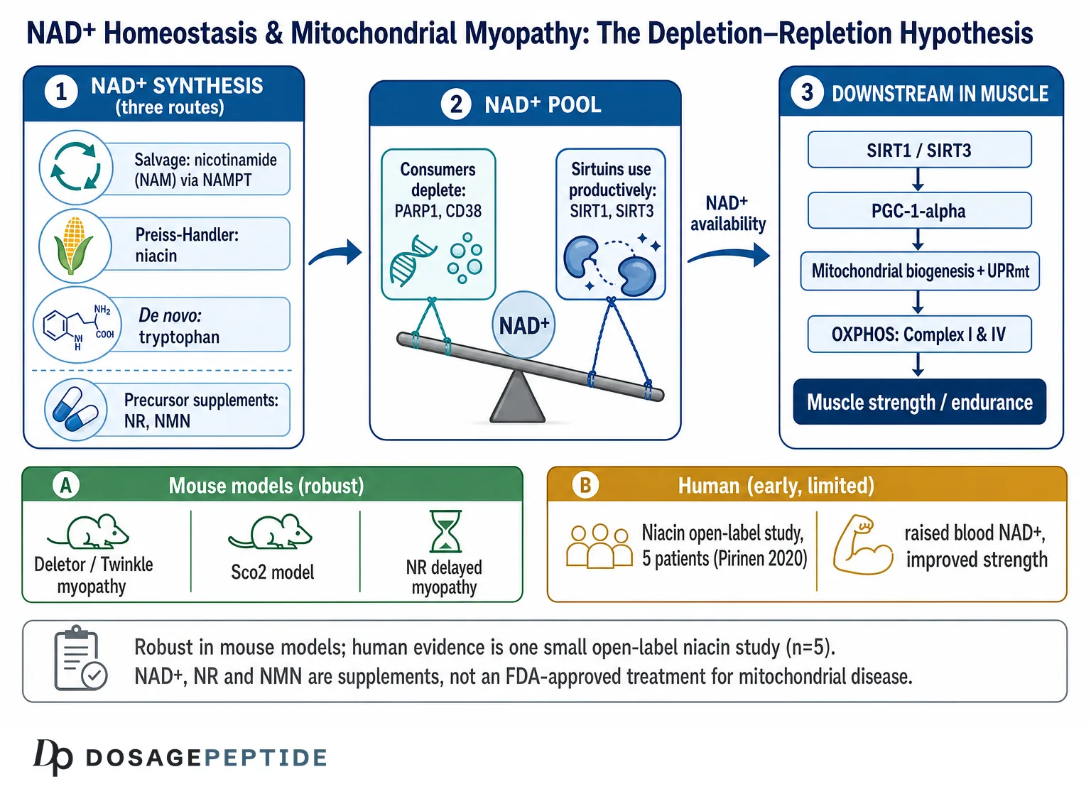
Yes — NAD+ is measurably depleted in myopathic muscle across several models, and restoring it improves mitochondrial function in animals. What has not followed is a matching human result: the clinical evidence is limited to small, early-stage work, and no NAD+ precursor is an approved drug for mitochondrial disease. Mitochondrial myopathies are inherited disorders in which skeletal muscle bears the brunt of a defect in oxidative phosphorylation (OXPHOS), and the NAD+ hypothesis is one of the few mechanistic stories in the field with both animal support and a plausible route to treatment. Below we separate the robust preclinical findings from the thin human data, explain how NAD+ is actually measured, and set out what remains unknown. Dosing conventions used in research are summarised in our NAD+ 500 mg vial protocol.
What Are Mitochondrial Myopathies, and Why Does Cellular Energy Fail?
Mitochondrial diseases are, collectively, the most common group of inherited metabolic disorders, with a conservative minimum prevalence estimated at roughly 1 in 5,000 adults when both mitochondrial DNA (mtDNA) and nuclear DNA (nDNA) causes are counted.[1] They arise from mutations in genes — encoded by either the small circular mitochondrial genome or the nucleus — that build or maintain the respiratory chain, the set of protein complexes on the inner mitochondrial membrane that convert the energy of electron transfer into a proton gradient and, ultimately, adenosine triphosphate (ATP). When this machinery is faulty, tissues with the highest energy demand — muscle, brain, heart, and the retina — are affected first and most severely.
The term “mitochondrial myopathy” describes presentations in which skeletal muscle involvement dominates: exercise intolerance, fatigability, muscle weakness, and, in many adult-onset forms, drooping eyelids (ptosis) and progressive external ophthalmoplegia (PEO), a paralysis of the eye muscles. Clinically recognized syndromes that fall under, or overlap with, the mitochondrial myopathy umbrella include mitochondrial encephalomyopathy with lactic acidosis and stroke-like episodes (MELAS), myoclonic epilepsy with ragged-red fibers (MERRF), Kearns–Sayre syndrome (KSS), and chronic progressive external ophthalmoplegia (CPEO). The classic histological hallmark is the “ragged-red fiber,” a muscle fiber crammed with abnormal, proliferating mitochondria that stain red with the modified Gomori trichrome method — a visible attempt by the cell to compensate for failing energy production.[1]
How do mtDNA mutations and deletions cause muscle disease?
Each mitochondrion carries multiple copies of mtDNA, and each cell carries hundreds to thousands of mitochondria, so a mutation is rarely present in every copy. Instead, mutant and normal mtDNA coexist in a state called heteroplasmy, and disease emerges only once the mutant fraction crosses a tissue-specific threshold, often above 60–90%. Post-mitotic tissue such as muscle is especially vulnerable because it cannot dilute mutant genomes through cell division. Two broad genetic mechanisms recur across mitochondrial myopathies: point mutations in mtDNA genes (for example, the m.3243A>G mutation associated with MELAS) and the accumulation of large-scale mtDNA deletions, which is the pattern seen in many adult-onset PEO cases.
Deletions frequently trace back to defects in the nuclear-encoded machinery that replicates and maintains mtDNA. A prominent example is the mitochondrial replicative helicase Twinkle (encoded by TWNK, formerly PEO1/C10orf2): dominant mutations in Twinkle cause autosomal-dominant PEO with multiple mtDNA deletions that build up in skeletal muscle over decades.[3] This is important background for the NAD+ story, because the most influential mouse model of adult-onset mitochondrial myopathy — the “Deletor” mouse — carries exactly this kind of dominant Twinkle mutation and reproduces the slow, progressive accumulation of mtDNA deletions and ragged-red fibers seen in patients. For readers who want plain-language definitions of terms like OXPHOS, heteroplasmy, or ATP as they appear here, our peptide and metabolic research glossary collects them in one place.
What Is NAD+ and How Is Its Homeostasis Maintained?
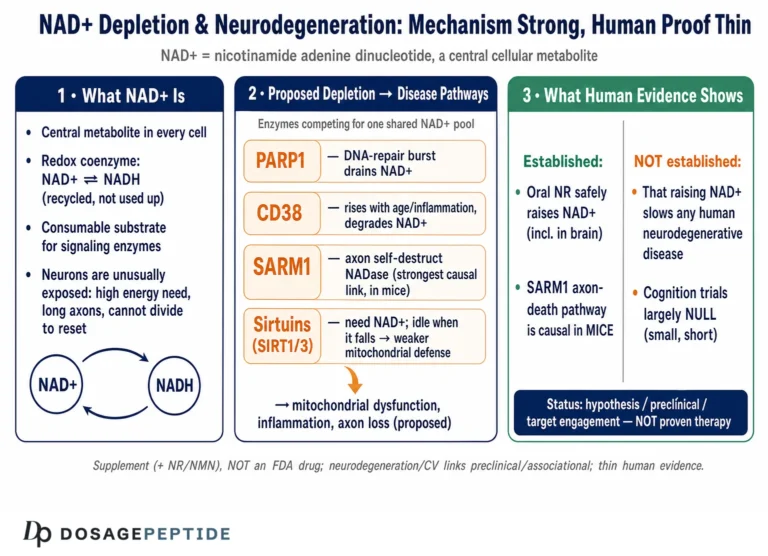
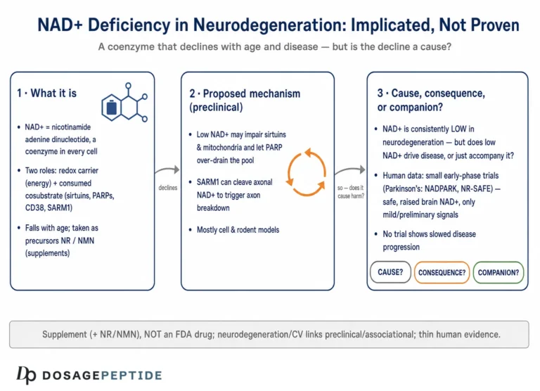
NAD+ is one of the most heavily trafficked small molecules in the cell. It plays two conceptually distinct roles. First, it is a redox coenzyme: the NAD+/NADH couple ferries electrons through glycolysis, the tricarboxylic acid (TCA) cycle, and fatty-acid oxidation, feeding the respiratory chain. In this role NAD+ is not consumed — it cycles endlessly between its oxidized (NAD+) and reduced (NADH) forms. Second, and more relevant to disease signaling, NAD+ is a substrate that is physically broken apart by a family of NAD+-consuming enzymes. Every time one of these enzymes acts, it cleaves NAD+ and releases nicotinamide (NAM), meaning the cell must continuously resynthesize NAD+ to keep pace.[2]
“NAD+ homeostasis” therefore refers to the dynamic balance between biosynthesis and consumption, maintained separately across cellular compartments (cytosol, nucleus, and mitochondria each hold their own NAD+ pools). When consumption outpaces synthesis, the free NAD+ concentration falls, and the enzymes that depend on it — particularly the sirtuins — lose activity. It is precisely this imbalance that has been repeatedly documented in mitochondrial myopathy models.
Where does cellular NAD+ come from?
Cells build NAD+ through three converging routes, and the vocabulary of these pathways is essential for understanding why different vitamin B3 forms behave differently in research:
- The de novo pathway synthesizes NAD+ from the amino acid tryptophan through the kynurenine pathway. It is metabolically expensive and contributes a minority of the total NAD+ pool in most tissues.
- The Preiss–Handler pathway uses nicotinic acid (niacin, NA) — the vitamin B3 form used in the human mitochondrial myopathy study discussed later. Niacin is converted to nicotinic acid mononucleotide by the enzyme NAPRT, then onward to NAD+.
- The salvage pathway recycles nicotinamide (NAM) — the very byproduct released whenever an NAD+-consuming enzyme fires. NAM is converted to nicotinamide mononucleotide (NMN) by the rate-limiting enzyme NAMPT, and NMN is then adenylylated to NAD+ by NMNAT enzymes. Nicotinamide riboside (NR), the precursor used in the pivotal mouse studies, enters this pathway one step downstream by being phosphorylated to NMN by nicotinamide riboside kinases (NRK1/NRK2).[2]
The salvage pathway carries most of the moment-to-moment flux, which is why NAMPT activity is a critical determinant of NAD+ availability, and why a fall in NAMPT expression — as reported in degenerating muscle — can tip the balance toward depletion.[5]
What consumes NAD+, and why does that matter in disease?
Three enzyme families dominate NAD+ consumption, and each becomes relevant to mitochondrial myopathy:
- Sirtuins (SIRT1–SIRT7) are NAD+-dependent deacylases that read the NAD+/NADH ratio as a signal of energy status. SIRT1 (nuclear) and SIRT3 (mitochondrial) are central to the response. When NAD+ is abundant, sirtuins are active and promote mitochondrial biogenesis and oxidative metabolism; when NAD+ falls, they go quiet.
- Poly(ADP-ribose) polymerases (PARPs), especially PARP1, are activated by DNA damage and can consume NAD+ voraciously, competing with sirtuins for the shared pool.
- CD38 is a membrane NADase whose expression rises with age and inflammation and which is a major driver of tissue NAD+ decline.[8]
The key conceptual point is that sirtuins, PARPs, and CD38 all draw on the same finite NAD+ supply. If stress chronically activates PARPs or CD38, sirtuins are starved of substrate even when total NAD+ looks only modestly reduced — a “competition” model that recurs throughout the mechanistic discussion below.
Is NAD+ Depleted in Mitochondrial Myopathy Models?
This is the empirical heart of the question, and the honest answer is that NAD+ depletion has been observed with reasonable consistency across several distinct models of muscle mitochondrial dysfunction. The strength of the evidence varies sharply by model type, so it is worth separating them.
What did the Deletor mouse reveal?
The Deletor mouse expresses a dominant patient-derived mutation in the Twinkle helicase and develops adult-onset mitochondrial myopathy, with progressive accumulation of multiple mtDNA deletions and ragged-red-equivalent fibers appearing after roughly 12 months of age. In the 2014 study by Khan and colleagues, myopathic Deletor muscle showed evidence of disturbed NAD+ metabolism, and the investigators reasoned that boosting NAD+ might drive the compensatory mitochondrial biogenesis these cells were already attempting.[3] This model is valuable precisely because it mimics the slow, deletion-driven adult PEO seen in humans rather than an artificial acute insult.
What did the Sco2 knockout/knock-in mouse show?
A second, independent model — the Sco2 knockout/knock-in (Sco2KO/KI) mouse, which carries a defect in a nuclear-encoded assembly factor for cytochrome c oxidase (respiratory Complex IV) — also exhibits a respiratory-chain defect and exercise intolerance. Cerutti and colleagues reported in 2014 that raising NAD+ in this model, either by supplying a precursor or by blocking NAD+ consumption, improved the biochemical defect, again pointing to NAD+ availability as a limiting factor.[4] Because Sco2 and Twinkle sit in entirely different parts of mitochondrial biology — one a Complex IV assembly factor, the other a replication helicase — convergent results across the two models strengthen the association considerably.
Is NAD+ loss seen in degenerating and dystrophic muscle more broadly?
Ryu and colleagues extended the picture beyond primary mitochondrial disease into muscular dystrophy. Working in the mdx mouse (a model of Duchenne muscular dystrophy) and in C. elegans, they reported that degenerating muscle loses both mitochondria and NAD+, and that the depletion is worsened by a fall in NAD+ biosynthetic enzymes such as NAMPT alongside heightened PARP-driven consumption.[5] This matters for interpretation: it suggests NAD+ depletion is a shared downstream feature of muscle energetic failure across several etiologies, not a quirk of one genetic lesion — though it also means the finding is not specific to primary mitochondrial myopathy.
What about human muscle?
The most direct human observation comes from the 2020 study by Pirinen and colleagues, who measured NAD+ in skeletal muscle from adult patients with mtDNA-deletion PEO. They reported systemic NAD+ deficiency — lower NAD+ in the muscle and blood of patients compared with healthy controls — providing the first clear evidence that the association documented in mice also appears in human mitochondrial myopathy.[6] This is a genuinely important data point, but it is drawn from a very small cohort, and correlation in a handful of patients cannot establish causation on its own.
| Model / system | Defect | NAD+ finding | Evidence tier |
|---|---|---|---|
| Deletor mouse | Dominant Twinkle helicase mutation; mtDNA deletions | Disturbed NAD+ metabolism; NAD+ boosting drove biogenesis | Preclinical (robust) |
| Sco2KO/KI mouse | Complex IV assembly defect | NAD+ boosting improved respiratory defect | Preclinical (robust) |
| mdx mouse / C. elegans | Dystrophin loss; general muscle degeneration | NAD+ and NAMPT fall; PARP consumption rises | Preclinical (adjacent disease) |
| Human PEO muscle | mtDNA deletions | Systemic NAD+ deficiency vs. controls | Human (early, very small n) |
How Might NAD+ Loss Drive Mitochondrial Myopathy?
Establishing that NAD+ is low is only the first step; the more demanding question is mechanistic — how would a fall in NAD+ translate into worsening muscle energetics, and why would restoring it help? Several interlocking pathways have been proposed, and it is best to treat them as a connected network rather than competing explanations.
Does the sirtuin–PGC-1α axis link NAD+ to mitochondrial biogenesis?
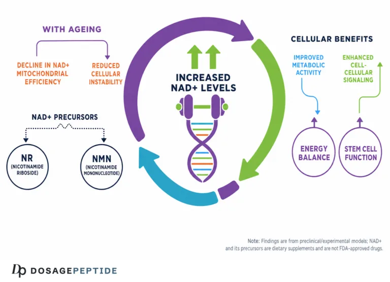
The central proposed mechanism runs through SIRT1. When NAD+ is plentiful, SIRT1 deacetylates and activates PGC-1α (peroxisome proliferator-activated receptor gamma coactivator 1-alpha), the master transcriptional coactivator of mitochondrial biogenesis. Activated PGC-1α drives expression of nuclear-encoded respiratory genes and stimulates the cell to build more, healthier mitochondria. In the myopathy models, the logic is that low NAD+ leaves SIRT1 under-active, so the muscle’s compensatory biogenesis program cannot run at full strength; supplying NAD+ precursors re-engages SIRT1–PGC-1α and boosts biogenesis. Khan and colleagues reported robust induction of mitochondrial biogenesis in skeletal muscle after NAD+ repletion, consistent with this axis.[3] Cerutti and colleagues framed their findings explicitly as NAD+-dependent activation of Sirt1 correcting the phenotype.[4] A general dosing and unit-conversion reference — our research dosage calculator — can help readers who want to see how the milligram-per-kilogram precursor doses used in these animal studies are expressed, purely as an educational aid and never as human guidance.
Do PARPs steal NAD+ away from mitochondrial repair?
The competition model is the second pillar. PARP1, activated by DNA damage and cellular stress, consumes NAD+ to build poly(ADP-ribose) chains (PARylation). If PARPs are chronically overactive, they draw down the shared NAD+ pool and effectively out-compete the sirtuins. Cerutti and colleagues tested this directly by reducing NAD+ consumption with a PARP inhibitor and observed improvement in the Sco2 model, mirroring the benefit of adding a precursor — two routes to the same endpoint of higher available NAD+.[4] Ryu and colleagues similarly reported that the benefits of NAD+ repletion in dystrophic muscle were accompanied by a reduction in global PARylation, tying the therapeutic effect back to the consumption side of the ledger.[5]
What is the mitochondrial unfolded protein response, and how does NAD+ engage it?
A third mechanism involves the mitochondrial unfolded protein response (UPRmt), a protective stress program that upregulates mitochondrial chaperones and proteases to restore protein-folding balance inside the organelle. Khan and colleagues reported that NR supplementation stimulated the UPRmt in myopathic muscle, suggesting that part of the benefit of NAD+ repletion is the activation of an endogenous mitochondrial quality-control and stress-buffering system rather than biogenesis alone.[3] Sirtuins — SIRT1 in particular — have been mechanistically linked to UPRmt induction, providing a plausible bridge between raising NAD+ and switching on this response.
Can NAD+ decline create a “pseudohypoxic” energy crisis?
Work on aging biology, though not conducted in myopathy models, offers a complementary mechanism relevant to any state of NAD+ scarcity. Gomes and colleagues showed that a decline in nuclear NAD+ can disrupt communication between the nucleus and mitochondria: with SIRT1 inactive, the transcription factor HIF-1α accumulates even under normal oxygen, producing a “pseudohypoxic” state in which the cell behaves as if starved of oxygen and specifically loses mtDNA-encoded OXPHOS subunits.[7] Raising NAD+ in aged mice restored mitochondrial function in a SIRT1-dependent manner. While this study addressed aging rather than inherited myopathy, it illustrates how NAD+ loss can, on its own, degrade the very respiratory subunits that are already compromised in mitochondrial disease.
Does CD38 accelerate NAD+ loss?
Finally, the consumption side of homeostasis is shaped by CD38. Camacho-Pereira and colleagues demonstrated that rising CD38 expression is a major driver of NAD+ decline and mitochondrial dysfunction, acting through an SIRT3-dependent mechanism.[8] In inflamed or aging muscle, elevated CD38 would deepen any pre-existing NAD+ deficit, adding a third consumer — alongside PARPs and the sirtuins themselves — that must be balanced by synthesis. Taken together, the mechanistic picture is coherent: low NAD+ silences the sirtuin–PGC-1α biogenesis program while excess consumption by PARPs and CD38 keeps the pool depleted, and precursor supplementation is proposed to restore the balance from the synthesis side.
How Does the NAD+/NADH Redox Balance Itself Affect Failing Mitochondria?
So far the discussion has focused on NAD+ as a signaling substrate that gets consumed. But there is a second, more fundamental way that mitochondrial disease disturbs NAD+ — one rooted in the redox chemistry of the respiratory chain, and it is important because it can create NAD+ scarcity even before any signaling enzyme fires. Understanding it clarifies why mitochondrial myopathy and NAD+ imbalance are so tightly linked.
Why does a broken respiratory chain skew the NADH:NAD+ ratio?
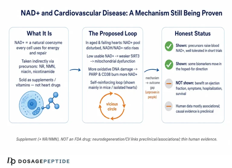
The redox couple NAD+/NADH is the primary electron carrier feeding the respiratory chain. Fuel oxidation in glycolysis, the TCA cycle, and fatty-acid oxidation reduces NAD+ to NADH; the electrons in NADH are then handed to Complex I (NADH:ubiquinone oxidoreductase), which oxidizes NADH back to NAD+ and pushes the electrons down the chain toward oxygen. In healthy mitochondria this cycle keeps a high ratio of NAD+ to NADH, ensuring a ready supply of oxidized NAD+ for both metabolism and signaling. When the respiratory chain is defective — whether from an mtDNA deletion crippling Complex I subunits or a Complex IV assembly failure as in the Sco2 model — electrons back up. NADH cannot be efficiently reoxidized, the NADH:NAD+ ratio rises, and the pool of free, oxidized NAD+ available to sirtuins and other reactions shrinks.[1] In other words, the same OXPHOS defect that defines the disease also directly promotes a functional NAD+ deficit, quite apart from the consumption by PARPs and CD38 discussed earlier.
Why does this make NAD+ imbalance a plausible amplifier rather than a bystander?
This redox angle helps explain why NAD+ disturbance appears so consistently across otherwise dissimilar mitochondrial defects: any lesion that impairs NADH reoxidation will tend to lower the effective NAD+ pool. It also frames a genuine open question in the field — whether adding NAD+ precursors simply provides more total nucleotide, or whether the meaningful therapeutic variable is the ratio of oxidized to reduced forms in a specific compartment. Because the redox couple and the signaling pool are chemically the same molecules but functionally distinct, a precursor could in principle raise total NAD+ while the local oxidized fraction that sirtuins actually need remains constrained by the failing chain. This nuance is one reason the animal-to-human translation is not straightforward and why measuring NAD+ well — not just raising it — matters so much.
What Does the Animal Evidence Show for NAD+ Repletion?
The preclinical intervention data are the strongest part of this entire literature, and they are worth reviewing study by study, because the quality and directness of the evidence differ.
Khan 2014: nicotinamide riboside in the Deletor mouse
Khan and colleagues administered nicotinamide riboside to Deletor mice at 400 mg/kg/day for four months, treating animals both before symptoms emerged (presymptomatic) and after myopathy was established (postsymptomatic). NR robustly induced mitochondrial biogenesis in skeletal muscle and brown adipose tissue, prevented mitochondrial ultrastructural abnormalities, reduced the formation of new mtDNA deletions, and stimulated the UPRmt. Critically, NR delayed disease progression at both early and late stages, which the authors interpreted as evidence that NAD+ repletion is a viable strategy to slow adult-onset mitochondrial myopathy in this model.[3] This remains the single most cited preclinical demonstration that boosting NAD+ can meaningfully alter a primary mitochondrial myopathy phenotype.
Cerutti 2014: precursor supply versus consumption blockade
Cerutti and colleagues took a complementary two-pronged approach in the Sco2KO/KI mouse. They showed that either supplementing NR or inhibiting the NAD+-consuming PARPs improved the respiratory-chain defect and exercise intolerance, and they attributed the benefit to NAD+-dependent activation of SIRT1 and downstream mitochondrial biogenesis.[4] The elegance of showing that two mechanistically opposite manipulations — adding substrate versus blocking its destruction — converge on the same functional improvement is what makes this study particularly persuasive about the causal role of NAD+ availability.
Ryu 2016: NAD+ repletion in dystrophic and degenerating muscle
Ryu and colleagues broadened the concept to muscular dystrophy. Restoring NAD+ in mice and worms with degenerating muscle improved muscle function, an effect they traced to increased mitochondrial content, greater expression of structural proteins such as α-dystrobrevin and δ-sarcoglycan, and reductions in PARylation, inflammation, and fibrosis.[5] This study is best read as adjacent evidence — it concerns dystrophy rather than primary mitochondrial disease — but it reinforces the general principle that NAD+ availability is a lever on muscle mitochondrial health.
| Study | Model | Intervention | Principal outcome |
|---|---|---|---|
| Khan 2014[3] | Deletor mouse (Twinkle) | NR 400 mg/kg/day, 4 months | Induced biogenesis; delayed myopathy; stimulated UPRmt |
| Cerutti 2014[4] | Sco2KO/KI mouse | NR or PARP inhibition | Improved respiratory defect and exercise tolerance via SIRT1 |
| Ryu 2016[5] | mdx mouse; C. elegans | NAD+ repletion | Improved muscle function; less PARylation, inflammation, fibrosis |
Across these studies the direction of effect is consistent, the models are diverse, and the mechanisms proposed are mutually reinforcing. That is a strong preclinical foundation. It is not, however, the same as evidence that NAD+ precursors treat human mitochondrial myopathy — a distinction the human data make painfully clear.
What Does the Human Evidence Show?
Here the honesty framing becomes non-negotiable. The human evidence that NAD+ boosting affects mitochondrial myopathy is limited to a single small, early study, and it must not be read as proof of efficacy.
The Pirinen 2020 niacin study
Pirinen and colleagues conducted an open-label study in which adult patients with mtDNA-deletion progressive external ophthalmoplegia received niacin (nicotinic acid) in a dose-escalating fashion — starting at 250 mg/day and increasing to 750–1,000 mg/day — alongside matched healthy controls. The study enrolled only five patients, with two age- and sex-matched controls per patient, and treatment continued for up to about 10 months in patients (4 months in controls).[6] The reported results were striking within this small sample: patients began with systemic NAD+ deficiency, and niacin raised skeletal-muscle NAD+ to the level seen in healthy controls. Alongside the biochemical normalization, the authors reported increased muscle strength, increased mitochondrial biogenesis, a muscle metabolome that shifted toward the control pattern, and a roughly 50% decrease in liver fat.[6] This was the first human demonstration that the NAD+ deficiency and its correction, so well described in mice, could be recapitulated in patients.
Why must these human results be read cautiously?
The limitations are substantial and were acknowledged by the field. First, the sample is tiny — five patients — so the strength and biogenesis findings, while encouraging, carry wide uncertainty and cannot be generalized. Second, the design was open-label without a placebo arm in the patients, so expectation effects and the natural variability of a slowly progressive disease cannot be excluded. Third, the study measured NAD+ correction and physiological markers over months, not long-term clinical outcomes such as disability, survival, or quality of life. An accompanying commentary in the same journal captured the appropriate posture — describing the work as providing “hope” and a rationale for larger trials rather than establishing a treatment.[10] The study was registered as a formal clinical investigation, underscoring that this is research, not established care.[11]
What do human NAD+ precursor trials in general tell us?
Separately from mitochondrial disease, several human trials have established that NAD+ precursors can raise blood NAD+ and are generally well tolerated in healthy people. Martens and colleagues ran a randomized, double-blind, placebo-controlled crossover trial of NR (1,000 mg/day) in 30 healthy middle-aged and older adults and found it was well tolerated and raised average NAD+ by roughly 60%, with hints of lower blood pressure and aortic stiffness that require confirmation.[9] These pharmacokinetic and safety data are reassuring for feasibility, but they were conducted in healthy volunteers, not mitochondrial myopathy patients, and raising blood NAD+ is not the same as improving disease. The broader in vivo evidence base for NAD+-boosting molecules has been reviewed comprehensively, and that review is candid that translation from robust animal data to proven human benefit remains incomplete across nearly every indication.[2]
What would a definitive human trial need to establish?
The gap between the Pirinen signal and a validated therapy is a specific one, and naming it clarifies why the honest verdict is “associated and biologically plausible” rather than “proven.” A confirmatory study would need to be randomized and placebo-controlled rather than open-label, so that expectation effects and the spontaneous fluctuation inherent to a slowly progressive disease can be separated from a genuine drug effect. It would need to be large enough to detect a clinically meaningful change against the genetic and phenotypic heterogeneity of mitochondrial myopathy — patients differ in the causative mutation, the tissue distribution of heteroplasmy, the age of onset, and the rate of decline, all of which widen the variance that any trial must overcome. It would need to run long enough to capture outcomes that matter to patients — walking distance, fatigue, disability, and quality of life — rather than only the biochemical surrogate of muscle NAD+, because normalizing a laboratory value is not the same as changing the course of a disease. And it would need to prespecify which precursor, at which dose, reaching which tissue compartment is being tested, since the animal work and the human pilot used different molecules — nicotinamide riboside in the mice, niacin in the patients — that enter NAD+ synthesis by different routes and may distribute differently across muscle, brain, and heart. The registration of the pilot as a formal clinical investigation was the first rung on this ladder, not the last,[11] and the commentary that accompanied the study framed the result explicitly as a rationale for larger trials rather than as a treatment already in hand.[10] Until such trials report, the broader NAD+ literature’s own candid conclusion holds: robust animal data have repeatedly outrun confirmed human benefit.[2]
How Do Researchers Measure NAD+, and What Does “Depletion” Mean?
A claim that NAD+ is “depleted” in mitochondrial myopathy is only as strong as the measurement behind it, so it is worth understanding what these studies actually quantified and where the methodological caveats lie. This is part of reading the evidence honestly rather than accepting a headline number at face value.
How is NAD+ quantified in tissue and blood?
Two families of methods dominate. Enzymatic cycling assays use NAD+-dependent dehydrogenases in a coupled reaction that amplifies a colorimetric or fluorescent signal, giving total NAD+ (and, with variations, the NAD+/NADH split). Modern studies increasingly rely on liquid chromatography–tandem mass spectrometry (LC-MS/MS), which can resolve NAD+ from its precursors and breakdown products — nicotinamide, NMN, NR, and ADP-ribose — in the same sample, giving a fuller picture of flux through the pathway. The human trials cited here used such approaches to show, for example, that chronic NR raised blood NAD+ by roughly 60%,[9] and the Pirinen study measured NAD+ and its splitting products in both muscle biopsies and blood to demonstrate correction of the deficit.[6] Definitions of these analytes and the biochemistry linking them appear in our research glossary for readers who want to follow the terminology.
Why can “depletion” be a slippery term?
Several caveats deserve emphasis. First, blood NAD+ does not necessarily mirror muscle NAD+; a supplement can raise circulating NAD+ substantially while tissue-level change is smaller or slower, which is why the muscle-biopsy data in the myopathy work are more informative than blood alone. Second, most assays report total NAD+, whereas the biologically decisive quantity may be the free, compartment-specific, oxidized pool available to a given enzyme — a number that is technically hard to isolate. Third, NAD+ is labile and sensitive to how quickly a sample is frozen after collection, so pre-analytical handling can influence results. Finally, the magnitude of “depletion” reported in different models is not uniform; it is often a relative reduction versus matched controls rather than a total collapse. None of this negates the consistent finding of lower NAD+ in myopathic muscle, but it does mean the word “depletion” should be read as a measured relative deficit, not an absolute switch that is either on or off.
How Do the NAD+ Precursors Compare in Research?
Because different studies used different vitamin B3 forms — NR in the mouse work, niacin in the human study — it is worth clarifying how these precursors differ, since they are not interchangeable in their biochemistry, tolerability, or the pathway they feed.
| Precursor | Pathway entered | Notable feature in research | Common tolerability note |
|---|---|---|---|
| Nicotinic acid (niacin, NA) | Preiss–Handler (via NAPRT) | Used in the Pirinen human PEO study | Prostaglandin-mediated skin flushing at higher doses |
| Nicotinamide (NAM) | Salvage (via NAMPT) | Direct salvage substrate; can inhibit sirtuins at high levels | Generally low flushing; hepatic caution at very high intake |
| Nicotinamide riboside (NR) | Salvage (via NRK to NMN) | Used in Khan and Cerutti mouse studies | Well tolerated in healthy-adult trials |
| Nicotinamide mononucleotide (NMN) | Salvage (one step from NAD+) | Widely studied precursor; requires conversion/transport | Well tolerated in short human studies |
Two research-relevant distinctions stand out. First, niacin uniquely enters through the Preiss–Handler route via NAPRT, which may matter in tissues where salvage-pathway enzymes are limiting — a possible reason the Pirinen team chose it for human muscle. Its main drawback is the well-known flushing reaction, which shaped the dose-escalation design. Second, NAM, while a direct salvage substrate, is a product-inhibitor of sirtuins at high concentrations, so more NAM does not automatically mean more sirtuin activity — a nuance that complicates the naive assumption that any NAD+ precursor is equivalent. NR and NMN were the precursors of choice in the biogenesis-focused animal work because they raise NAD+ without the flushing of niacin or the sirtuin-inhibition concern of high-dose NAM. None of these forms is an approved treatment for mitochondrial disease, and the comparison here is mechanistic, not a recommendation.
What Are the Limitations and Open Questions in This Research?
A responsible reading of this literature requires holding the promising signal and the serious caveats in mind simultaneously. The following limitations define the current frontier.
Preclinical strength does not equal human proof
The animal evidence is genuinely strong — multiple models, convergent mechanisms, reproducible biogenesis induction — but mouse mitochondrial myopathy is not human mitochondrial myopathy. Mice were often treated at defined disease stages under controlled genetics; human disease is heterogeneous in genotype, tissue distribution of heteroplasmy, age of onset, and rate of progression. The single human study to date is small, open-label, and short relative to the disease course.[6] Larger, randomized, placebo-controlled trials with clinically meaningful endpoints have not yet reported definitive results, and until they do, NAD+ boosting for mitochondrial myopathy remains an investigational metabolic hypothesis rather than a validated therapy.[10]
Association versus causation in patient tissue
The finding that human PEO muscle has low NAD+ establishes an association. It does not, by itself, prove that NAD+ depletion causes the myopathy rather than being a consequence of it — degenerating, energy-starved muscle might lose NAD+ secondarily. The mouse intervention studies argue for a causal contribution because raising NAD+ improved function, but that causal chain has not been demonstrated with the same rigor in humans. In people, the usual tools for disentangling cause from consequence are limited here: a low patient count precludes the kind of statistical adjustment that might separate NAD+ depletion from the broader metabolic wreckage of failing muscle, and the redox coupling described earlier means an OXPHOS defect can lower the effective NAD+ pool directly, so a measured deficit is at least partly expected as a downstream signature of the disease rather than its origin. The honest reading is therefore that low NAD+ is a robust, reproducible correlate of mitochondrial myopathy whose causal weight in humans remains to be quantified.
Dose, tissue delivery, and precursor choice are unsettled
It is not established which precursor, at which dose, delivered how, best raises NAD+ in the specific tissues that matter in a given patient. Muscle, brain, and heart may respond differently; salvage-pathway enzyme availability varies by tissue and disease; and the animal doses (for example, 400 mg/kg/day NR in mice) do not translate directly to human regimens. The milligram-per-kilogram framing used in preclinical work is a common source of confusion when readers try to map it onto people, which is one reason we keep such conversions in neutral educational tools rather than as guidance.
Potential harms and biological trade-offs
NAD+ metabolism is not a simple “more is better” system. High-dose niacin causes flushing and, at pharmacological doses, has hepatic and glycemic considerations. Because NAD+ and its consumers intersect with cell proliferation and DNA-damage responses, there are theoretical concerns about the effects of chronically elevated NAD+ in the context of pre-existing malignancy, and these have not been resolved in long-term human studies. The competition between PARPs and sirtuins also means the optimal intervention might sometimes be reducing consumption rather than adding substrate — the two strategies are not always equivalent in a diseased tissue. These uncertainties are precisely why the research framing, rather than a therapeutic one, is appropriate.
Handling of research-grade compounds
NAD+ precursors used in laboratory settings are research chemicals when supplied for in vitro or animal experimentation, and their preparation, storage, and concentration standardization are handled under laboratory protocols. Readers interested in the general laboratory practices behind reconstituting and standardizing lyophilized research compounds can consult our research reconstitution reference, and definitions of the biochemical terms used throughout this article are collected in the research glossary. These resources are educational only and do not constitute instructions for human use.
What Is the Current Regulatory and Safety Status?
Clarity on regulatory status is essential to the honest framing of this topic. NAD+ itself and its precursors nicotinamide riboside and nicotinamide mononucleotide are regulated as dietary ingredients and supplements in the United States, not as approved drugs for mitochondrial disease. No NAD+ precursor has been approved by the U.S. Food and Drug Administration (FDA) as a treatment for mitochondrial myopathy or any primary mitochondrial disease.
The regulatory history of NMN is instructive about how unsettled this space is: the FDA determined in late 2022 that NMN was excluded from the dietary-supplement definition because it had been investigated as a drug, then, after industry petitions, reversed that interpretation in 2025 and confirmed that NMN is not excluded — while still classifying it as a new dietary ingredient that requires premarket notification. Nicotinamide riboside, by contrast, has been marketed as a dietary ingredient supported by regulatory notifications. Niacin (nicotinic acid), the form used in the human myopathy study, is an old vitamin that is also an approved pharmaceutical for dyslipidemia at high doses — but it is not approved for, and its dyslipidemia approval says nothing about, mitochondrial myopathy. The essential point for readers is that regulatory availability as a supplement is not evidence of efficacy for mitochondrial disease, and none of these compounds should be understood as an approved therapy for it. Any use of NAD+ precursors in the context of a diagnosed mitochondrial disorder is a matter for physicians and, ideally, controlled clinical trials — not self-directed experimentation.
Frequently Asked Questions
Is impaired NAD+ homeostasis actually associated with mitochondrial myopathies?
Yes, an association is documented. Mouse models of mitochondrial myopathy — including the Twinkle-mutant Deletor and the Sco2 knockout/knock-in — show disturbed NAD+ metabolism, and a small human study found systemic NAD+ deficiency in patients with mtDNA-deletion progressive external ophthalmoplegia. The association is reproducible across models, but whether NAD+ loss is a primary driver or a downstream consequence of failing muscle energetics is not fully resolved in humans.
Does NAD+ or nicotinamide riboside treat mitochondrial disease?
No. NAD+ precursors are not approved treatments for mitochondrial disease, and it would be inaccurate to say they “treat” it. In mouse models, nicotinamide riboside delayed myopathy and boosted mitochondrial biogenesis, and one small open-label human study of niacin reported improved muscle strength. That preclinical and early human signal is a rationale for larger controlled trials, not proof of efficacy or a basis for self-treatment.
What did the Khan 2014 mouse study actually find?
Khan and colleagues gave nicotinamide riboside (400 mg/kg/day for four months) to Deletor mice with adult-onset mitochondrial myopathy. NR robustly induced mitochondrial biogenesis, prevented ultrastructural damage, reduced new mtDNA deletions, stimulated the mitochondrial unfolded protein response, and delayed disease progression at both early and late stages. It remains the most influential preclinical demonstration that raising NAD+ can alter a primary mitochondrial myopathy phenotype in animals.
How strong is the human evidence?
It is early and limited. The main human study (Pirinen 2020) enrolled only five patients with progressive external ophthalmoplegia in an open-label design without a patient placebo arm. Niacin normalized muscle NAD+ and was associated with increased muscle strength and mitochondrial biogenesis over about ten months. These are encouraging but preliminary results from a tiny sample; they cannot establish efficacy and await confirmation in larger randomized trials.
How does low NAD+ impair mitochondria mechanistically?
The leading model is that low NAD+ under-activates the sirtuin SIRT1, which normally activates PGC-1α to drive mitochondrial biogenesis. Simultaneously, NAD+-consuming enzymes such as PARPs and CD38 compete for the shared pool, deepening the deficit. Additional proposed mechanisms include impaired activation of the mitochondrial unfolded protein response and a “pseudohypoxic” state in which HIF-1α accumulates and mtDNA-encoded respiratory subunits are lost.
Are niacin, NR, and NMN interchangeable?
Not exactly. Niacin enters NAD+ synthesis through the Preiss–Handler pathway and can cause skin flushing; nicotinamide riboside and NMN feed the salvage pathway and were preferred in the biogenesis-focused animal studies; nicotinamide is a direct salvage substrate but can inhibit sirtuins at high levels. They differ in the pathway they use, tissue handling, and tolerability, so results with one precursor do not automatically transfer to another.
Are NAD+ precursors approved by the FDA for this use?
No. NAD+, nicotinamide riboside, and nicotinamide mononucleotide are regulated as dietary ingredients in the United States, not as approved drugs for mitochondrial myopathy. NMN’s supplement status has been contested and revised by the FDA in recent years. Niacin is an approved drug for high cholesterol but not for mitochondrial disease. Supplement availability does not indicate proven efficacy for mitochondrial myopathy.
Why is this framed as research rather than medical advice?
Because the evidence base does not support clinical recommendations. The robust data are from mice; the human data are limited to a single very small study. Mitochondrial disease is genetically and clinically heterogeneous, optimal precursor, dose, and tissue targeting are unknown, and there are unresolved safety questions around long-term NAD+ elevation. Decisions about any intervention in diagnosed mitochondrial disease belong with clinicians and, ideally, within controlled trials.
References
- Gorman GS, Chinnery PF, DiMauro S, et al. Mitochondrial diseases. Nat Rev Dis Primers. 2016;2:16080. https://www.nature.com/articles/nrdp201680
- Rajman L, Chwalek K, Sinclair DA. Therapeutic Potential of NAD-Boosting Molecules: The In Vivo Evidence. Cell Metab. 2018;27(3):529–547. https://pubmed.ncbi.nlm.nih.gov/29514064/
- Khan NA, Auranen M, Paetau I, et al. Effective treatment of mitochondrial myopathy by nicotinamide riboside, a vitamin B3. EMBO Mol Med. 2014;6(6):721–731. https://pubmed.ncbi.nlm.nih.gov/24711540/
- Cerutti R, Pirinen E, Lamperti C, et al. NAD+-dependent activation of Sirt1 corrects the phenotype in a mouse model of mitochondrial disease. Cell Metab. 2014;19(6):1042–1049. https://pubmed.ncbi.nlm.nih.gov/24814483/
- Ryu D, Zhang H, Ropelle ER, et al. NAD+ repletion improves muscle function in muscular dystrophy and counters global PARylation. Sci Transl Med. 2016;8(361):361ra139. https://pubmed.ncbi.nlm.nih.gov/27798264/
- Pirinen E, Auranen M, Khan NA, et al. Niacin Cures Systemic NAD+ Deficiency and Improves Muscle Performance in Adult-Onset Mitochondrial Myopathy. Cell Metab. 2020;31(6):1078–1090.e5. https://www.cell.com/cell-metabolism/fulltext/S1550-4131(20)30190-X
- Gomes AP, Price NL, Ling AJY, et al. Declining NAD+ induces a pseudohypoxic state disrupting nuclear-mitochondrial communication during aging. Cell. 2013;155(7):1624–1638. https://pmc.ncbi.nlm.nih.gov/articles/PMC4076149/
- Camacho-Pereira J, Tarragó MG, Chini CCS, et al. CD38 Dictates Age-Related NAD Decline and Mitochondrial Dysfunction through an SIRT3-Dependent Mechanism. Cell Metab. 2016;23(6):1127–1139. https://www.cell.com/cell-metabolism/fulltext/S1550-4131(16)30224-8
- Martens CR, Denman BA, Mazzo MR, et al. Chronic nicotinamide riboside supplementation is well-tolerated and elevates NAD+ in healthy middle-aged and older adults. Nat Commun. 2018;9:1286. https://pubmed.ncbi.nlm.nih.gov/29599478/
- Fang EF (commentary). Of Mice and Men: NAD+ Boosting with Niacin Provides Hope for Mitochondrial Myopathy Patients. Cell Metab. 2020;31(6):1069–1070. https://pubmed.ncbi.nlm.nih.gov/32492387/
- ClinicalTrials.gov. Niacin Supplementation in Healthy Controls and Mitochondrial Myopathy Patients. NCT03973203. https://clinicaltrials.gov/study/NCT03973203