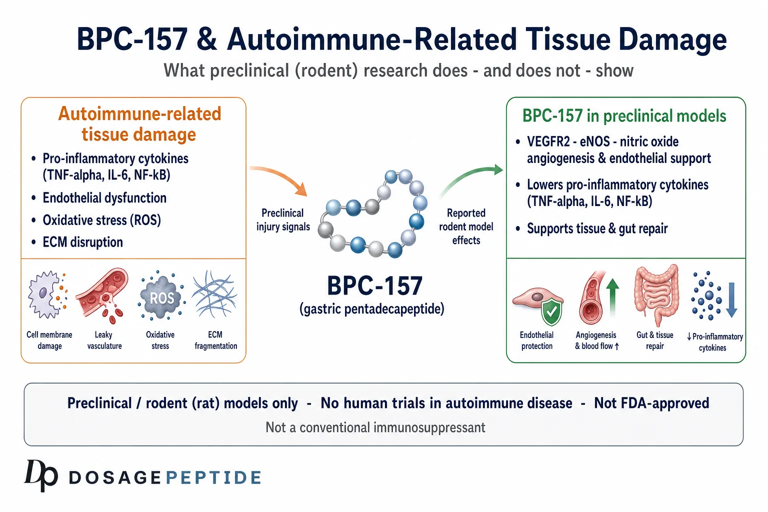
BPC-157 has attracted intense interest as a putative regenerative and anti-inflammatory agent, and a recurring question in the research literature is whether it might blunt the inflammatory tissue damage that characterizes autoimmune disease. The honest answer, based on the current evidence, is that this remains a hypothesis drawn almost entirely from rodent and in-vitro models — there are no rigorous published human clinical trials of BPC-157 in any autoimmune condition, and none establishing efficacy or safety for that use. This article examines what the peptide is, how its mechanisms plausibly intersect inflammation and immune signaling, what the autoimmune-adjacent preclinical models actually show, and where the evidence gap begins.
The material below is provided for educational and research purposes only. BPC-157 is an investigational compound that is not approved by the FDA or any comparable regulator for human therapeutic use, and nothing here should be read as medical advice or as a dosing recommendation for people.
Key points at a glance
- BPC-157 is a synthetic 15-amino-acid peptide derived from a fragment of a human gastric protein (BPC), studied primarily by the Zagreb (Sikiric) research group.
- Its proposed mechanisms center on nitric-oxide-system modulation, VEGFR2–Akt–eNOS-driven angiogenesis, FAK–paxillin cell migration, growth-factor sensitization, and gut–brain-axis signaling.
- In animal models, it reduces markers of inflammation — TNF-α, IL-6, NF-κB activity, neutrophil and macrophage infiltration — and supports tissue repair.
- The most autoimmune-adjacent evidence is from chemically induced rodent colitis, which is an acute injury model, not spontaneous human autoimmune disease.
- There are no rigorous published human clinical trials of BPC-157 in any autoimmune disease; a 2025 systematic review found 35 of 36 studies were preclinical and identified no clinical safety data.
- BPC-157 is not FDA-approved; its Section 503A compounding status has shifted (placed in Category 2 in 2023, removed from that list in April 2026 and referred for advisory-committee review, which is not approval), and it is prohibited in sport under WADA.
What is BPC-157 and where does it come from?
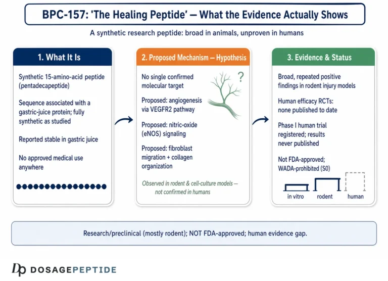
BPC-157 (Body Protection Compound-157) is a synthetic peptide composed of 15 amino acids — a pentadecapeptide — with the sequence Gly-Glu-Pro-Pro-Pro-Gly-Lys-Pro-Ala-Asp-Asp-Ala-Gly-Leu-Val. It is a partial sequence derived from, and stabilized relative to, a larger cytoprotective protein called Body Protection Compound (BPC) that was originally isolated from human gastric juice. In other words, the peptide is a laboratory construct modeled on a fragment of a naturally occurring gastric protein, and much of its early characterization grew out of research into the stomach’s own defensive and regenerative machinery.[1]
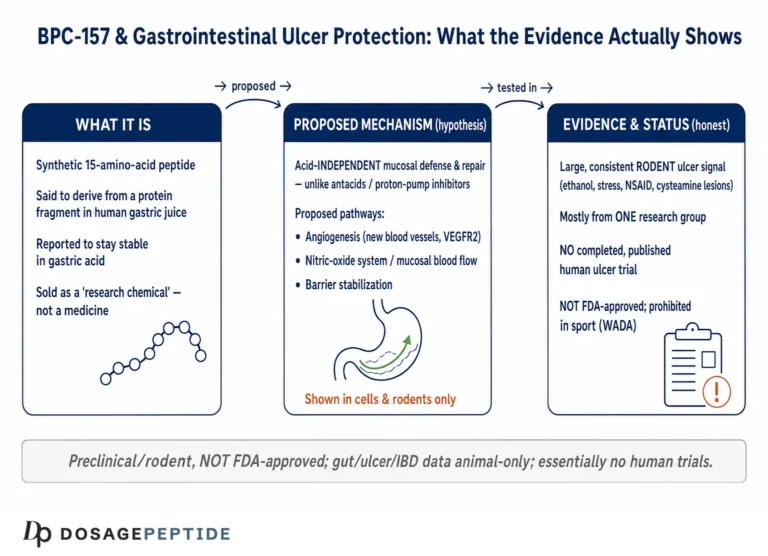
The molecule is frequently described in the literature as the “stable gastric pentadecapeptide” because, unlike many peptides, it is reported to remain intact in human gastric juice for extended periods and to resist hydrolysis under conditions that would rapidly degrade less stable sequences. This relative stability is scientifically important: it is one reason investigators have been able to test the compound not only by injection but also via oral and intragastric routes in animals, and it distinguishes BPC-157 from peptides that require careful protection from enzymatic breakdown. For researchers, that stability profile is also a practical handling consideration — the reconstituted material still requires appropriate storage, but the parent molecule is comparatively robust.
Historical development from a cytoprotection concept
The intellectual lineage of BPC-157 matters because it frames how the compound is described in the primary literature. The peptide emerged from a research tradition centered on gastric cytoprotection — the observation, associated with the work of Andre Robert and later extended by Hans Selye’s stress-coping framework, that the stomach lining possesses intrinsic defensive mechanisms that protect and rapidly repair the mucosa against noxious insults. The Zagreb group positioned BPC-157 as a mediator and pharmacological embodiment of this endogenous cytoprotective and “organoprotective” response, and much of the review literature explicitly frames the peptide within Robert’s cytoprotection and Selye’s adaptation concepts rather than as a targeted receptor agonist.[1] This framing has two consequences for the reader: it explains the peptide’s reported “pleiotropy” (many effects across many organs), and it signals that the mechanism is described as adaptive and homeostatic — nudging stressed tissue back toward balance — rather than as a single, cleanly defined pharmacological action.
Why stability and structure matter for interpretation
The 15-residue sequence is notable for its proline-rich core, which contributes to conformational rigidity and is one structural explanation for the reported resistance to enzymatic degradation. From a research-interpretation standpoint, stability is a double-edged property: it makes the compound tractable to study across multiple administration routes, but it also means that comparisons between studies must account for route, formulation, and species, since a peptide that survives the gastric environment in a rat may behave differently in another model system. None of these structural features, it should be stressed, tell us anything about clinical efficacy in humans — they are properties of the molecule, not evidence of therapeutic benefit.
Naming, abbreviations, and what BPC-157 is not
Because the peptide is discussed under several labels, it is worth clarifying terminology. “BPC-157,” “PL-14736,” “PL-10,” and “PLD-116” are development codes that appear across the Zagreb-group and Pliva (Croatia) literature for the same or closely related pentadecapeptide preparations.[2] BPC-157 is not a growth hormone, not a growth-hormone secretagogue, and not a corticosteroid or a conventional immunosuppressant. It does not fit neatly into any single established drug class, which is part of why its mechanism has been the subject of so much investigation and why claims about it should be read with care. A useful orientation to peptide nomenclature and abbreviations of this kind is available in the peptide research glossary.
What is the molecular mechanism of BPC-157?
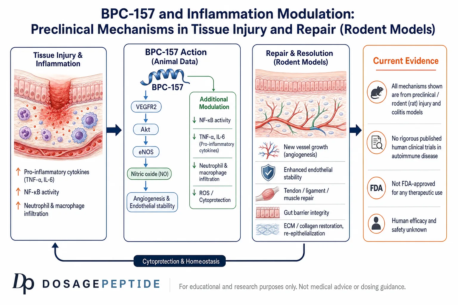
The most consistent theme across the preclinical literature — the great majority of which comes from Predrag Sikiric’s group at the University of Zagreb — is that BPC-157 does not appear to act through a single dedicated receptor but instead modulates several interlocking signaling systems that converge on vascular integrity, angiogenesis, and cytoprotection.[1] Understanding these pathways is essential context for any discussion of inflammation, because the same machinery that drives blood-vessel formation and endothelial stability also shapes how inflamed tissue is supplied, repaired, and resolved.
The nitric oxide (NO) system
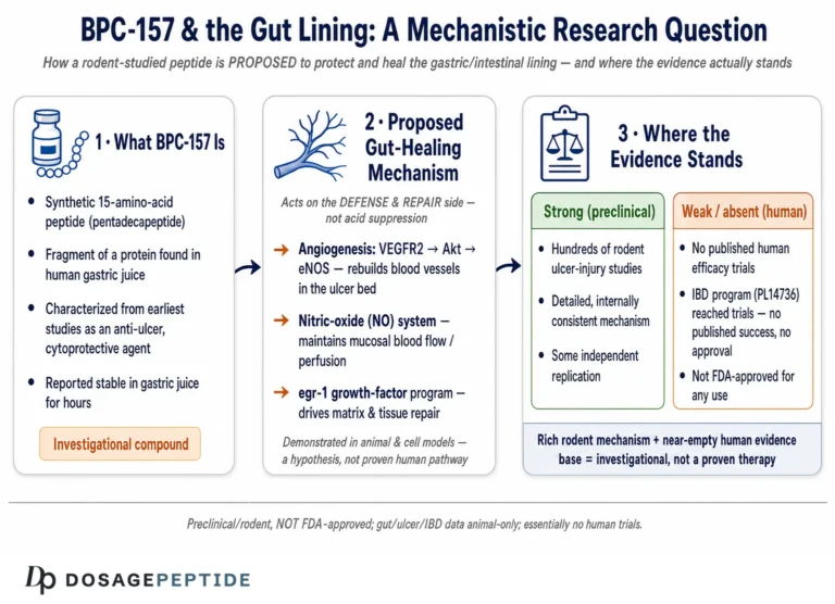
A central and repeatedly emphasized element of BPC-157’s proposed mechanism is its interaction with the nitric oxide system. Across a range of experimental models, the peptide is reported to counteract the effects of both NO-synthase blockade (for example with L-NAME) and NO-substrate overload (with L-arginine), behaving less like a simple NO donor and more like a modulator that pushes a dysregulated NO system back toward balance.[1] Nitric oxide is a master regulator of vascular tone, endothelial permeability, and leukocyte adhesion, so an agent that stabilizes NO signaling has a plausible, mechanistically coherent route to influencing inflammation — although “plausible route” is not the same as “demonstrated clinical effect.”
eNOS, VEGFR2, and angiogenesis
The angiogenic arm of BPC-157’s biology has been characterized in more molecular detail than most other aspects. Experimental work links the peptide to the VEGFR2–Akt–eNOS signaling axis: BPC-157 is reported to upregulate expression of vascular endothelial growth factor receptor 2 (VEGFR2), promote its internalization, and thereby activate downstream Akt and endothelial nitric oxide synthase (eNOS), driving endothelial-cell tube formation and new vessel growth in ischemia models.[3] A separate line of work has implicated the Src–Caveolin-1–eNOS pathway in the peptide’s effects on vasomotor tone, offering a second endothelial mechanism by which BPC-157 appears to influence blood flow and nitric-oxide-dependent signaling.[1] Because inflamed and autoimmune-injured tissues frequently suffer from microvascular dysfunction, an angiogenic and endothelium-stabilizing signal is one of the more direct conceptual bridges between BPC-157’s core biology and the pathology of inflammatory tissue damage.
FAK–paxillin and cell migration
Beyond the vasculature, the Zagreb reviews list the focal adhesion kinase (FAK)–paxillin pathway among the signaling routes engaged by BPC-157.[1] FAK and paxillin are core components of focal adhesions — the structures that couple a cell’s cytoskeleton to the extracellular matrix — and their activity governs cell spreading, migration, and the coordinated movement of fibroblasts and endothelial cells into a wound bed. Effects on this axis are consistent with the peptide’s reported acceleration of tendon, ligament, muscle, and epithelial repair, all of which depend on directed cell migration into damaged tissue.
Growth-factor and receptor upregulation
BPC-157 has also been associated with upregulation of growth-factor signaling and, notably, of the growth hormone receptor in tendon fibroblasts, alongside effects on early growth response and EGF-related pathways in various tissues.[1] Rather than supplying a growth factor directly, the peptide appears in these models to increase tissue sensitivity to endogenous regenerative signals. This “sensitization” framing helps explain why its effects are often described as adaptive and context-dependent rather than as blanket stimulation.
Dopaminergic, serotonergic signaling and the gut–brain axis
A distinctive feature of the BPC-157 literature is the claim that a peptide originating from gastric cytoprotection also modulates central neurotransmitter systems. The reviews describe interactions with the dopaminergic and serotonergic systems and frame the compound’s activity within the gut–brain axis — presenting gastric cytoprotection as a peripheral node that can influence central nervous system function.[1] These reports come from behavioral and neuropharmacological rodent experiments. They are mechanistically interesting and relevant to the idea that gut inflammation and systemic/neural signaling are coupled, but they remain preclinical and should not be over-interpreted.
The gut–brain axis framing is more than a curiosity for the inflammation question. Autoimmune and chronic inflammatory conditions of the gut are increasingly understood as involving bidirectional communication between mucosal immune activity, the enteric nervous system, and central circuits. If — and this remains a large “if” grounded in animal data — BPC-157 stabilizes gut mucosal integrity while also touching central monoaminergic signaling, that would position it at exactly the interface where gut inflammation and systemic regulation meet. The Zagreb reviews describe rodent experiments in which BPC-157 counteracted behavioral and neurological disturbances associated with gastrointestinal or systemic insult, consistent with this peripheral-to-central framing.[1] The appropriate scientific posture is to treat this as a generative hypothesis about mechanism, not as a demonstrated effect in human disease.
How the mechanisms fit together
It is worth pausing to synthesize, because the individual pathways are most informative when viewed as a system. The reported picture is of a compound that (1) stabilizes the nitric-oxide system and endothelial function, (2) drives organized angiogenesis through VEGFR2–Akt–eNOS signaling, (3) engages FAK–paxillin-mediated cell migration to populate the repair zone, (4) increases tissue sensitivity to endogenous growth factors, and (5) touches central monoaminergic signaling through the gut–brain axis. In injured or inflamed tissue, these actions are complementary: better perfusion and new vessels supply an inflamed area, endothelial stabilization limits leak and edema, and coordinated cell migration and growth-factor sensitivity accelerate structural repair. This integrated, homeostasis-restoring profile is what makes the anti-inflammatory hypothesis mechanistically attractive — and it is precisely why disciplined skepticism about human translation is warranted, since attractive mechanisms in rodents routinely fail to reproduce in people.
How does BPC-157 intersect inflammation and immune signaling?
To understand the autoimmune question, it helps first to recall what autoimmune tissue damage involves. Immune-mediated injury across musculoskeletal, gastrointestinal, and vascular systems is characterized by persistent pro-inflammatory cytokine activation, endothelial dysfunction, oxidative stress, and disruption of the extracellular matrix.[4] Cytokines are the central orchestrators of this process, driving the induction, amplification, and — when balanced — the regulation of autoimmune responses.[5] Any peptide proposed to influence autoimmune damage must be evaluated against these specific mechanisms.
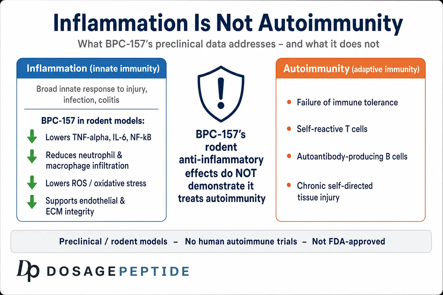
A critical distinction underlies everything that follows: inflammation is not the same as autoimmunity. Inflammation is a broad, largely innate response to injury or insult, common to a cut, a sprain, an infection, or a chemical burn. Autoimmunity is a specific failure of immune tolerance in which the adaptive immune system — T cells and autoantibody-producing B cells — mistakenly targets the body’s own tissues, producing chronic, self-sustaining injury. A compound that dampens acute inflammation in a rodent injury model has demonstrated an anti-inflammatory action; it has not thereby demonstrated that it can restore immune tolerance or interrupt an autoantigen-driven attack. This distinction is the single most important reason to be cautious in extrapolating BPC-157’s animal-model anti-inflammatory data to human autoimmune disease.
Cytokine modulation in animal models
Across rodent injury and colitis models, BPC-157 has been reported to reduce expression of key pro-inflammatory cytokines, particularly tumor necrosis factor-alpha (TNF-α) and interleukin-6 (IL-6), in inflamed tissue.[6] A 2025 systematic review of the musculoskeletal literature found that improved functional, structural, and biomechanical outcomes were consistently accompanied by growth-factor upregulation, organized angiogenesis, and reductions in inflammatory cytokines across the included animal studies.[6] Because TNF-α and IL-6 are precisely the cytokines targeted by approved biologic therapies in several human autoimmune diseases, a preclinical signal in the same direction is why the autoimmune hypothesis is raised at all — but it must be stressed that these are animal-model readouts, not evidence of clinical anti-cytokine efficacy in people.
NF-κB, macrophages, and neutrophils
Mechanistically, the anti-inflammatory effects observed in rodent models have been linked to attenuation of NF-κB–driven signaling, together with the peptide’s NO-system modulation and its angiogenic actions.[1] NF-κB is a master transcriptional switch for inflammatory gene expression — it sits upstream of TNF-α, IL-6, adhesion molecules, and numerous other inflammatory mediators — so suppression or restraint of this pathway offers a coherent upstream explanation for the downstream cytokine reductions described above. It is worth noting that NF-κB is not a purely pathological signal; it is essential for normal host defense, and so an agent that restrains it would need careful characterization to distinguish beneficial modulation from immunosuppression. The BPC-157 literature frames the effect as a restoration of balance in an over-activated inflammatory state rather than as global immune shutdown, but this nuance rests on animal data.
In colitis models specifically, an independent early study reported that BPC-157 (studied under its earlier designation BPC-15) dose-dependently reduced trinitrobenzene-sulfonic-acid (TNBS)-induced colonic damage in rats, and that this protection was accompanied by a statistically significant, dose-dependent fall in colonic myeloperoxidase (MPO) activity — a biochemical marker of neutrophil infiltration.[9] The wider Zagreb-group review literature similarly describes dampened innate-immune cell recruitment, with decreased neutrophil and macrophage accumulation in inflamed gut mucosa across gut-injury models.[1] Neutrophils and macrophages are principal drivers of tissue damage in active mucosal inflammation, so reducing their accumulation is a plausible route to limiting collateral injury. The macrophage point deserves emphasis because macrophage behavior is not simply “on or off”: these cells occupy a spectrum from pro-inflammatory to reparative phenotypes, and effective resolution of inflammation involves a shift toward the reparative end. Whether BPC-157 influences that phenotypic balance, or simply reduces total infiltrate, is not fully resolved in the available preclinical work.
Oxidative stress and matrix preservation
Preclinical work also describes BPC-157 limiting reactive-oxygen-species accumulation and helping to preserve extracellular matrix integrity in inflamed and injured tissue.[1] Oxidative stress and matrix breakdown are shared features of many autoimmune lesions, so these observations round out a picture in which the peptide appears — in animals — to act simultaneously on several arms of the inflammatory response rather than on one isolated target. Whether this multi-pronged profile is an advantage or simply a reflection of nonspecific pleiotropy is an open scientific question.
The angiogenesis–inflammation connection
One reason BPC-157’s angiogenic and anti-inflammatory profiles are difficult to separate is that vascular biology and inflammation are mechanistically intertwined. Inflamed tissue is frequently hypoxic and poorly perfused; resolving inflammation requires clearing debris, delivering reparative cells, and re-establishing a functional microvasculature. The VEGFR2–Akt–eNOS axis that BPC-157 engages sits at the center of this process, and endothelial nitric oxide itself has anti-adhesive and anti-inflammatory properties that limit leukocyte sticking and vascular leak.[3] In the BPC-157 literature, angiogenesis is therefore not framed merely as “growing new vessels” but as part of a coordinated resolution program in which improved perfusion, endothelial stabilization, and reduced inflammatory signaling reinforce one another. This is a coherent model — and, again, one established in animals and cell systems rather than in human autoimmune tissue.
What “immune modulation” does and does not mean here
It is important to characterize BPC-157’s reported immune effects accurately. The animal data describe dampening of innate inflammatory readouts — fewer infiltrating neutrophils and macrophages, lower cytokine levels, reduced NF-κB activity — in the context of tissue injury. This is a modulatory, largely local effect on the inflammatory response to damage. The literature does not characterize BPC-157 as a classical immunosuppressant that broadly blunts adaptive immunity, nor as an agent shown to reprogram autoreactive T cells or eliminate autoantibodies. Conflating “reduces local inflammation in an injury model” with “suppresses autoimmune attack” would misrepresent the evidence. The accurate statement is narrower and more honest: in animal models of tissue injury and chemically induced colitis, BPC-157 reduces markers of the inflammatory response and supports repair.
| Inflammatory feature | Reported effect of BPC-157 in preclinical models | Relevance to autoimmune tissue damage |
|---|---|---|
| TNF-α / IL-6 expression | Reduced in inflamed tissue (rodent injury and colitis models) | These cytokines are validated human autoimmune therapeutic targets |
| NF-κB signaling | Attenuated (proposed upstream mechanism) | Central switch for inflammatory gene transcription |
| Neutrophil / macrophage infiltration | Decreased; lower myeloperoxidase activity | Innate-immune recruitment drives tissue injury |
| Endothelial / NO-system function | Stabilized; NO signaling normalized in both directions | Vascular dysfunction is common in autoimmune lesions |
| Angiogenesis (VEGFR2–Akt–eNOS) | Promoted; increased vessel formation | Supports repair of hypoperfused, damaged tissue |
| Oxidative stress / ECM | ROS reduced; matrix integrity preserved | Both are shared features of autoimmune injury |
What does the autoimmune-adjacent preclinical evidence actually show?
This is the crux of the article, and it requires a blunt framing statement up front: there are no published, rigorous, randomized, placebo-controlled human trials of BPC-157 in any autoimmune disease. What exists is a body of animal and in-vitro work in inflammation and tissue-injury models that share features with — but are not the same as — autoimmune pathology. Reading that literature as if it demonstrated a human autoimmune therapy would be a serious over-interpretation.
Colitis and inflammatory bowel disease models
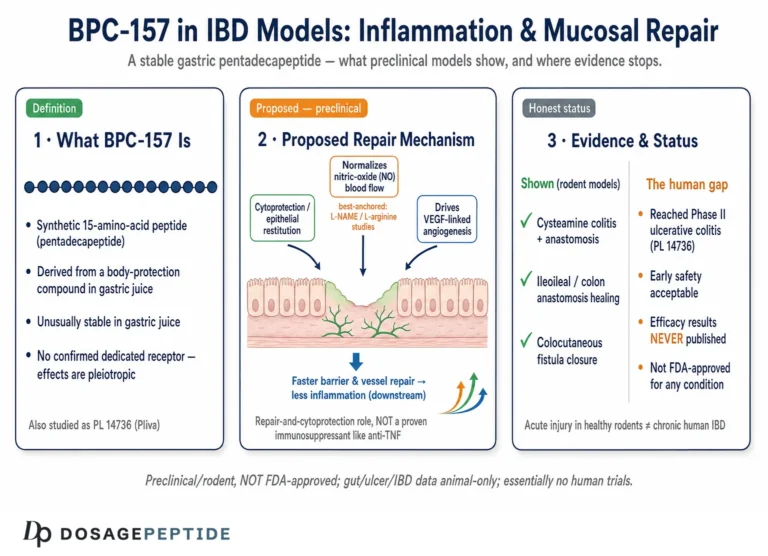
The most autoimmune-adjacent evidence comes from experimental colitis. In the trinitrobenzene-sulfonic-acid (TNBS) colitis model in rats, BPC-157 has been reported to reduce the extent of colonic damage in a dose-dependent manner, with a parallel reduction in colonic myeloperoxidase activity reflecting lower neutrophil infiltration.[9] More broadly, the Zagreb-group review literature describes the peptide lowering inflammatory infiltration, preserving mucosal architecture, and accelerating healing across experimental gut-injury models.[1] The peptide was itself developed under codes including PL-14736 as a candidate for inflammatory bowel disease, and Zagreb-group work has shown that it promotes healing of intestinal anastomoses in the rat.[2]
It is important to be precise about what these models represent. Ulcerative colitis and Crohn’s disease are complex, chronic, immune-mediated conditions in humans; chemically induced rodent colitis reproduces some inflammatory and mucosal features but is an acute, artificially triggered injury, not a spontaneous autoimmune disease. A protective effect in TNBS or DSS colitis is genuinely informative about mucosal cytoprotection and repair, and it is the strongest available preclinical bridge to the autoimmune question — but it does not establish that the peptide would modify the natural history of human IBD. Researchers interested in the mucosal and gut-barrier angle can compare the two structured dosing frameworks that pair the peptide with a co-agent in the TB-500 + BPC-157 combination protocol.
There is one further wrinkle worth surfacing honestly. Development codes associated with BPC-157 (PL-14736 and related designations) reflect that the compound was advanced as an IBD candidate, and the surrounding literature references early-phase trial activity in that indication.[2] However, no rigorous, peer-reviewed, controlled human efficacy results in inflammatory bowel disease have been published in the accessible literature. The absence of published positive human trial data — despite the compound having a development pedigree pointing toward IBD — is itself informative, and it should temper enthusiasm rather than feed it. Unpublished or incompletely reported trial activity is not evidence of efficacy.
The gut immune interface and mucosal barrier
The reason the colitis data are the most autoimmune-adjacent is that the intestinal mucosa is where the immune system and the external environment meet most intensively. A healthy gut barrier depends on tight junctions between epithelial cells, a mucus layer, and a tightly regulated local immune population; when that barrier fails, luminal contents provoke immune activation that can perpetuate inflammation. BPC-157’s reported ability to preserve mucosal integrity and accelerate epithelial healing — as summarized in the Zagreb-group cytoprotection review literature — is mechanistically relevant precisely because barrier dysfunction is implicated in inflammatory bowel disease and in the broader concept of immune dysregulation at mucosal surfaces.[1] It should be noted that these accounts describe mucosal restitution and cytoprotection rather than direct, quantified measurement of tight-junction proteins, so the barrier framing is an interpretive bridge rather than a fully characterized molecular finding. Combined with the angiogenic support of the mucosal microvasculature, this offers a plausible, layered account of why the peptide protects the gut in animal models. As with every claim in this article, the account is preclinical: barrier-protective effects in rodents do not demonstrate disease modification in human IBD.
Tissue-damage and repair models
The broader preclinical dataset — spanning tendon, ligament, muscle, nerve, gastric, hepatic, and vascular injury — consistently reports faster and more organized repair with BPC-157, tied to the angiogenic and cytokine-modulating mechanisms described earlier.[6] Where these models intersect the autoimmune theme is in their shared endpoints: reduced inflammatory cell infiltration, preserved collagen and matrix organization, and stabilized microvasculature. In connective tissue and joints, studies describe reduced inflammatory infiltration and preservation of collagen organization; in gastrointestinal tissue, improved mucosal integrity and reduced ulceration; and at the vascular endothelium, stabilized barriers with attenuated inflammatory leakage.
A recurring and genuinely interesting observation in this literature is that some anti-inflammatory and structural effects are reported to persist beyond the active dosing window, suggesting durable remodeling rather than a purely transient pharmacological response. Animal experiments have used dosing periods ranging from roughly one week to two months, and several report sustained readouts after cessation.[1] This durability is provocative from a mechanistic standpoint — but it also raises exactly the kind of long-term safety question (what are the consequences of durable tissue and vascular remodeling?) that has never been addressed in controlled human study.
Rheumatoid arthritis, systemic lupus, and multiple sclerosis: what is and is not there
Prospective readers often ask specifically about the classic systemic autoimmune diseases — rheumatoid arthritis, systemic lupus erythematosus, multiple sclerosis, psoriasis, or type 1 diabetes. Here the honest accounting is stark: there is no rigorous published human trial of BPC-157 in any of these conditions, and the disease-specific preclinical evidence in validated autoimmune models is thin to absent. The joint and connective-tissue data come predominantly from mechanical or chemical injury models, not from established autoimmune-arthritis models such as collagen-induced arthritis, and they should not be represented as rheumatoid arthritis evidence. Where the pathology of rheumatoid arthritis is well characterized — a synovial process driven by cytokine networks and immune-cell infiltration[4] — BPC-157’s cytokine-modulating profile is at most a mechanistic point of interest, not evidence of efficacy. Any claim that BPC-157 “treats” or “modulates” a named human autoimmune disease outruns the data.
The human-evidence statement, stated plainly
The 2025 HSS Journal systematic review is the most rigorous recent synthesis, and its findings are instructive. Of 36 included studies, 35 were preclinical and only one was clinical — a small retrospective series of intra-articular injections for chronic knee pain — and the authors reported that no clinical safety data were identified and that the human evidence base was inadequate to support clinical recommendations.[6] None of that single clinical reference concerned autoimmune disease. In short: for autoimmune indications specifically, the human clinical evidence for BPC-157 is effectively nonexistent, and every anti-inflammatory claim in this domain rests on animal and cell-based work whose translational relevance is unproven.
How does BPC-157 compare with other regenerative peptides?
BPC-157 is frequently discussed alongside TB-500, and the comparison is instructive because it illustrates how “regenerative peptide” encompasses genuinely different mechanisms.
TB-500 and thymosin beta-4
TB-500 is a synthetic peptide corresponding to the active, actin-binding region of thymosin beta-4 (Tβ4), a naturally occurring 43-amino-acid peptide. Tβ4’s defining biochemical property is sequestration of monomeric G-actin, which regulates actin-cytoskeleton dynamics and thereby promotes cell migration; it also has documented anti-inflammatory activity and promotes angiogenesis and re-epithelialization in dermal wound and cardiac ischemia models.[7] Where BPC-157 works largely through NO-system modulation, VEGFR2-driven angiogenesis, and growth-factor sensitization, Tβ4/TB-500 works primarily through cytoskeletal regulation that mobilizes cells into the injury zone. The mechanisms are complementary rather than redundant.
An important scientific caveat applies specifically to TB-500 as opposed to full-length thymosin beta-4: much of the rigorous peer-reviewed literature — including the classic wound-healing work — concerns native Tβ4, while “TB-500” as sold for research typically refers to a shorter synthetic fragment. Effects demonstrated for full-length Tβ4 cannot be assumed to transfer identically to the fragment, and this distinction is frequently blurred in non-academic sources. As with BPC-157, the human clinical evidence for TB-500 in autoimmune disease is absent; the anti-inflammatory and angiogenic data are preclinical.
The combination rationale — and its limits
The mechanistic complementarity is the stated rationale for pairing the two peptides in many research protocols: BPC-157 is framed as providing angiogenic and growth-factor-receptor-mediated proliferative and cytoprotective signals, while TB-500 supplies the cytoskeletal machinery for cell migration into the repair zone. It is essential to be clear that this combination rationale is a mechanistic hypothesis built on separate preclinical datasets, not a conclusion from head-to-head or combination clinical trials in autoimmune disease — no such trials exist. Researchers modeling the combination can reference the paired TB-500 and BPC-157 combination dosing protocol for a structured framework, with the strong caveat that it is a research-handling reference, not therapeutic guidance.
| Property | BPC-157 | TB-500 (thymosin beta-4 fragment) |
|---|---|---|
| Origin | Fragment of gastric protein BPC (synthetic pentadecapeptide) | Active region of naturally occurring thymosin beta-4 |
| Primary mechanism | NO-system modulation; VEGFR2–Akt–eNOS angiogenesis; growth-factor sensitization | G-actin sequestration; cytoskeletal regulation of cell migration |
| Reported anti-inflammatory action | Cytokine (TNF-α/IL-6) and NF-κB modulation in models | Documented anti-inflammatory activity in wound models |
| Angiogenesis | Central and well characterized | Present; promotes endothelial recruitment |
| Human autoimmune trials | None | None |
What research models and methodology are used to study BPC-157?
Because the entire evidence base is preclinical, the methodology behind these studies determines how much weight the findings can bear. Being literate in the models is part of reading the claims critically.
Common experimental designs
The BPC-157 literature draws on several standard model families: chemically induced injury models (TNBS/DSS colitis, ethanol or NSAID gastric lesions), surgical models (tendon transection, ligament and muscle injury, intestinal anastomosis), ischemia–reperfusion models (hind-limb ischemia, vascular occlusion), and in-vitro assays (endothelial tube formation, tenocyte and fibroblast migration). Typical animal experiments administer the peptide at nanogram-to-microgram per-kilogram doses over periods ranging from days to several weeks, using intraperitoneal, intragastric, or topical routes.[1]
Outcome measures and their limits
Endpoints commonly include histological damage scoring, myeloperoxidase and cytokine quantification, biomechanical testing of repaired tissue, vessel density measurements, and functional recovery assays.[6] These are legitimate, quantifiable measures — but the systematic-review literature notes that much of the primary work carries methodological limitations characteristic of an emerging field: many studies originate from a single research group, sample sizes are often small, blinding and randomization are inconsistently reported, and negative results may be underrepresented in the published record.[6] These are exactly the conditions under which promising preclinical signals frequently fail to translate to humans, which is why methodological transparency and independent replication matter so much here.
What a rigorous autoimmune study would require
Framing what is missing clarifies the size of the evidence gap. To move BPC-157 from “interesting in animal inflammation models” toward “plausibly relevant to human autoimmune disease” would require, at minimum: validated autoimmune-specific animal models (for example, collagen-induced arthritis for RA-like disease, or experimental autoimmune encephalomyelitis for MS-like disease) rather than acute chemical-injury models; independent replication across multiple laboratories; dose–response characterization with pharmacokinetic data; and eventually well-designed, randomized, blinded, placebo-controlled human trials with standardized immune biomarkers, imaging, and predefined clinical endpoints. None of these later stages has been completed. A single small retrospective human case series in a non-autoimmune orthopaedic context — the one clinical study identified in the 2025 systematic review — does not begin to close this gap.[6]
Why single-lineage evidence is a specific concern
The concentration of the mechanistic literature within one research program is not a moral criticism — it reflects that a single group pioneered and sustained this field. But it is a methodological concern with a well-understood remedy: independent replication. Effects that are large, real, and reproducible tend to be confirmed by unaffiliated laboratories once a field matures. The gradual appearance of independent mechanistic work — for example, the endothelial Src–Caveolin-1–eNOS characterization[1] — is encouraging, but the autoimmune-relevant efficacy data have not yet been broadly replicated or advanced into rigorous human study. Until they are, confident claims are premature.
How is BPC-157 handled and reconstituted in a research context?
This section is framed strictly as research-handling information, not medical or dosing advice. BPC-157 is not an approved therapeutic, and the doses used in animal studies do not translate into human recommendations.
Reconstitution and stability considerations
Lyophilized research peptides such as BPC-157 are typically reconstituted with bacteriostatic or sterile water, with the target concentration determined by the mass of peptide in the vial and the volume of diluent added. Accurate concentration math is the single most consequential step in reproducible peptide work: an error in reconstitution volume propagates through every subsequent measurement. General principles of aseptic handling, appropriate diluents, and refrigerated storage of reconstituted material are covered in the peptide reconstitution guide, and concentration and volume calculations can be worked through with the reconstitution and dosage calculator.
Research-dosing frameworks and vial context
Peptide research is typically organized around a defined vial size and a target working concentration, which together set how a given quantity of reconstituted material is measured out for a model system. Structured, vial-specific frameworks — again as research-handling references, not human dosing guidance — are documented in the BPC-157 5 mg vial dosage protocol and the BPC-157 10 mg vial dosage protocol. These pages exist to support consistent, documented laboratory practice; they are not clinical instructions and carry no implication that the compound is safe or appropriate for human use.
Why animal doses do not become human recommendations
It bears repeating in concrete terms why the nanogram-to-microgram-per-kilogram doses reported in rodent studies cannot be read as human guidance. Interspecies dose translation is not a simple body-weight multiplication; it depends on metabolic scaling, differences in pharmacokinetics and clearance, route of administration, and target-tissue exposure, none of which have been characterized for BPC-157 in humans. A dose that produces a clean anti-inflammatory readout in a rat colitis model provides no validated basis for a human dose, and because no controlled human pharmacokinetic or safety studies exist, any “human protocol” circulating for this compound is extrapolation rather than evidence. Treating research-handling frameworks as clinical dosing is a category error the responsible literature is careful to avoid.
Material quality as a source of experimental variability
For the preclinical work that is legitimate, material quality is a decisive and often underappreciated variable. Inflammation and immune-pathway experiments are sensitive to peptide purity, correct sequence, and the absence of endotoxin and process-related impurities — any of which can independently perturb inflammatory readouts and confound interpretation. This is one reason regulators cite peptide-related impurities and immunogenicity as concerns for unapproved peptides. In practice, reproducible inflammation research depends on analytically characterized material (for example, purity confirmed by HPLC and identity by mass spectrometry), documented batch records, and consistent reconstitution and storage — controls that reduce the experimental noise that would otherwise obscure genuine biological signal.
What are the limitations and the human-evidence gap?
Even a sympathetic reading of the BPC-157 literature must confront several structural limitations that bear directly on the autoimmune question.
- No human autoimmune trials. There are no randomized, blinded, or placebo-controlled human studies of BPC-157 in any autoimmune disease, and no standardized human data on inflammatory markers, immune profiling, or imaging outcomes for such use.[6]
- Concentration in one research lineage. A large fraction of the mechanistic and efficacy data derives from a single group, which raises the importance of independent replication before any of it can be considered robust.[6]
- Model-to-human translation gap. Acute chemically induced injury in rodents does not reproduce the chronic, self-perpetuating immune dysregulation of human autoimmune disease; protective effects in these models may not carry over.
- Absent long-term safety data. No controlled human data exist on the long-term consequences of chronic immune modulation with BPC-157, and the systematic review found no clinical safety data at all.[6]
- Theoretical concerns of pro-angiogenic activity. A compound that promotes angiogenesis warrants careful scrutiny in any context where unwanted vessel growth could be harmful — a consideration that has not been resolved in humans.
The overall conclusion is unambiguous: BPC-157’s anti-inflammatory profile in animals is real as an experimental observation, but its relevance to human autoimmune disease is an untested hypothesis. It remains an investigational compound with no approved clinical indication for autoimmune-related inflammation. The scientifically appropriate stance is neither dismissal nor endorsement, but calibrated interest: the preclinical signal is coherent enough to justify continued, independent, well-designed research, while the human evidence is too sparse to support any therapeutic claim or use. Holding both of those truths at once — genuine mechanistic promise alongside a genuine, unbridged evidence gap — is the honest reading of where BPC-157 and autoimmune inflammation currently stand.
What are the safety, regulatory, and handling considerations in research?
Regulatory status
BPC-157 is not approved by the FDA for human therapeutic use. Its status under Section 503A of the Federal Food, Drug, and Cosmetic Act — which governs which bulk drug substances may be used by compounding pharmacies — has moved over time and remains unsettled. In 2023 the FDA placed BPC-157 in the Category 2 list of bulk drug substances (a category identifying substances with significant safety concerns or insufficient data), which prohibited its use as a bulk substance in compounding. In April 2026 the FDA removed BPC-157 from that Category 2 list, and the compound is scheduled for review by the Pharmacy Compounding Advisory Committee.[8] Removal from Category 2 is not FDA approval and does not place BPC-157 on the approved 503A bulks list; it returns the substance to an evaluative state pending a formal recommendation and final decision, so the practical regulatory posture has not changed to endorsement. Cited concerns raised in the earlier action have included immunogenicity risk, peptide-related impurities, and limited human safety data. In sport, BPC-157 is prohibited at all times under the World Anti-Doping Agency (WADA) framework, listed under class S0 (Non-Approved Substances) — the category covering pharmacological substances not approved by any governmental regulatory health authority for human therapeutic use — meaning its presence in an athlete sample constitutes an anti-doping rule violation.[10]
The practical upshot of this regulatory picture is that BPC-157 exists, in most jurisdictions, only as a research-use-only material — not a medicine, not a supplement, and not a compounding ingredient. Its regulatory status remains fluid and has been the subject of ongoing debate, so anyone tracking the field should rely on primary regulatory sources rather than secondary commentary. For the autoimmune question specifically, the regulatory absence is consistent with the scientific absence: there is no approved indication because there is no adequate human evidence base to support one.
Research handling and ethical framework
Legitimate BPC-157 research is confined to preclinical settings under appropriate oversight. Studies involving immune modulation or autoimmune-related tissue injury must operate under institutional review board (IRB) or ethics-committee approval, use scientifically justified disease models with predefined humane endpoints, and follow standardized biomarkers, validated assays, and transparent, reproducible reporting so that results can be independently evaluated. For research-grade material, purity verification, batch testing, and analytical characterization (for example by mass spectrometry and HPLC) are the practical prerequisites for interpretable, reproducible inflammation and immune-pathway experiments — variability in material quality is a well-recognized source of experimental noise that can confound inflammatory readouts.
Frequently Asked Questions
Can BPC-157 treat autoimmune diseases in humans?
No. There are no published, rigorous, controlled human clinical trials of BPC-157 in any autoimmune disease, so its efficacy and safety for such use are unestablished. All anti-inflammatory findings come from rodent and in-vitro models. BPC-157 is an investigational compound with no approved clinical indication for autoimmune-related inflammation, and it should not be regarded as a treatment.
What inflammatory pathways does BPC-157 affect in animal studies?
In preclinical models, BPC-157 is reported to reduce pro-inflammatory cytokines such as TNF-α and IL-6, attenuate NF-κB signaling, decrease neutrophil and macrophage infiltration (lower myeloperoxidase activity), stabilize nitric-oxide-dependent vascular function, and limit oxidative stress. These effects are observed in animals and have not been confirmed to occur or to be clinically meaningful in humans with autoimmune disease.
Is the colitis research evidence that BPC-157 works for IBD?
Not in humans. BPC-157 reduces damage scores and inflammation in chemically induced rodent colitis (TNBS/DSS) models, and it was developed under codes such as PL-14736 as an IBD candidate. But these are acute, artificially triggered injuries, not spontaneous autoimmune disease, and no rigorous published human trial has demonstrated that BPC-157 modifies the course of ulcerative colitis or Crohn’s disease.
How does BPC-157 differ from TB-500?
They work through different mechanisms. BPC-157 acts largely via nitric-oxide-system modulation, VEGFR2-driven angiogenesis, and growth-factor sensitization. TB-500, a fragment of thymosin beta-4, works mainly by sequestering G-actin to regulate the cytoskeleton and promote cell migration. Their mechanisms are complementary, which is the stated rationale for combining them in research protocols — a hypothesis, not a clinically proven strategy.
Is BPC-157 approved or legal?
BPC-157 is not FDA-approved for human therapeutic use. Its Section 503A compounding status has changed over time: the FDA placed it in the Category 2 bulk-substances list in 2023 (barring its use in compounding due to safety and data concerns) and then removed it from that list in April 2026, referring it for advisory-committee review — a step that is not approval and does not place it on the approved bulks list. It is also prohibited at all times in sport under the WADA framework, listed as an S0 (Non-Approved) substance. It is generally available only as a research-use-only material, and its regulatory status continues to evolve, so verify against primary FDA sources.
Why is BPC-157 evidence considered weak despite many studies?
The volume of studies is large but the quality and independence are limited. Much of the data comes from a single research group, sample sizes are often small, blinding and randomization are inconsistently reported, and essentially all of it is preclinical. A 2025 systematic review found 35 of 36 studies were preclinical, with no clinical safety data — conditions under which promising animal signals frequently fail to translate to humans.
What does “modulating the NO system” actually mean?
Rather than simply raising nitric oxide, BPC-157 appears in animal models to counteract both the blockade and the overstimulation of nitric oxide synthase, pushing a dysregulated NO system back toward balance. Because nitric oxide governs vascular tone, endothelial permeability, and leukocyte adhesion, this modulatory behavior is one proposed route by which the peptide influences inflammation in experimental settings.
How should BPC-157 be handled in a laboratory?
As a research-only material, lyophilized BPC-157 is typically reconstituted with sterile or bacteriostatic water using accurate concentration math, handled aseptically, and stored refrigerated once reconstituted. Purity verification and batch testing support reproducibility. These are handling practices for controlled research, not instructions for human use; the compound is not approved for people.
For an immune-modulating peptide with a substantial clinical-research record, see our overview of what Thymosin Alpha-1 is and how it modulates immune signaling.
References
- Sikiric P, Seiwerth S, Rucman R, et al. Stable Gastric Pentadecapeptide BPC 157, Robert’s Stomach Cytoprotection/Adaptive Cytoprotection/Organoprotection, and Selye’s Stress Coping Response: Progress, Achievements, and the Future. Gut and Liver. 2020;14(2):153-167. https://pmc.ncbi.nlm.nih.gov/articles/PMC7096228/
- Vuksic T, Zoricic I, Brcic L, et al. Stable gastric pentadecapeptide BPC 157 in trials for inflammatory bowel disease (PL-10, PLD-116, PL14736, Pliva, Croatia) heals ileoileal anastomosis in the rat. Surgery Today. 2007;37(9):768-777. https://pubmed.ncbi.nlm.nih.gov/17713731/
- Sikiric P, et al. Stable Gastric Pentadecapeptide BPC 157 as a Therapy and Safety Key: A Special Beneficial Pleiotropic Effect Controlling and Modulating Angiogenesis and the NO-System. 2025. https://pmc.ncbi.nlm.nih.gov/articles/PMC12195719/
- Firestein GS, McInnes IB. Immunopathogenesis of Rheumatoid Arthritis. Immunity. 2017;46(2):183-196. https://pubmed.ncbi.nlm.nih.gov/28228278/
- Moudgil KD, Choubey D. Cytokines in Autoimmunity: Role in Induction, Regulation, and Treatment. Journal of Interferon & Cytokine Research. 2011;31(10):695-703. https://pmc.ncbi.nlm.nih.gov/articles/PMC3189547/
- Vasireddi N, Hahamyan H, Salata MJ, et al. Emerging Use of BPC-157 in Orthopaedic Sports Medicine: A Systematic Review. HSS Journal. 2025. https://journals.sagepub.com/doi/10.1177/15563316251355551
- Malinda KM, Sidhu GS, Mani H, et al. Thymosin beta4 accelerates wound healing. Journal of Investigative Dermatology. 1999;113(3):364-368. https://pubmed.ncbi.nlm.nih.gov/10469335/
- U.S. Food and Drug Administration. Bulk Drug Substances Used in Compounding Under Section 503A of the FD&C Act. https://www.fda.gov/drugs/human-drug-compounding/bulk-drug-substances-used-compounding-under-section-503a-fdc-act
- Veljaca M, Lesch CA, Pllana R, Sanchez B, Chan K, Guglietta A. BPC-15 reduces trinitrobenzene sulfonic acid-induced colonic damage in rats. Journal of Pharmacology and Experimental Therapeutics. 1995;272(1):417-422. https://pubmed.ncbi.nlm.nih.gov/7815358/
- U.S. Anti-Doping Agency (USADA). BPC-157: Experimental Peptide Creates Risk for Athletes. https://www.usada.org/spirit-of-sport/bpc-157-peptide-prohibited/