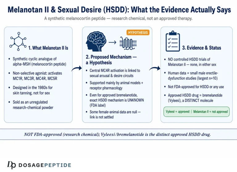
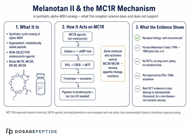
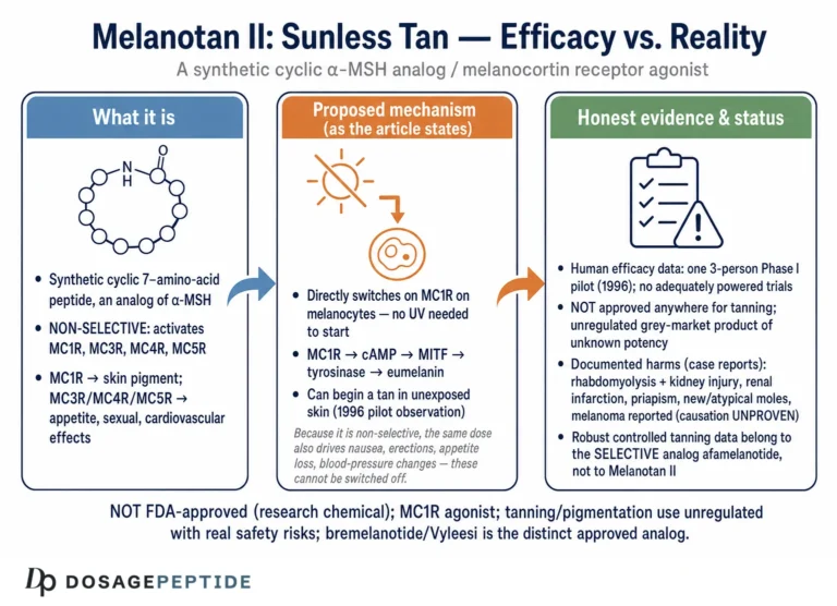
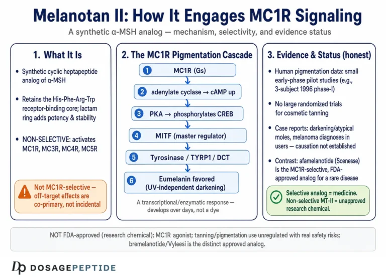
Short answer: Melanotan II is not selective. It activates four of the five melanocortin receptors — MC1R, MC3R, MC4R and MC5R — with high affinity, sparing only the adrenal MC2R.1 That single fact explains why the same injection darkens skin, blunts hunger, causes flushing and nausea, and affects sexual arousal: they are different receptors being hit at once. It also means no experiment in an intact animal can tell you what the compound did at MC1R alone.
So “which model best measures the MC1R effect?” has no one-assay answer. It is a design problem: isolating one receptor’s contribution from a pharmacologically noisy background. This page maps the toolkit researchers actually use — amphibian and reptile skin bioassays, recombinant cyclic-AMP reporter cells, pigment-cell and melanoma cultures, cryo-electron microscopy structures, and knockout and humanised-receptor animals — and says what each can and cannot show. One thing to fix before reading further: Melanotan II is not an approved medicine anywhere, its use as a tanning agent is unregulated and carries documented safety concerns, and it should not be confused with the two approved melanocortin drugs it is routinely muddled with, afamelanotide (Scenesse) and bremelanotide (Vyleesi).1112
What Melanotan II and MC1R Actually Are
Melanotan II is a synthetic cyclic heptapeptide, most precisely written as Ac-Nle-cyclo[Asp-His-D-Phe-Arg-Trp-Lys]-NH₂, an analogue of the core message sequence of α-MSH. Its lineage is instructive. In 1980, Sawyer, Hruby, Hadley and colleagues at the University of Arizona reported that replacing methionine at position 4 with norleucine and the L-phenylalanine at position 7 with its D-enantiomer produced [Nle⁴, D-Phe⁷]-α-MSH (NDP-α-MSH), a linear analogue with dramatically enhanced potency and “ultralong” biological activity.2 A subsequent line of work showed that cyclising the peptide through a lactam bridge locked the pharmacophore into its bioactive conformation and yielded superpotent cyclic melanotropins.3 Melanotan II is the best-known product of that design program. The same effort ultimately spun off bremelanotide (PT-141), essentially the C-terminal deamidated (free-acid) metabolite of Melanotan II — still a cyclic heptapeptide, not a linear or ring-opened molecule — which lost the pigmentary emphasis and was developed instead for its central effects on sexual desire.12
The receptor at the center of the tanning story is MC1R, a class A (rhodopsin-like) G protein-coupled receptor expressed on cutaneous melanocytes. When an agonist binds, MC1R couples predominantly to Gαs, raises intracellular cyclic AMP, activates protein kinase A, and drives the transcription factor MITF, which in turn up-regulates tyrosinase and the other enzymes of the melanogenic pathway. The functional consequence is a shift in pigment synthesis from the reddish-yellow pheomelanin toward the photoprotective brown-black eumelanin.67 This cAMP–MITF–tyrosinase cascade is the biochemical spine on which nearly every MC1R assay is built: whether a model reads out receptor occupancy, second-messenger accumulation, enzyme activity, or visible pigment, it is sampling one station along this single conserved line.
Two features of MC1R make it an unusually interesting — and unusually tricky — receptor to model. First, it is one of five melanocortin receptor subtypes (MC1R–MC5R) that share the same family of endogenous ligands derived from pro-opiomelanocortin, and that all recognise the conserved His-Phe-Arg-Trp (HFRW) tetrapeptide core.4 That shared pharmacophore is exactly why Melanotan II is non-selective and why isolating MC1R responses is hard. Second, MC1R is one of the most polymorphic genes in the human genome, with common loss-of-function variants that produce red hair, fair skin, poor tanning, and elevated skin-cancer risk.8 That natural allelic variation is both a complication for modelling and, as we will see, a powerful experimental resource.
There is a third feature that complicates modelling and is easy to overlook: MC1R signalling in a real melanocyte is not a simple on/off switch but a dial set against opposing inputs. The receptor has measurable constitutive (agonist-independent) activity, it is negatively regulated by endogenous inverse agonists and antagonists — agouti-signalling protein in the mouse and the human ASIP, and the antagonist β-defensin in some contexts — and its surface expression is modulated by trafficking, desensitisation, and post-translational modification such as palmitoylation. A faithful model of Melanotan II at MC1R therefore has to decide how much of this regulatory context to reproduce. A stripped-down recombinant assay deliberately discards it to gain a clean potency number; a native melanocyte retains it and pays for the realism with interpretive ambiguity. Neither choice is wrong, but the choice is consequential, and much confusion in the popular literature comes from reading a number obtained in one context as though it applied to the other.
Why Receptor Selectivity Is the Central Modelling Problem
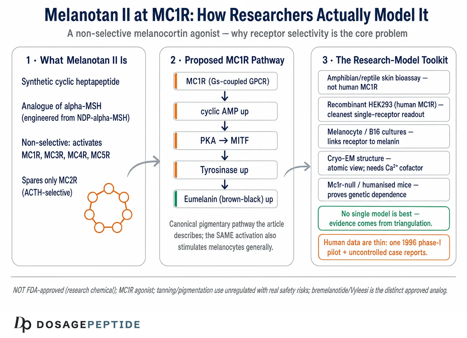
If there is one idea to carry through this entire article, it is that a model’s value for assessing Melanotan II at MC1R is determined almost entirely by how well it isolates MC1R from the other melanocortin receptors. Consider the binding data. Competitive radioligand studies using [¹²⁵I]-NDP-MSH report that Melanotan II displaces the tracer with affinities in the sub-nanomolar to low-nanomolar range not just at MC1R but also at MC3R, MC4R, and MC5R, with only MC2R (which is essentially ACTH-selective and poorly responsive to the HFRW core) showing negligible affinity.14 Functionally, cAMP-accumulation assays likewise show Melanotan II behaving as a high-efficacy agonist across MC1R, MC3R, MC4R, and MC5R.
This has a direct methodological consequence. In any intact system that expresses more than one melanocortin receptor — a whole animal, an organ explant, even some primary cell preparations — a response to Melanotan II cannot be attributed to MC1R without additional controls. The pro-erectile and appetite effects that surfaced in early human exposure to Melanotan II, for example, are mediated centrally through MC3R/MC4R, not MC1R, a dissociation explored in the site’s companion articles on how Melanotan II influences erectile performance mechanistically and whether it could play a role in hypoactive sexual desire disorder. Those effects are real, but they are the wrong receptor for a pigmentation question. A model that conflates them tells you about melanocortin biology in general, not MC1R in particular.
The corollary is that the “best” models for the MC1R question are the ones that engineer selectivity into the experiment rather than hoping for it. In practice this is achieved in three ways: (1) heterologous expression of MC1R alone in a null-background cell line, so the only melanocortin receptor present is the one under study; (2) counter-screening the same compound in parallel against MC3R, MC4R, and MC5R to build a full selectivity profile; and (3) genetic manipulation — knockout, knock-in, or humanised alleles — that removes or swaps MC1R while leaving the rest of the system intact. Each of the model classes below is, at bottom, an attempt to buy MC1R specificity in one of these three currencies.
The Classic Bioassays: Amphibian and Reptile Skin
The oldest tools for measuring melanotropic activity long predate the cloning of MC1R, and they remain conceptually clarifying. Vertebrate melanophores — the pigment cells of frog and lizard skin — disperse their intracellular melanosomes in response to α-MSH, darkening the skin visibly and reversibly. The classic assays use isolated skins from frogs (Rana species, Xenopus) or the lizard Anolis carolinensis, exposing the tissue to graded doses of a melanotropin and quantifying darkening by reflectance.5 The Anolis preparation was prized because a single animal’s skin could be used repeatedly in one day with high precision, making it an efficient dose-response platform in the pre-recombinant era.
These bioassays were the crucible in which the melanotropin structure-activity relationship was forged. It was in frog and lizard skin that investigators established the HFRW core as the minimal active sequence and demonstrated that the Nle⁴/D-Phe⁷ substitutions of NDP-MSH conferred superpotency — the very findings that made Melanotan II possible.25 As drug-discovery platforms, melanophore-based systems retain appeal because pigment dispersion is a robust, amplified, whole-cell readout of Gs/cAMP signalling.
But for the specific question of Melanotan II at human MC1R, the amphibian assays have a decisive limitation that must be stated plainly: the receptor being activated is not human MC1R. Amphibian and reptile melanophore responses are mediated by the species’ own melanocortin receptors, whose sequence, pharmacology, and — critically — ligand selectivity differ from the human receptor. A frog skin darkening in response to Melanotan II confirms melanotropic bioactivity in a beautifully integrated tissue, but it cannot resolve whether that activity would occur specifically through human MC1R versus a related subtype, nor can it capture the effects of human MC1R polymorphisms. The classic bioassays are best understood today as historically foundational and useful for cross-species SAR, not as definitive models of the human MC1R interaction.
Recombinant Cell Systems: the Workhorse of MC1R Pharmacology
If a single class of model deserves to be called the modern standard for assessing a compound at MC1R, it is the heterologous recombinant cell system — and the reason is exactly the selectivity argument above. By transfecting the cloned human MC1R gene into a cell line that expresses no endogenous melanocortin receptors (HEK293 is the canonical choice; COS and CHO cells are also used), the experimenter creates a background in which MC1R is the only relevant target. Any Melanotan II–driven signal in that dish is, by construction, an MC1R signal.6
Two complementary readouts dominate. The first is radioligand binding: membranes from MC1R-expressing cells are incubated with a labelled high-affinity tracer, classically [¹²⁵I]-NDP-MSH, and unlabelled Melanotan II is titrated in to compete it off. The resulting displacement curve yields an IC₅₀ or, after correction, a Kₜ, giving a quantitative measure of binding affinity independent of what the receptor does downstream. The second is functional signalling: because MC1R couples to Gs and raises cyclic AMP, activity is read as cAMP accumulation. Historically this was measured with a CRE (cAMP-response-element)-driven reporter such as β-galactosidase or luciferase, in which agonist-driven cAMP switches on an easily quantified enzyme; more recent laboratories use direct competitive-immunoassay or biosensor cAMP kits, which shorten the assay and improve dynamic range.67 From the functional curve one extracts an EC₅₀ (potency) and an Eₔ₄ₓ (efficacy) relative to a reference agonist such as α-MSH or NDP-MSH.
The great strength of this platform is that it lets a compound be profiled across all melanocortin subtypes under identical conditions. Running Melanotan II in parallel dishes of MC1R-, MC3R-, MC4R-, and MC5R-expressing cells generates the selectivity table that intact systems cannot — and it is that comparative panel, more than any single MC1R number, that constitutes a rigorous assessment of the compound at MC1R.
It is worth being concrete about what the numbers from these assays do and do not mean, because the distinction is frequently blurred in secondary sources. A Kₜ from a binding assay quantifies how tightly Melanotan II occupies the receptor; it says nothing about whether that occupancy switches the receptor on. An EC₅₀ from a functional assay quantifies the concentration producing half-maximal signalling, and the accompanying Eₔ₄ₓ (relative efficacy) captures how fully the receptor is activated at saturation — the difference between a full agonist and a partial one. A compound can bind avidly yet activate weakly, or bind modestly yet activate fully; only the pairing of binding and functional data describes the pharmacology. For Melanotan II the available data place it as a high-affinity, high-efficacy agonist at MC1R, but the same descriptors apply at MC3R, MC4R, and MC5R, which is precisely the point — the receptor-level numbers are impressive and nearly interchangeable across subtypes, so the individual MC1R value, quoted alone, is easy to over-interpret.14
Recombinant systems also make it possible to interrogate signalling beyond cAMP. MC1R can recruit β-arrestin and, in some contexts, activate ERK/MAP-kinase pathways; assays that separate G-protein signalling from arrestin recruitment can reveal whether a ligand is “biased.” The study of the naturally occurring E92K MC1R mutant is a useful illustration: it showed constitutive cAMP production and arrestin recruitment but not ERK activity, evidence that these arms can be uncoupled and that a full pharmacological description of a ligand at MC1R may need more than a cAMP number.9 This matters for interpreting variant receptors too: the common red-hair alleles are not uniformly “dead” but hypomorphic to varying degrees, some retaining partial cAMP coupling while losing others, so a single-pathway assay can over- or under-state how much function a given variant preserves. A model chosen to assess Melanotan II should be matched to the question — cAMP for the canonical pigmentary pathway, multiplexed readouts if signalling texture or variant behaviour is the object of study.
The honest caveats are also worth stating. Heterologous overexpression can push receptor density far above physiological levels, which inflates apparent potency and can make a partial agonist look full; results are sensitive to the reporter, the cell background, and the expression construct. And a transfected HEK293 cell is not a melanocyte — it lacks the pigment machinery, the native regulatory partners, and the trafficking context of a real pigment cell. The recombinant system answers the question “does Melanotan II engage and activate MC1R, and how selectively?” with great rigour; it does not by itself tell you what that engagement does to pigmentation.
Melanocyte and Melanoma Cell Models: from Receptor to Pigment
To connect MC1R activation to its biological output — melanin — the field turns to pigment cells themselves. Two categories matter. The murine B16 melanoma line has been a melanogenesis workhorse for decades because it expresses functional Mc1r and responds to melanotropins with measurable increases in tyrosinase activity and melanin content; darkening can be quantified spectrophotometrically or the cells simply pellet darker. Human systems include primary or immortalised normal human epidermal melanocytes and human melanoma lines, which carry human MC1R with its authentic downstream apparatus.67
These models add what recombinant cells cannot: an integrated, physiologically meaningful endpoint. A rise in cAMP is a proximal signal; an increase in MITF, tyrosinase transcription and activity, and finally total melanin is the phenotype that actually matters for a tanning agent. Melanocyte models therefore close the loop between receptor engagement and pigment output, and human melanocytes carrying defined MC1R genotypes can reveal how loss-of-function variants blunt the response — directly relevant to why fair-skinned, red-haired individuals tan poorly regardless of stimulus.78
A refinement that has strengthened these models is the use of more physiological formats: co-cultures of melanocytes with keratinocytes (which normally take up transferred melanosomes), reconstructed human epidermis, and organotypic skin equivalents that reproduce the melanin unit’s architecture. These systems can report not just how much melanin a melanocyte makes in response to an MC1R agonist but whether that melanin is transferred and distributed as it would be in skin — a downstream step entirely invisible to a monolayer culture. For a compound whose entire proposed cosmetic rationale is visible skin darkening, the distinction between intracellular melanin synthesis and its delivery to the epidermis is not academic.
Their limitation, again, is selectivity and context. Melanocytes may express more than one melanocortin receptor and sit within regulatory feedback (for example, the antagonist agouti-signalling protein) absent from a transfected dish. Attributing a pigmentary response purely to MC1R still requires genetic or pharmacological controls — for instance, confirming that a selective MC1R antagonist abolishes the response, or that melanocytes from an Mc1r-null source fail to respond. And because Melanotan II’s clinical notoriety involves melanocytic lesions, it bears emphasis that a cell darkening in culture says nothing about the safety of stimulating melanocytes in vivo — a point developed in the safety section and in the site’s review of what the current evidence says about Melanotan II and dermatologic disease. Indeed, one of the more sobering uses of melanocyte and melanoma models has been to probe how MC1R signalling intersects with the DNA-damage response and oxidative stress: variant, loss-of-function receptors appear less able to support the protective, pigment-independent responses that guard melanocytes against ultraviolet injury, which is part of why MC1R genotype tracks with melanoma risk. That literature is a reminder that MC1R biology is entangled with cancer-relevant pathways, and that pharmacologically flogging the receptor is not a consequence-free intervention.
Structural Models: Cryo-EM and the Atomic View of Ligand Recognition
A qualitatively different class of model does not measure a response at all but instead resolves how ligand and receptor physically fit together. In 2021, cryo-electron microscopy delivered the first near-atomic structures of the human MC1R–Gs complex bound to three different agonists: the endogenous hormone α-MSH, the approved drug afamelanotide (NDP-MSH), and the synthetic probe SHU9119.10 These structures were a milestone for the whole melanocortin field.
They matter for the Melanotan II question in two ways. First, they visualise the orthosteric pocket that accommodates the conserved HFRW motif shared by α-MSH, afamelanotide, and Melanotan II, explaining at a mechanistic level why the Nle⁴/D-Phe⁷ substitutions and the cyclic constraint of these analogues enhance potency: they pre-organise the message sequence for a snug fit and resist the proteolysis that degrades the native hormone. Second, and unexpectedly, the structures revealed that MC1R requires a calcium ion as a cofactor for high-affinity agonist binding, with the divalent cation bridging acidic receptor residues and the ligand’s conserved glutamate and arginine.10 This calcium dependence is a genuine mechanistic insight and also a methodological warning: binding and functional assays run in calcium-depleted buffers can misreport affinity, so the ionic composition of an in-vitro model is not a trivial detail when the target is MC1R.
The structures also illuminate the selectivity problem at the atomic level, which is where it ultimately originates. Because the HFRW-binding orthosteric pocket is highly conserved across MC1R through MC5R, a ligand built around that core — as Melanotan II is — is essentially reading a message that every subtype can interpret. Subtype selectivity, when it exists, comes from peripheral contacts outside the conserved core; the reason Melanotan II is non-selective is that its cyclic scaffold optimises engagement of the shared pocket without exploiting those subtype-distinguishing peripheral differences. Comparative structures of the related MC4R bound to NDP-MSH and to the selective agonist setmelanotide have made this logic explicit, showing how selective and non-selective melanocortin ligands differ in exactly which receptor regions they touch. This gives the structural work real predictive value: it suggests where a chemist would have to modify Melanotan II to make it MC1R-preferring, and by the same token why the parent compound is not.
Structural models excel at explaining and predicting — they rationalise SAR and can guide the design of more selective ligands — but they are static snapshots of a purified, often stabilised complex, typically captured with a reference agonist rather than with Melanotan II itself. They tell you how the HFRW core is read by MC1R; they do not, on their own, quantify Melanotan II’s potency or its selectivity margin over MC4R. Structure and pharmacology are complementary, not interchangeable, and a structure obtained with afamelanotide is a strong but not perfect proxy for how the cyclic Melanotan II scaffold sits in the same pocket.
Genetic and Animal Models: Knockouts, Variants, and Humanised Receptors
The most powerful way to prove that a response depends on MC1R specifically is to remove or alter the gene and watch the response disappear or change — the logic of genetic models. MC1R is exceptionally well served here because its biology is written across coat and hair colour in many species.
In the mouse, the extension locus is Mc1r. Loss-of-function alleles produce yellow fur (pheomelanin), while constitutively active alleles produce dark coats; the null phenotype is yellow.6 This makes coat colour a visible genetic readout of receptor function and gives loss-of-function mice as a built-in negative control: a melanotropic response that persists in an Mc1r-null animal cannot be attributed to MC1R. In humans, the corresponding natural experiment is the panel of common loss-of-function variants — R151C, R160W, D294H, and others — strongly associated with red hair, fair skin, and impaired tanning. Beaumont and colleagues showed that red hair behaves essentially as the null phenotype of human MC1R, cementing the gene’s role as the master switch of the tanning response and providing a graded series of naturally “detuned” receptors for functional study.8
Because mouse and human MC1R differ pharmacologically, investigators built humanised transgenic mice that express the human receptor, revealing human-specific aspects of receptor function that a native mouse model would miss.6 Non-mammalian genetics contribute too: melanocortin signalling shapes pigment patterning across vertebrates, and models from zebrafish to large domesticated animals (whose coat-colour loci repeatedly map to MC1R) corroborate the receptor’s conserved role.8 A useful orientation to the receptor family and its ligand vocabulary is collected in the site’s peptide glossary.
Genetic models also let investigators run the cleanest possible causal experiment for a compound like Melanotan II: dose the drug in a wild-type animal and in an Mc1r-null littermate, and compare. Any pigmentary response that survives in the null cannot be an MC1R effect, and any that vanishes is, by subtraction, MC1R-dependent. This loss-of-function logic is far more persuasive than a correlation in an intact system, because it removes the receptor rather than inferring its involvement. The converse gain-of-function experiment — expressing a constitutively active or humanised MC1R allele — tests whether the receptor is sufficient to produce the phenotype. Together these approaches convert the selectivity problem from an interpretive worry into an experimentally answerable question, which is exactly why genetic models sit at the apex of evidence for receptor attribution even though they are the least convenient for routine screening.
Animal and genetic models supply what every dish lacks: an intact organism with real skin, immune surveillance, pharmacokinetics, and feedback. Their price is complexity and, for Melanotan II specifically, the return of the selectivity problem — a systemically dosed animal exposes every melanocortin receptor at once, so pigmentation endpoints must be paired with genetic controls to keep the readout MC1R-specific. There is also an unavoidable translational gap: rodent pigmentation biology, skin architecture, and melanocyte distribution differ from human skin, which is why humanised alleles and, ultimately, human genetic epidemiology remain necessary complements. Animal models are indispensable for validation and causal attribution, and least suited to clean, isolated potency measurement — the reverse of the recombinant systems, which is precisely why the two are used together rather than in place of one another.
A Comparative Map of the Models
The models are best understood not as competitors but as a layered toolkit, each answering a different sub-question and each with a characteristic blind spot. The table summarises how they stack up specifically for assessing Melanotan II at MC1R.
| Model class | Primary readout | MC1R specificity | Key limitation for the MT-II–MC1R question |
|---|---|---|---|
| Amphibian/reptile skin bioassay | Visible melanophore darkening (Gs/cAMP) | Low — non-human receptor, subtype ambiguous | Not human MC1R; cannot model human variants5 |
| Recombinant HEK293/COS/CHO expressing human MC1R | Radioligand binding (Kₜ); cAMP/CRE-reporter (EC₅₀, efficacy) | High — single defined receptor in null background | Overexpression artefacts; not a pigment cell6 |
| B16 / human melanocyte / melanoma cultures | Tyrosinase activity, melanin content, MITF | Moderate — native but may express other MCRs | Needs genetic controls to isolate MC1R7 |
| Cryo-EM structural model | Atomic ligand–receptor geometry; Ca²⁺ cofactor | High — purified human MC1R complex | Static; usually a reference agonist, not MT-II; no potency10 |
| Mc1r-null / variant / humanised mice; zebrafish | Coat/skin pigment; genetic dependence | Definitive by genetics, but whole-organism exposure | Systemic dosing hits all MCRs; needs KO controls68 |
Read across the table and a clear principle emerges: no single model is “best” in isolation, because the strengths and weaknesses are complementary. A defensible assessment of Melanotan II at MC1R triangulates — recombinant systems to quantify affinity, potency, and selectivity across the receptor panel; pigment-cell models to confirm the functional consequence; structural data to explain the molecular basis; and genetic models to prove causal dependence on MC1R in a living system. The recombinant selectivity panel is the analytical core, but it is the convergence across levels that constitutes real evidence.
Designing a Rigorous Selectivity Assessment
Given all of the above, what would a well-designed program to assess Melanotan II at MC1R actually look like? The through-line is that potency at MC1R is close to meaningless without the accompanying selectivity margin, because the compound is intrinsically promiscuous.
A rigorous design would proceed in tiers. Tier one establishes the pharmacological fingerprint: full concentration-response curves for Melanotan II run in parallel against human MC1R, MC3R, MC4R, and MC5R in the same null-background cell line, using both binding (competition against [¹²⁵I]-NDP-MSH) and function (cAMP), with α-MSH and NDP-MSH as reference agonists and calcium-replete buffers to respect the cofactor requirement.110 The deliverable is not a single MC1R EC₅₀ but a selectivity ratio — how much, if at all, does MC1R activity separate from MC4R? For Melanotan II the honest expectation, based on existing data, is that the separation is small; it is a broad agonist, not an MC1R-selective one.14
Tier two tests signalling texture: does Melanotan II engage MC1R’s cAMP arm, arrestin arm, and ERK arm proportionately, or is there bias?9 Tier three moves to native pigment cells (human melanocytes of defined MC1R genotype, B16) to confirm that receptor activation translates into tyrosinase induction and melanin, and to quantify how loss-of-function variants blunt the response.78 Tier four, where justified, uses genetic animal models to prove MC1R dependence in vivo, always with Mc1r-null controls to catch off-target melanocortin contributions.
A final design consideration is reproducibility across laboratories. Because apparent potency at MC1R is sensitive to receptor expression level, cell background, buffer calcium, tracer choice, and reporter linearity, results are only comparable when normalised to a shared reference agonist run in the same plate. Reporting a Melanotan II EC₅₀ as a bare number, divorced from the reference and the assay conditions, is one of the commonest ways the literature becomes internally inconsistent — a caution that applies with special force to values quoted in non-peer-reviewed vendor material, where the assay context is usually omitted entirely.
The reason to spell this out is that most public claims about Melanotan II “working through MC1R” rest on the intuitive but incomplete first half of tier one — it binds MC1R, therefore the tan is an MC1R effect — while ignoring that the same molecule binds MC3R/MC4R/MC5R just as avidly and that the systemic effects users actually report (nausea, flushing, priapism, appetite change) are substantially non-MC1R. A model program that measured only MC1R would be structurally incapable of detecting the compound’s defining feature, which is its lack of selectivity.
Approved Melanocortin Drugs as Reference Points
Two approved drugs anchor the MC1R and broader melanocortin space, and they are the correct comparators against which any Melanotan II claim should be read — not because they are interchangeable with it, but precisely because they are not.
Afamelanotide (Scenesse) is the closest approved analogue to Melanotan II’s pigmentary arm. It is, in fact, NDP-α-MSH — the same linear [Nle⁴, D-Phe⁷] superagonist from the 1980 Arizona work — formulated as a subcutaneous implant. The U.S. Food and Drug Administration approved it in October 2019 to increase pain-free light exposure in adults with erythropoietic protoporphyria (EPP), a rare photodermatosis; the European Medicines Agency had approved it in 2014.11 Note what the approval is and is not: it is a narrow orphan indication in which MC1R-driven eumelanin provides photoprotection, granted after controlled trials and under strict pharmacovigilance. It is not an approval for cosmetic tanning, and it does not validate Melanotan II, which is a different (cyclic, non-selective) molecule sold without any of that oversight.
Bremelanotide (Vyleesi) is the melanocortin drug most genealogically tied to Melanotan II: it emerged directly from observations that Melanotan II produced unexpected sexual effects in early human exposure. The FDA approved it in June 2019 for acquired, generalised hypoactive sexual desire disorder in premenopausal women; it acts centrally at MC3R/MC4R with reduced MC1R activity relative to its Melanotan II parent.12 Bremelanotide is therefore an MC4R-oriented drug — the opposite selectivity emphasis from a pigmentation agent — and its existence is a concrete reminder that the melanocortin receptors carry distinct functions that a non-selective compound cannot separate. Readers interested in that pathway can consult the site’s coverage of how PT-141 (bremelanotide) affects libido and fatigue.
The honest lesson from both drugs is about evidentiary standards. Where a melanocortin agent has a legitimate use, it has been characterised across exactly the model tiers described above, taken through controlled human trials, and approved for a defined indication under monitoring. Melanotan II has none of that for tanning. It is an investigational compound whose human record is limited to a handful of small studies and a large, uncontrolled body of case reports from unregulated use.
Safety, Regulatory Status, and the Limits of Model Data
No discussion of experimental models for Melanotan II is complete without confronting what happens when the compound leaves the model and enters a person — because the gap between an elegant cAMP curve and real-world harm is where the honest reader’s attention belongs.
The controlled human data are thin. The foundational clinical exposure was a 1996 pilot phase-I study by Dorr and colleagues in three male volunteers, which documented increased pigmentation but also dose-limiting nausea, facial flushing, and spontaneous penile erections accompanied by a stretching-and-yawning complex — the last two being classic MC3R/MC4R effects, again underscoring the compound’s non-selectivity.13 Beyond such small studies, the human record comes overwhelmingly from unregulated recreational use, where the picture is more concerning.
Reviews of melanotan use catalogue a consistent set of adverse effects: nausea and vomiting, facial flushing, spontaneous erections, appetite suppression, blood-pressure changes, and injection-site reactions in the near term, plus a dermatological signal that dominates the case literature — darkening and change of existing melanocytic nevi, eruptive new atypical nevi, and isolated reports of melanoma diagnosed during or after use, alongside rarer serious events such as posterior reversible encephalopathy syndrome, priapism, and rhabdomyolysis.1415 Case series report the sudden eruption of multiple dysplastic nevi and rapid transformation of existing moles following courses of Melanotan II, with histopathology confirming dysplasia.16 The mechanistic plausibility is uncomfortable: MC1R activation is precisely what stimulates melanocytes, and stimulating melanocytes indiscriminately in a person — especially one with atypical-mole predisposition — is not obviously benign. This is the sharpest illustration of the article’s central caution: a model that shows “MC1R activation → melanin” is measuring the desired effect and the feared effect through the same receptor.
| Compound | Chemical class | Receptor emphasis | Regulatory status |
|---|---|---|---|
| Melanotan II | Cyclic heptapeptide α-MSH analogue | Non-selective (MC1R, MC3R, MC4R, MC5R) | Not approved anywhere; unregulated tanning use; safety warnings issued14 |
| Afamelanotide (Scenesse) | Linear NDP-α-MSH (13-mer) | MC1R-oriented (broad melanocortin agonist) | FDA-approved 2019 for EPP; EMA 201411 |
| Bremelanotide (Vyleesi) | Cyclic heptapeptide (MT-II–derived) | Central MC3R/MC4R; reduced MC1R | FDA-approved 2019 for HSDD in premenopausal women12 |
Regulatory bodies have acted accordingly. The U.K. Medicines and Healthcare products Regulatory Agency and the U.S. FDA have both warned that melanotan tanning injections and sprays are unlicensed, of unverified quality, and potentially unsafe; they are not approved products, and material sold as “Melanotan II” is not manufactured to pharmaceutical standards nor tested for purity or potency by any regulator.14 This is a crucial coda to the modelling discussion. Even a perfectly characterised MC1R pharmacology — a clean Kₜ, a confirmed structure, a validated genetic dependence — would say nothing about the safety of injecting an unregulated peptide of unknown purity into human skin. Model data describe the molecule’s action at a receptor; they do not license its use. Anyone genuinely investigating Melanotan II should do so within properly authorised research, and general handling considerations for research peptides — reconstitution, storage, sterility — are described for educational reference in the site’s peptide reconstitution guide and broader dosages index, neither of which is a recommendation for human use.
Frequently Asked Questions
Which single model best measures Melanotan II’s effect on MC1R?
There isn’t one, and treating any single assay as definitive is the most common methodological error. The closest thing to an analytical core is a recombinant cell system — human MC1R expressed in a null background such as HEK293 — read out by radioligand binding and a cAMP/CRE-reporter, because it isolates MC1R cleanly.6 But that must be paired with a parallel selectivity panel against MC3R/MC4R/MC5R, a pigment-cell model to confirm the functional consequence, and ideally genetic controls. The “best” assessment is a convergence across these levels, not any one number.
Why is receptor selectivity such a big deal for Melanotan II specifically?
Because Melanotan II is intrinsically non-selective. It binds MC1R, MC3R, MC4R, and MC5R with high affinity, sparing only MC2R.14 In any intact system a response could reflect several receptors at once, so isolating the MC1R contribution requires either single-receptor expression or genetic controls. A program that measured only MC1R would miss the compound’s defining property.
Do frog and lizard skin bioassays tell us about human MC1R?
Only indirectly. These classic melanophore assays were essential for working out the melanotropin structure-activity relationship, including the substitutions that gave rise to Melanotan II.25 But they activate the animal’s own melanocortin receptors, not human MC1R, so they cannot resolve human receptor subtype selectivity or the effects of human MC1R variants. They are historically foundational, not definitive for the human question.
What did the cryo-EM structures of MC1R reveal?
The 2021 cryo-EM structures of the human MC1R–Gs complex, solved with α-MSH, afamelanotide, and SHU9119, visualised the orthosteric pocket that reads the conserved HFRW motif and revealed that a calcium ion is a required cofactor for high-affinity agonist binding.10 That explains why the Nle⁴/D-Phe⁷ analogues are potent and warns that in-vitro assays run without calcium can misreport affinity. The structures are static snapshots, usually with a reference agonist rather than Melanotan II itself.
Are genetic mouse models useful here, and how?
Very. In mice the extension locus is Mc1r, so coat colour is a visible genetic readout — loss-of-function gives yellow fur, and the null is a built-in negative control.6 Human loss-of-function variants (R151C, R160W, D294H) cause red hair and poor tanning, and red hair behaves as the null phenotype of MC1R.8 Humanised transgenic mice express the human receptor to capture human-specific pharmacology. Genetic dependence is the strongest proof that a response is truly MC1R-mediated.
Is Melanotan II the same as the approved drugs afamelanotide or bremelanotide?
No. Afamelanotide (Scenesse) is a different molecule — linear NDP-α-MSH — approved only for the rare photodermatosis erythropoietic protoporphyria, not for cosmetic tanning.11 Bremelanotide (Vyleesi) is a Melanotan II–derived analogue approved for hypoactive sexual desire disorder that acts centrally at MC3R/MC4R.12 Melanotan II itself is not approved anywhere and is sold and used illegally as an unregulated tanning agent.
Does an MC1R cell assay tell us whether Melanotan II is safe?
No. Model data describe how a molecule engages a receptor — affinity, potency, selectivity, structure — but say nothing about the safety of injecting an unregulated peptide of unknown purity into human skin. The same MC1R activation that drives a tan also stimulates melanocytes, and the case literature links Melanotan II to eruptive and atypical nevi and to isolated melanoma reports, alongside nausea, flushing, and blood-pressure effects.1416 Regulators have warned against its use.
What safety concerns are best documented for Melanotan II?
The controlled data (a 1996 phase-I pilot) noted nausea, flushing, and spontaneous erections as dose-limiting.13 The larger, uncontrolled case literature adds darkening and transformation of moles, eruptive atypical nevi, and rare serious events including posterior reversible encephalopathy syndrome, priapism, and rhabdomyolysis.141516 Products are unregulated and untested for purity, compounding the risk.
References
- Mun Y, Kim W, Shin D. Melanocortin 1 Receptor (MC1R): Pharmacological and Therapeutic Aspects. Int J Mol Sci. 2023;24(15):12152. PMCID: PMC10419527. https://www.mdpi.com/1422-0067/24/15/12152
- Sawyer TK, Sanfilippo PJ, Hruby VJ, et al. 4-Norleucine, 7-D-phenylalanine-alpha-melanocyte-stimulating hormone: a highly potent alpha-melanotropin with ultralong biological activity. Proc Natl Acad Sci USA. 1980;77(10):5754-5758. PMID: 6777774. https://www.pnas.org/doi/10.1073/pnas.77.10.5754
- Sawyer TK, Hruby VJ, Darman PS, Hadley ME. [half-Cys4,half-Cys10]-alpha-Melanocyte-stimulating hormone: a cyclic alpha-melanotropin exhibiting superagonist biological activity. Proc Natl Acad Sci USA. 1982;79(6):1751-1755. PMID: 6281785. https://www.pnas.org/doi/10.1073/pnas.79.6.1751
- Yuan XC, Tao YX. Ligands for Melanocortin Receptors: Beyond Melanocyte-Stimulating Hormones and Adrenocorticotropin. Biomolecules. 2022;12(10):1407. PMCID: PMC9599618. https://pmc.ncbi.nlm.nih.gov/articles/PMC9599618/
- Salim S, Ali SA. Vertebrate melanophores as potential model for drug discovery and development: a review. Cell Mol Biol Lett. 2011;16(2):162-200. PMID: 21225472. https://link.springer.com/article/10.2478/s11658-010-0044-y
- Wolf Horrell EM, Boulanger MC, D’Orazio JA. Melanocortin 1 Receptor: Structure, Function, and Regulation. Front Genet. 2016;7:95. PMCID: PMC4886693. https://www.frontiersin.org/journals/genetics/articles/10.3389/fgene.2016.00095/full
- Doedens L, Opperer F, Cai M, et al. NMR Insights into the Structure-Function Relationships in the Binding of Melanocortin Analogues to the MC1R Receptor. Molecules. 2017;22(7):1189. PMCID: PMC6152105. https://www.ncbi.nlm.nih.gov/pmc/articles/PMC6152105/
- Beaumont KA, Shekar SN, Cook AL, et al. Red hair is the null phenotype of MC1R. Hum Mutat. 2008;29(8):E88-E94. PMID: 18484624. https://pubmed.ncbi.nlm.nih.gov/18484624/
- Cai M, Stankova M, Muthu D, et al. The E92K Melanocortin 1 Receptor Mutant Induces cAMP Production and Arrestin Recruitment but Not ERK Activity Indicating Biased Constitutive Signaling. PLoS One (PMC). 2011. PMCID: PMC3172247. https://www.ncbi.nlm.nih.gov/pmc/articles/PMC3172247/
- Ma S, Yin Y, Yang Y, et al. Structural mechanism of calcium-mediated hormone recognition and Gβ interaction by the human melanocortin-1 receptor. Cell Res. 2021;31(10):1061-1071. DOI: 10.1038/s41422-021-00557-y. https://www.nature.com/articles/s41422-021-00557-y
- U.S. Food and Drug Administration. SCENESSE (afamelanotide) implant, for subcutaneous use — full prescribing information. NDA 210797, approved October 8, 2019 for erythropoietic protoporphyria. https://www.accessdata.fda.gov/drugsatfda_docs/label/2019/210797s000lbl.pdf
- U.S. Food and Drug Administration. VYLEESI (bremelanotide injection), NDA 210557, prescribing information / approval 2019. https://www.accessdata.fda.gov/drugsatfda_docs/label/2019/210557s000lbl.pdf
- Dorr RT, Lines R, Levine N, et al. Evaluation of melanotan-II, a superpotent cyclic melanotropic peptide in a pilot phase-I clinical study. Life Sci. 1996;58(20):1777-1784. PMID: 8637402. https://pubmed.ncbi.nlm.nih.gov/8637402/
- Habbema L, Halk AB, Neumann M, Bergman W. Risks of unregulated use of alpha-melanocyte-stimulating hormone analogues: a review. Int J Dermatol. 2017;56(10):975-980. DOI: 10.1111/ijd.13585. https://onlinelibrary.wiley.com/doi/10.1111/ijd.13585
- Brennan R, Wells JG, Van Hout MC. An unhealthy glow? A review of melanotan use and associated clinical outcomes. Perform Enhanc Health. 2014;3(2):78-92. DOI: 10.1016/j.peh.2015.06.001. https://www.sciencedirect.com/science/article/abs/pii/S2211266915000055
- Eruptive Dysplastic Nevi Following Melanotan Use (case report). Actas Dermosifiliogr. 2012. https://actasdermo.org/en-eruptive-dysplastic-nevi-following-melanotan-articulo-S1578219012001357
Educational and research-use disclaimer: This article is provided solely for scientific and educational purposes. Melanotan II is not approved by the FDA, EMA, or any comparable regulator for tanning, pigmentation, or any other use, and it should not be confused with the distinct, approved melanocortin drugs afamelanotide (Scenesse) and bremelanotide (Vyleesi). Its unregulated use as a tanning agent carries documented safety concerns, including nausea and blood-pressure effects and, most seriously, the darkening and transformation of melanocytic nevi with monitoring implications for melanoma risk; regulatory agencies have warned against it. Nothing here is medical advice or a recommendation for human use. Any legitimate investigation of this compound should occur within properly authorized preclinical or clinical research under appropriate oversight. Readers should consult qualified professionals and applicable regulations before making any decisions.