Two of the most discussed molecules in the regenerative-peptide literature, TB-500 and BPC-157, are frequently grouped together as generic “healing peptides” — yet the experimental record hints that they may act on nervous tissue through genuinely different biology. This article asks a narrow, testable question: does the published experimental literature suggest distinct neuroregenerative functions for these two compounds, or are they interchangeable? To answer it honestly, we have to separate what has actually been measured in animals and cells from what is merely inferred, extrapolated, or marketed.
The short version, stated up front so nothing is misread: both TB-500 and BPC-157 are research chemicals, not approved medicines, and essentially all of the neuroregeneration data comes from rodent injury models and in-vitro systems. There are no controlled human trials showing that either peptide regenerates nerves or treats any neurological disease. Within that important limitation, the preclinical mechanisms do diverge in interesting and reasonably well-characterized ways — and that divergence is the real subject of this review.
What are TB-500 and BPC-157, and why is neuroregeneration even a question?
BPC-157 is a synthetic 15-amino-acid peptide (a “pentadecapeptide,” sequence Gly-Glu-Pro-Pro-Pro-Gly-Lys-Pro-Ala-Asp-Asp-Ala-Gly-Leu-Val). It is derived from — and represents a partial, stabilized fragment of — a protein found in human gastric juice, sometimes referred to as Body Protection Compound. The laboratory version is engineered for stability in gastric acid, which is why so many rodent studies administer it orally or intraperitoneally and still report systemic effects. Crucially, BPC-157 as studied in the literature is the same 15-mer sold as a research chemical, so the animal data map reasonably directly onto the molecule people refer to by that name.
TB-500 is a different story, and the distinction matters enormously for how the evidence is read. The name “TB-500” is used commercially for a synthetic peptide marketed as the active fragment of thymosin beta-4 (Tβ4) — a naturally occurring 43-amino-acid protein that is the principal actin-sequestering peptide in mammalian cells. The fragment most often associated with the TB-500 label is the seven-residue actin-binding motif Ac-LKKTETQ (residues 17–23 of Tβ4), although some research-chemical material sold under that name is actually the full 43-mer.[15] This is a recurring source of confusion: the overwhelming majority of the neurological studies were performed with full-length thymosin beta-4, not with the short LKKTETQ fragment. So when the literature describes Tβ4 improving outcomes after brain injury, that is evidence about the parent molecule, and only indirect, component-level evidence about the fragment sold as TB-500.
The reason neuroregeneration became a question at all is that both peptides emerged from wound-healing and cytoprotection research, and the nervous system is one of the tissues in which repair is slowest and least complete. Peripheral nerves regenerate poorly and slowly; central nervous system (CNS) axons barely regenerate at all after injury. So it is a legitimate scientific question — and a heavily hyped commercial one — whether molecules that accelerate skin, tendon, and vascular repair also nudge nervous tissue toward recovery. The honest answer requires grading the evidence, not just collecting positive headlines.
A note on how the two names are used in this article
To keep the evidence honest, this review uses “BPC-157” for the pentadecapeptide throughout, because the studied molecule and the named molecule are the same. For the thymosin family, it uses “thymosin beta-4” (or Tβ4) when describing what the experiments actually administered, and reserves “TB-500” for the research-chemical label — flagging wherever a claimed TB-500 effect actually rests on full-length Tβ4 data. Readers who want precise definitions of terms such as oligodendrogenesis, angiogenesis, or actin sequestration can consult our peptide research glossary, which is referenced throughout.
How strong is the evidence, and what does “neuroregeneration” actually mean?
Before comparing the two compounds, it helps to fix two things: an evidence hierarchy, and a working definition of neuroregeneration. Both are routinely blurred in popular write-ups, which is how preclinical signals get inflated into implied cures.
“Neuroregeneration” is not one process. In the studies discussed below it is an umbrella term for several distinct, separately measurable events:
- Neurogenesis — the birth of new neurons from neural progenitor cells, typically assessed with markers such as BrdU incorporation and doublecortin.
- Angiogenesis — the growth of new blood vessels, which supports metabolically demanding repair and is often a prerequisite for the other processes.
- Axonal sprouting and outgrowth — the extension of new or damaged axons, measured morphometrically or with tract-tracing.
- Oligodendrogenesis and remyelination — the generation of myelin-forming oligodendrocytes from oligodendrocyte progenitor cells (OPCs) and the re-wrapping of demyelinated axons.
- Neuroprotection — strictly, the prevention of cell death rather than the regrowth of tissue; frequently reported alongside regeneration but mechanistically separate.
A molecule can score on one of these axes and not the others, and — as we will see — that is precisely where TB-500/Tβ4 and BPC-157 appear to differ. Conflating “reduced cell death” with “regenerated nerve” is one of the most common errors in the secondary literature.
The evidence hierarchy is equally important. From weakest to strongest for making claims about humans: in-vitro/cell-culture work; component-level or parent-molecule inference (relevant to TB-500); small-animal (rodent) injury models; large-animal models; human observational data; and finally randomized controlled human trials. For neuroregeneration, both peptides sit at the lower rungs of this ladder — rodent models and in-vitro systems — with no randomized controlled trials in any neurological indication for either. Every claim in this article should be read against that ceiling.
| Evidence tier | BPC-157 (neuro) | Thymosin β4 / TB-500 (neuro) |
|---|---|---|
| In-vitro / cell culture | Present (endothelial, neuronal survival signals) | Present (OPC differentiation, neurite outgrowth, endothelial) |
| Rodent injury models | Present (sciatic, spinal cord, TBI, cerebral ischemia) | Present (TBI, embolic stroke, EAE demyelination) |
| Large-animal models | Essentially absent | Essentially absent for neuro |
| Human trials, any neuro indication | None | None |
| Human trials, non-neuro | None completed and published to approval | Yes — dry-eye and dermal wound (ophthalmology/dermatology) |
| FDA-approved neurological use | No | No |
Which experimental models generate this evidence, and what can each actually show?
Because the entire case for either peptide rests on animal and cell models, it is worth pausing on what those models can and cannot demonstrate. A reader who does not know the difference between a sciatic-crush model and an embolic-stroke model cannot judge whether a positive result reflects true regeneration, mere tissue-sparing, or an artifact of the assay. The models below recur throughout both literatures, and each carries its own interpretive ceiling.
Peripheral-nerve transection and crush models
Here a rodent sciatic (or facial) nerve is cut or crushed, then repair is tracked with the sciatic functional index (a walking-track measurement of toe spread), electromyography, and histomorphometry counting the number, diameter, and myelination of regenerating axons. These models measure genuine axonal regeneration, which is their strength. Their limitation is that the peripheral nervous system already regenerates thanks to Schwann-cell support, so a positive result demonstrates acceleration or improved organization, not the creation of a capacity that was absent — a much stronger claim that no peripheral model can support.
Spinal-cord compression and contusion models
A laminectomy exposes the cord, which is then compressed, contused, or transected; recovery is scored with locomotor rating scales and tail-function or evoked-potential measures. These models straddle two phenomena that are easy to conflate: sparing of tissue that would otherwise die (neuroprotection) and regrowth of lost connections (regeneration). Most reported peptide benefits in spinal models are better read as tilted toward the protective end, because the endpoints rarely isolate new axonal bridging from preserved existing tissue.
Traumatic-brain-injury models
Controlled cortical impact or fluid-percussion injury creates a reproducible cortical lesion; outcomes include lesion volume, a modified neurological severity score, and spatial-learning tasks such as the Morris water maze. The most informative pattern in this class — and the one seen with thymosin beta-4 — is unchanged lesion volume but improved function, because it rules out “the drug simply prevented the initial damage” and points instead to remodeling of surviving tissue.
Cerebral ischemia and embolic stroke models
Middle-cerebral-artery occlusion, by suture or embolus, produces an ischemic core surrounded by a salvageable penumbra. Readouts include infarct size, vessel density in the ischemic boundary, and markers of myelination and progenitor activity. These models are notoriously poor predictors of human stroke-drug success, which is the single most important reason to treat even elegant positive results with caution.
Demyelination and autoimmune models
Experimental autoimmune encephalomyelitis (a multiple-sclerosis analog) and toxin-induced demyelination let investigators watch oligodendrocyte progenitor cells proliferate and mature using BrdU labeling and myelin markers such as myelin basic protein. These are the models in which thymosin beta-4’s remyelination signature is clearest, precisely because they are built to visualize myelin turnover.
In-vitro and endothelial systems
Cell-culture assays — endothelial tube formation, the chick chorioallantoic membrane assay, OPC differentiation cultures, and neurite-outgrowth assays — establish direct molecular actions without the confounds of a whole animal. Their weakness is the mirror image: a molecule that reshapes a cell in a dish may do nothing useful in an injured brain. In-vitro data are best treated as mechanism-generating, not efficacy-proving.
The through-line across all of these is that a positive finding is only as meaningful as the model is faithful, and none of these models is a human. With that scaffolding in place, the compound-specific evidence below can be read for what it actually is.
What do peripheral-nerve studies suggest about BPC-157?
The most direct nerve-regeneration evidence for BPC-157 comes from a transected-sciatic-nerve model in rats. In that work, BPC-157 was applied shortly after the nerve was cut — intraperitoneally, intragastrically, locally at the anastomosis site, or delivered into a tube bridging a resected 7 mm nerve gap — and outcomes were tracked over one to two months.[1] The reported picture was consistent across the levels of analysis the investigators used: clinical (reduced autotomy, i.e. less self-mutilation of the denervated limb, a proxy for restored sensation), microscopic/morphometric (faster axonal regeneration, better-organized neural fascicles, increased density and size of regenerating fibers, and epineural/perineural regeneration), and functional (electromyography and walking recovery via the sciatic functional index).
What makes this study useful for the “distinct functions” question is that it measured actual axonal architecture and walking recovery, not merely a survival marker. In the peripheral nervous system, Schwann cells and a permissive environment allow real regeneration to occur even without intervention, so the claim being tested is acceleration and improved organization, not the creation of regeneration from nothing. Within a rodent model, BPC-157 appeared to shift the trajectory in that direction. That is a meaningful preclinical signal — but it is a single research group’s model, in one species, without the independent multi-lab replication that would be needed before any human inference.
Why the peripheral-nerve result is easier to interpret than CNS claims
Peripheral nerve is the “friendly” case for regeneration: it has an intrinsic capacity to regrow that the CNS lacks. A compound that accelerates a process that already happens is a more modest and more plausible claim than one that restarts a process the adult CNS has largely switched off. This is worth holding onto as we move to the central nervous system, where the bar for “regeneration” is far higher and where much of what is reported for both peptides is more accurately described as neuroprotection or remodeling rather than true regrowth of lost circuits.
What do central-nervous-system models suggest about BPC-157?
Beyond peripheral nerve, several rodent CNS-injury studies — predominantly from the Zagreb pharmacology group that first characterized BPC-157 — report benefit. The consistency of the source is itself a limitation worth naming early.
Spinal cord injury
In a rat spinal-cord-injury model, BPC-157 was given intraperitoneally (200 or 2 µg/kg) shortly after a compression injury, with outcomes followed out to 360 days.[2] Treated animals showed progressive recovery of tail motor function, absence of autotomy, and resolution of spasticity, alongside electromyographic changes and a microscopic profile in which the vacuolization, white-matter axon loss, gray-matter edema, and motoneuron loss typical of the injury were attenuated. The authors framed the result as an effect spanning the stages of secondary injury rather than a single mechanism. Importantly, “improved healing course” here blends genuine tissue-sparing (neuroprotection) with claimed regenerative change; disentangling the two from the published endpoints is difficult, and readers should not treat “functional recovery in rats” as evidence of spinal-cord regeneration in humans.
Traumatic brain injury and cerebral ischemia
In a mouse traumatic-brain-injury model, BPC-157 — including when given prophylactically before injury — was associated with less intense traumatic lesions (subarachnoid and intraventricular hemorrhage, laceration), reduced consecutive brain edema, and an improved conscious/unconscious/death distribution.[3] Separately, in a rat model of transient hippocampal ischemia/reperfusion, BPC-157 was reported to counteract neuronal injury in the vulnerable hippocampus.[4] Both of these are, mechanistically, more about limiting damage (neuroprotection and vascular/edema effects) than about regenerating lost neurons — a distinction that matters for the central comparison, because it suggests BPC-157’s CNS story is weighted toward protection and vascular support rather than the progenitor-driven remodeling seen with thymosin beta-4.
The brain–gut axis angle
A distinctive strand of the BPC-157 literature ties its CNS effects to the gut. Reviews from the originating group and a more recent synthesis argue that peripherally administered BPC-157 can influence brain function through bidirectional brain–gut signaling, with reported effects on serotonergic and dopaminergic systems and on behavioral models.[5][6] This is biologically interesting and consistent with the peptide’s gastric origin, but the behavioral and neurotransmitter readouts are the softest kind of neuro evidence — easily confounded and far removed from “regeneration.” They belong in a mechanistic discussion, not in any claim that BPC-157 treats depression, neurodegeneration, or any human condition.
What do brain-injury and stroke models suggest about thymosin beta-4?
The thymosin beta-4 neuro literature has a notably different character: it comes from multiple groups (prominently Chopp, Xiong, Morris and colleagues), spans several distinct disease models, and — most importantly for the “distinct functions” question — is explicitly framed around neurorestoration driven by endogenous progenitor cells, not merely tissue-sparing.
Traumatic brain injury
In a rat traumatic-brain-injury model, thymosin beta-4 was administered intraperitoneally at 6 mg/kg starting 24 hours after injury and repeated every third day for several additional doses.[8] Compared with saline, delayed Tβ4 did not shrink the lesion volume — a telling detail, because it means the benefit was not simply “less initial damage” — yet it significantly reduced hippocampal cell loss, enhanced angiogenesis and neurogenesis in the injured cortex and hippocampus, increased oligodendrogenesis, and improved sensorimotor recovery and spatial learning. A follow-up study initiating treatment as late as 6 hours post-injury reported similar neuroprotective-and-neurorestorative effects.[9] The recurring signature — unchanged lesion size but improved function through new vessels, new neurons, and new myelin — is the hallmark of a restorative rather than a purely protective agent.
Ischemic stroke
In a rat embolic-stroke model, thymosin beta-4 given 24 hours after middle-cerebral-artery occlusion improved neurological outcome, with benefits detectable as early as 14 days.[10] Histologically, treatment increased myelinated axons and vessel density in the ischemic boundary zone and augmented remyelination, associated with more oligodendrocyte progenitor cells and myelinating oligodendrocytes. A later dose-response study explored the relationship between Tβ4 dose and functional recovery after stroke, reinforcing that the effect is dose-dependent rather than all-or-nothing.[11] The emphasis on OPC-driven remyelination in the ischemic penumbra is a mechanistic theme that essentially does not appear in the BPC-157 literature — an early hint that the two peptides are not doing the same thing.
What do demyelination models suggest about thymosin beta-4?
If there is one place where the thymosin beta-4 story is most clearly “regenerative” in a specific, mechanistic sense, it is myelin. In a mouse model of multiple sclerosis (experimental autoimmune encephalomyelitis, EAE), Tβ4 given every three days from the time of immunization improved neurological function.[12] The benefit was associated with an anti-inflammatory effect together with increased numbers of OPCs and mature oligodendrocytes in the CNS, with BrdU labeling indicating that the peptide stimulated OPC proliferation and their maturation into myelinating cells. Subsequent work reported that Tβ4 promotes oligodendrogenesis in the demyelinating CNS and drives OPC differentiation, with p38 MAPK signaling and myelin-basic-protein synthesis implicated in the pathway.[13]
This convergent finding across stroke, TBI, and EAE models — that thymosin beta-4 pushes oligodendrocyte progenitors toward maturation and remyelination — is arguably the most reproducible neuro-specific claim in the entire two-peptide literature. It is still preclinical, still mostly rodent, and still not translated to a human neurological trial, but it is mechanistically coherent across independent models. For readers unfamiliar with the cell biology, the glossary entry on oligodendrocyte progenitor cells and myelination gives the background needed to interpret these endpoints.
How might these peptides act on neural tissue — and where do the mechanisms diverge?
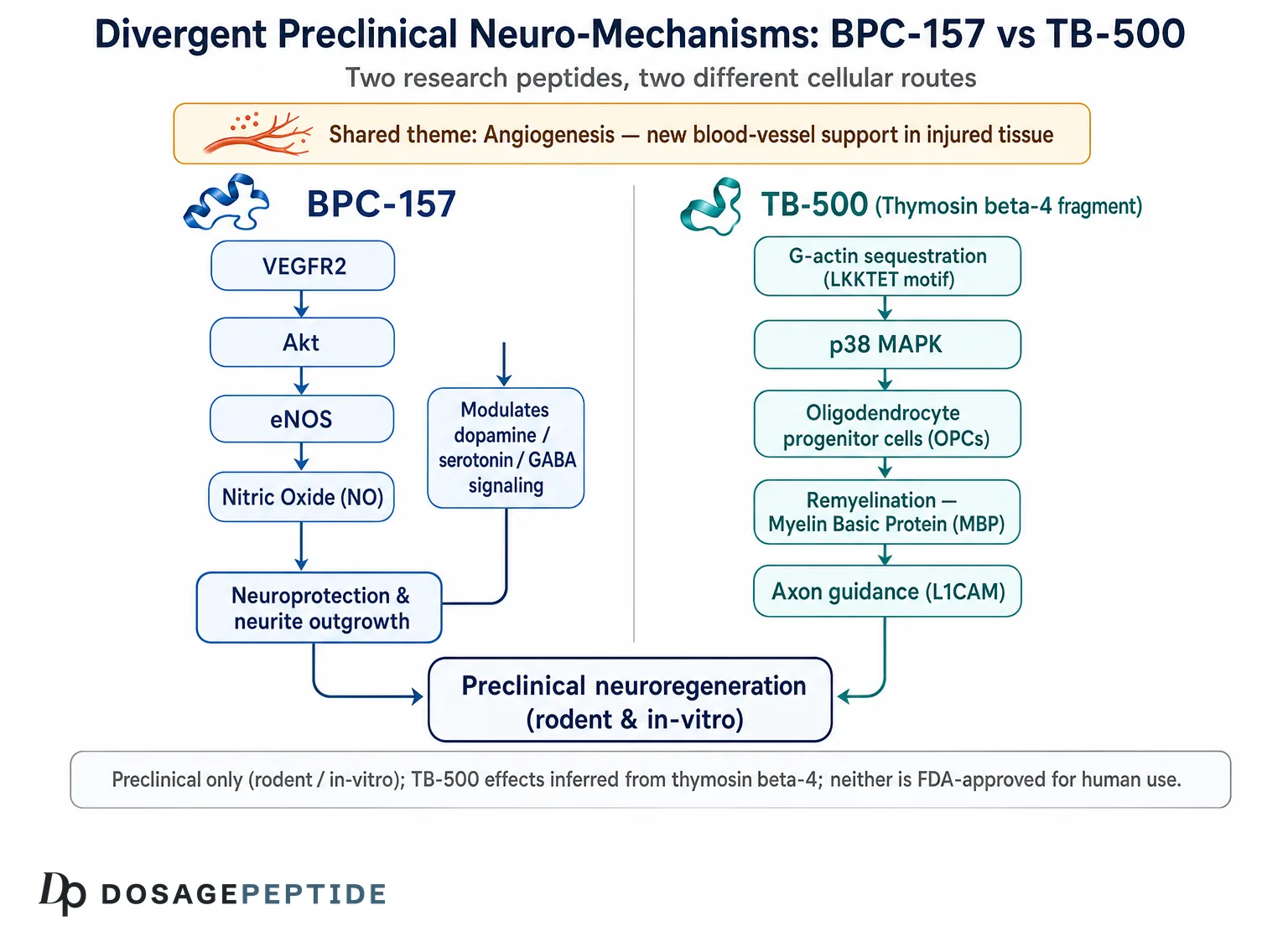
This is the mechanistic heart of the article, and the place where a “distinct functions” answer starts to take shape. The two peptides reach overlapping outcomes — both are pro-angiogenic and both improve functional scores in some injury models — but the routes are largely different. Understanding those routes is what separates an informed reading from a marketing one.
BPC-157: a nitric-oxide and VEGFR2 story with neuromodulatory extensions
The best-characterized molecular handle on BPC-157 is vascular. In human endothelial cells, BPC-157 upregulates VEGF receptor 2 (VEGFR2) and drives its internalization, activating the downstream VEGFR2–Akt–eNOS axis to promote angiogenesis; the same study showed increased vessel density in vitro and in vivo and faster blood-flow recovery in a rat hind-limb ischemia model.[7] Notably, BPC-157 raised VEGFR2 (the receptor) without necessarily raising VEGF-A (the ligand), suggesting it sensitizes the angiogenic machinery rather than simply flooding it with growth factor. Because new blood vessels underpin almost every repair process, this vascular action plausibly contributes to its effects in nerve, spinal cord, and brain models — but it is a general repair mechanism, not a nerve-specific one.
BPC-157 is also repeatedly linked to the nitric-oxide (NO) system more broadly, appearing to counteract both NO-blockade and NO-overstimulation states — a “buffering” or homeostatic interaction rather than a one-directional push.[5] On top of the vascular/NO layer sits the neuromodulatory extension discussed earlier: reported interactions with dopaminergic, serotonergic, and GABAergic systems, largely interpreted through the brain–gut axis.[6] The key mechanistic point for the comparison: BPC-157’s CNS actions read as vascular support plus neurotransmitter/homeostatic modulation and neuroprotection, with little direct evidence that it commands neural progenitor or oligodendrocyte-progenitor fate.
Thymosin beta-4: actin sequestration, progenitor differentiation, and neurite growth
Thymosin beta-4’s mechanism starts from a completely different place: it is the principal intracellular G-actin–sequestering peptide, binding monomeric actin and holding it in a polymerization-incompetent pool until the cell needs it — a fundamental role in cytoskeletal dynamics, cell migration, and shape change. The actin-binding motif itself — the LKKTET sequence at the core of the fragment marketed as TB-500 — is directly implicated in the peptide’s pro-angiogenic activity; mutating that motif substantially reduces Tβ4’s ability to promote blood-vessel formation.[14] Short peptide sequences within Tβ4 recapitulate several of its biological activities, which is the rationale behind fragment products in the first place.[15]
Where thymosin beta-4 becomes genuinely nerve-relevant is in two mechanisms with little BPC-157 counterpart. First, cytoskeletal remodeling supports neurite outgrowth and neuronal survival, and Tβ4 has been shown to promote these by upregulating the cell-adhesion molecule L1.[16] Second, and most distinctively, Tβ4 stimulates oligodendrocyte-progenitor differentiation and remyelination, apparently via p38 MAPK signaling, across stroke, TBI, and EAE models.[13] Add its documented anti-inflammatory activity in the demyelinating CNS,[12] and thymosin beta-4’s neuro profile reads as progenitor-driven remodeling — new myelin, new vessels, extended neurites — layered on immune quieting.
The critical fragment caveat
One honest qualification threads through this entire mechanism section. The myelination, neurogenesis, and L1/neurite findings were generated with full-length thymosin beta-4, not with the short Ac-LKKTETQ fragment that the TB-500 label usually denotes. The LKKTET motif carries the actin-binding and much of the angiogenic activity,[14] but whether the fragment reproduces the full molecule’s progenitor-differentiation effects in the CNS has not been established in the same way. So the strongest neuroregenerative claims attributed to “TB-500” are, strictly, claims about its parent protein — component-level evidence, not direct evidence for the fragment.
Where do the two mechanisms overlap, and where do they part ways?
The overlap is real and worth stating plainly, because it is what allows both peptides to be marketed as generic “healing” agents. Both are pro-angiogenic: BPC-157 through VEGFR2 sensitization and the Akt–eNOS axis,[7] thymosin beta-4 through its actin-binding LKKTET motif and downstream VEGF-linked signaling.[14] Both therefore support the vascular bed that any repairing tissue requires, both intersect the nitric-oxide/eNOS system, and both improve functional scores in at least some injury models. If the question were only “do they help injured tissue heal in animals,” the answer would look similar for the two.
The divergence appears the moment the question narrows to how nervous tissue is rebuilt. Thymosin beta-4 carries two mechanisms with essentially no BPC-157 equivalent in the literature: it directs oligodendrocyte-progenitor differentiation and remyelination, and it promotes neurite outgrowth via L1. BPC-157, conversely, carries a neurotransmitter/brain–gut modulatory layer and an apparently bidirectional nitric-oxide-buffering behavior that thymosin beta-4 is not described as having. Put compactly: they converge on vascular and cytoprotective support and diverge on cellular remodeling of the nervous system — and it is the divergence, not the overlap, that answers this article’s title.
Do TB-500 and BPC-157 have distinct neuroregenerative functions?
Pulling the threads together, the experimental literature does suggest a real functional distinction — provided one keeps the honesty caveats in view. They are not simply two names for the same “healing peptide.”
The clearest way to see the divergence is by the kind of neuro-outcome each is best evidenced for:
- BPC-157 is best supported for peripheral-nerve regeneration and CNS neuroprotection: accelerating axonal regrowth in transected peripheral nerve, sparing tissue and limiting edema after brain and spinal-cord injury, and supporting repair through VEGFR2/NO-mediated angiogenesis, with an additional brain–gut neuromodulatory layer.
- Thymosin beta-4 (the molecule behind TB-500) is best supported for central neurorestoration and remyelination: driving oligodendrocyte-progenitor differentiation and remyelination, stimulating neurogenesis and angiogenesis in injured cortex and hippocampus, and promoting neurite outgrowth — in models where lesion size does not change but function still improves.
In other words, the mechanistic centers of gravity differ. BPC-157’s neuro benefits lean toward protecting and revascularizing injured nervous tissue and accelerating the peripheral nerve’s own repair program. Thymosin beta-4’s lean toward rebuilding — specifically the progenitor-driven regeneration of myelin and neurons in the CNS. These are genuinely distinct functions, not redundant ones, which is a more interesting conclusion than either “they’re the same” or “they both cure nerves.”
| Dimension | BPC-157 | Thymosin β4 / TB-500 |
|---|---|---|
| Molecular class | 15-aa synthetic gastric-derived peptide | 43-aa actin-sequestering protein (fragment Ac-LKKTETQ marketed as TB-500) |
| Named molecule = studied molecule? | Yes | Often no — neuro data use full-length Tβ4 |
| Core molecular mechanism | VEGFR2–Akt–eNOS angiogenesis; NO-system modulation | G-actin sequestration; cytoskeletal remodeling |
| Strongest neuro evidence | Peripheral-nerve regeneration; CNS neuroprotection | Oligodendrogenesis/remyelination; neurogenesis |
| Effect on OPCs / myelin | Not a documented primary action | Central, reproducible across models |
| Neurotransmitter modulation | Reported (dopamine, serotonin, GABA via gut–brain) | Not a primary reported action |
| Anti-inflammatory / immune | Cytoprotective, secondary | Documented in EAE demyelination |
| Predominant research groups | Largely one originating group | Multiple independent groups |
| Human neuro trials | None | None |
Two honesty markers belong right next to this table. First, the BPC-157 CNS literature leans heavily on a single research lineage, which weakens confidence pending independent replication; the thymosin beta-4 neuro literature is more diversified across groups. Second, none of this distinction implies that either compound is a treatment for anything in humans — the distinction is between preclinical mechanistic profiles, full stop.
What are the limits of this evidence?
A responsible reading of the “distinct functions” conclusion has to foreground its limitations, because they are large enough to change how much weight the conclusion can bear.
The species and translation gap
Every regenerative outcome discussed here was measured in rodents or cells. Rodent nerve, brain, and immune biology differ from human biology in ways that have repeatedly defeated neurological drug candidates that looked excellent preclinically. The graveyard of neuroprotection and neurorestoration is full of compounds that improved rat stroke scores and then failed in humans. Preclinical success is necessary but nowhere near sufficient, and nothing in this literature has cleared even a Phase I neurological trial.
No human neuroregeneration data at all
It bears repeating because it is so often elided: there are no completed, published, controlled human trials showing that either BPC-157 or TB-500/thymosin beta-4 regenerates nerves or treats a neurological disease. Thymosin beta-4 has reached human trials — but in ophthalmology (dry-eye disease, e.g. Phase II ocular-surface studies)[18] and in dermal wound healing (e.g. venous stasis and pressure ulcers),[19] not for any neurological indication. Those trials establish that the parent molecule has been given to humans and studied for safety in other tissues; they say nothing about neuroregeneration and should never be cited as if they did. BPC-157 has no comparable body of completed, published human efficacy trials.
The fragment-versus-parent problem, again
Because so much “TB-500” neuro evidence is really thymosin beta-4 evidence, the actual research chemical people call TB-500 is, for neuroregeneration specifically, supported mostly by inference from its parent molecule and its shared actin-binding motif. Whenever you see a strong neuro claim for TB-500, ask whether the underlying study used the fragment or the full 43-mer — it is usually the latter.
Dose, route, timing, and model artifacts
Preclinical doses (for example, Tβ4 at 6–30 mg/kg in rats, or BPC-157 at microgram-to-nanogram-per-kilogram ranges) do not translate to humans by simple scaling, and route and timing strongly shaped the reported outcomes. Some BPC-157 brain-injury benefits were seen with prophylactic dosing, which is not how any real-world injury is encountered. These design choices make the findings informative about biology but poor guides to any practical protocol — and this article is emphatically not a protocol.
Publication and source concentration
The BPC-157 CNS literature is dominated by one originating group; single-source bodies of evidence are more vulnerable to systematic bias and demand independent replication before confident generalization. Positive-result publication bias affects both literatures. None of this makes the findings wrong — it makes them provisional.
Research-chemical quality and identity
Material sold as “TB-500” may be the 7-mer fragment, the full-length protein, or a mixture, and purity/identity are not guaranteed in the research-chemical market. That uncertainty compounds the fragment-versus-parent issue: two vials with the same label may not even contain the same molecule that the studies used. Any reader handling these compounds for laboratory work should treat identity and purity as open questions, and can consult our neutral peptide reconstitution guide for how such materials are typically prepared and handled in research settings, and the reconstitution and concentration calculator for the associated arithmetic — both provided strictly as educational references, not as encouragement to use these substances.
How do the safety and research-handling considerations compare?
Any discussion of neuroregeneration eventually collides with a simpler question — what is actually known about the safety of these compounds — and the honest answer is: far less than the enthusiasm implies. Neither peptide has an established human safety profile for neurological use, and the safety data that do exist are narrow, short-term, and drawn from other tissues.
For thymosin beta-4, the most relevant human safety information comes from its non-neurological trials. Small Phase II ocular-surface studies of a pharmaceutical-grade formulation reported tolerability without notable adverse events in the treated populations,[18] and dermal-wound trials in venous stasis and pressure ulcers proceeded through early phases.[19] That is genuinely more human exposure than BPC-157 has, but it is topical or short-course dosing for eye and skin conditions, not systemic dosing aimed at the nervous system, and it cannot be extrapolated to chronic neurological use. BPC-157, for its part, has an extensive rodent safety record within its originating literature but no comparable body of completed, published human safety trials at all.
The pro-angiogenic double edge
Both peptides share a mechanism that is a benefit for wound repair and a theoretical liability elsewhere: they promote angiogenesis. The same capacity to grow new blood vessels that aids injured tissue could, in principle, support the vascularization that tumors require. This is a theoretical concern raised by the biology, not a demonstrated harm in humans for either compound, and it should be described as exactly that — an unresolved question that the absence of long-term human data leaves open, not a proven risk and not a reason for false reassurance either.
Identity, purity, and formulation uncertainty
Beyond intrinsic pharmacology sit the practical hazards of the research-chemical market. As already noted, “TB-500” may denote the 7-mer fragment or the full 43-mer, so identity is not guaranteed. Synthetic peptides can carry synthesis-related impurities, endotoxin, or incorrect sequences, and none of this is quality-controlled to pharmaceutical standards in research-grade material. Peptide fragments can also be immunogenic. These are handling and identity questions that exist entirely independently of whether the underlying biology is promising, and they further widen the gap between the published studies (which used characterized material) and anything obtained as a research chemical. The practical mechanics of preparing such material for laboratory work — solvent choice, concentration, and storage — are covered neutrally in the reconstitution references linked earlier, purely so that readers can interpret the methods sections of the studies, not as any suggestion to administer these compounds.
What is the regulatory and research-use status of TB-500 and BPC-157?
This is where the honesty framing becomes a hard requirement rather than a stylistic preference. Neither BPC-157 nor TB-500/thymosin beta-4 is approved by the FDA for any human therapeutic use, and neither is an approved treatment for any neurological condition. They are handled and sold as research chemicals, and everything in this article is educational and mechanistic, not medical advice.
For BPC-157 specifically, the regulatory posture tightened in 2023, when the FDA placed BPC-157 in Category 2 of its interim 503A bulk-drug-substances list — the category for substances that the agency judges to raise significant safety concerns — a placement that in practice barred licensed compounding pharmacies from preparing it under that pathway.[17] The status has since remained unsettled rather than resolved in the compound’s favor: in 2026 the nomination behind that listing was withdrawn and BPC-157 was removed from Category 2, yet the FDA did not move it to the permitted (Category 1) list, and at its July 2026 Pharmacy Compounding Advisory Committee review the agency proposed against adding BPC-157 to the 503A bulk-substances list at all.[20] The practical upshot is a regulatory gray zone in which the substance is neither an approved drug nor an authorized compounding ingredient. Peptides in the thymosin family have similarly been the subject of FDA compounding scrutiny. The bottom line for a reader is unchanged through every twist of this process — these are not medicines, and the neuroregeneration evidence does not begin to support treating them as such.
How to read the research responsibly
The constructive way to hold all of this is as a genuinely interesting preclinical mechanistic question with a tentative answer — yes, the literature suggests distinct neuro-mechanisms — wrapped in a hard boundary: no human efficacy, no approval, no clinical claim. Readers who want to go deeper into how each compound is described in the wider dosing and research literature can consult our neutral reference summaries for BPC-157 and TB-500, which collate the reconstitution and handling conventions reported in laboratory studies. Those pages, like this article, are informational references for understanding the research — not endorsements, protocols, or commercial recommendations of any kind.
Frequently Asked Questions
Are TB-500 and BPC-157 the same type of peptide?
No. BPC-157 is a synthetic 15-amino-acid peptide derived from a gastric protein, and the studied molecule is the same one sold under that name. TB-500 is a commercial label for a fragment (Ac-LKKTETQ) of the 43-amino-acid protein thymosin beta-4, though some material sold as TB-500 is the full-length protein. They differ in origin, structure, and mechanism, and most neurological studies used full-length thymosin beta-4 rather than the short fragment.
Is there human evidence that either peptide regenerates nerves?
No. There are no completed, published, controlled human trials showing that BPC-157 or TB-500 regenerates nerves or treats any neurological disease. All neuroregeneration data come from rodent injury models and cell-culture experiments. Thymosin beta-4 has entered human trials for dry-eye disease and dermal wounds, but those are non-neurological indications and do not support any neuroregeneration claim.
What is the main mechanistic difference between them for the nervous system?
In preclinical work, BPC-157’s neuro effects center on angiogenesis (via the VEGFR2–Akt–eNOS pathway), nitric-oxide-system modulation, and neuroprotection, plus brain–gut neurotransmitter effects. Thymosin beta-4 centers on cytoskeletal remodeling through actin sequestration, driving oligodendrocyte-progenitor differentiation, remyelination, neurogenesis, and neurite outgrowth. In short, BPC-157 leans toward protecting and revascularizing; thymosin beta-4 leans toward progenitor-driven rebuilding.
Does BPC-157 help peripheral nerves specifically?
In a rat transected-sciatic-nerve model, BPC-157 was associated with faster axonal regeneration, better-organized nerve fascicles, and improved walking recovery. Peripheral nerve has an intrinsic capacity to regenerate, so this represents claimed acceleration of an existing process rather than regrowth from nothing. It is a single-model, single-group animal finding and has not been tested in controlled human studies.
Why is thymosin beta-4 linked to myelin repair?
Across rat stroke, rat traumatic-brain-injury, and mouse multiple-sclerosis (EAE) models, thymosin beta-4 consistently increased oligodendrocyte progenitor cells and mature oligodendrocytes and augmented remyelination, apparently through p38 MAPK signaling and myelin-basic-protein synthesis. This convergent, multi-model, multi-group signal is the most reproducible neuro-specific finding for the compound — though it remains preclinical, with no human neurological trials.
Is BPC-157 FDA-approved or legal to prescribe?
BPC-157 is not FDA-approved for any human use. The FDA placed it in Category 2 of its interim 503A bulk-drug-substances list in 2023 (the category for substances raising significant safety concerns); in 2026 that nomination was withdrawn and BPC-157 was removed from Category 2, but the FDA has proposed against adding it to the 503A bulks list, so it remains unauthorized for compounding rather than cleared for it. It is handled as a research chemical, and no neurological use is approved or supported by human evidence.
Can the “TB-500” fragment do everything full-length thymosin beta-4 does?
Not established. The Ac-LKKTETQ fragment carries the actin-binding motif and much of the angiogenic activity of the parent protein, but the strongest neuroregenerative findings — oligodendrogenesis, neurogenesis, L1-mediated neurite outgrowth — were generated with full-length thymosin beta-4. Whether the short fragment reproduces those CNS effects has not been demonstrated equivalently, so neuro claims for TB-500 rest largely on parent-molecule inference.
Does this research mean I should consider using either peptide?
No. This article is an educational review of preclinical mechanisms, not medical advice or a protocol. Neither compound is an approved treatment, the safety data in humans are limited, and no neurological benefit has been demonstrated in people. Any decision about compounds like these belongs with a qualified clinician and within applicable law; nothing here should be read as encouragement to obtain or use them.
References
- Gjurasin M, et al. Peptide therapy with pentadecapeptide BPC 157 in traumatic nerve injury. Regul Pept. 2010. https://pubmed.ncbi.nlm.nih.gov/19903499/
- Perovic D, et al. Stable gastric pentadecapeptide BPC 157 can improve the healing course of spinal cord injury and lead to functional recovery in rats. J Orthop Surg Res. 2019. https://www.ncbi.nlm.nih.gov/pmc/articles/PMC6604284/
- Tudor M, et al. Traumatic brain injury in mice and pentadecapeptide BPC 157 effect. Regul Pept. 2010. https://www.sciencedirect.com/science/article/abs/pii/S0167011509002341
- Vukojevic J, et al. The effect of pentadecapeptide BPC 157 on hippocampal ischemia/reperfusion injuries in rats. Brain Behav. 2020. https://www.ncbi.nlm.nih.gov/pmc/articles/PMC7428500/
- Sikiric P, et al. Brain-gut Axis and Pentadecapeptide BPC 157: Theoretical and Practical Implications. Curr Neuropharmacol. 2016. https://pubmed.ncbi.nlm.nih.gov/27138887/
- Sikiric P, et al. Stable Gastric Pentadecapeptide BPC 157 May Recover Brain–Gut Axis and Gut–Brain Axis Function. Pharmaceuticals (Basel). 2023. https://pmc.ncbi.nlm.nih.gov/articles/PMC10224484/
- Hsieh MJ, et al. Therapeutic potential of pro-angiogenic BPC157 is associated with VEGFR2 activation and up-regulation. J Mol Med (Berl). 2017. https://link.springer.com/article/10.1007/s00109-016-1488-y
- Xiong Y, et al. Treatment of traumatic brain injury with thymosin β4 in rats. J Neurosurg. 2011. https://pubmed.ncbi.nlm.nih.gov/20486893/
- Xiong Y, et al. Neuroprotective and neurorestorative effects of thymosin β4 treatment following experimental traumatic brain injury. Ann N Y Acad Sci. 2012. https://pmc.ncbi.nlm.nih.gov/articles/PMC3547647/
- Morris DC, et al. Thymosin β4 improves functional neurological outcome in a rat model of embolic stroke. Neuroscience. 2010. https://pmc.ncbi.nlm.nih.gov/articles/PMC2907184/
- Morris DC, et al. A dose-response study of thymosin β4 for the treatment of acute stroke. J Neurol Sci. 2014. https://pmc.ncbi.nlm.nih.gov/articles/PMC4177939/
- Zhang J, et al. Neurological functional recovery after thymosin beta4 treatment in mice with experimental auto encephalomyelitis. Neuroscience. 2009. https://pmc.ncbi.nlm.nih.gov/articles/PMC2784109/
- Santra M, et al. Thymosin beta4 promotes oligodendrogenesis in the demyelinating central nervous system. Neurobiol Dis. 2016. https://www.sciencedirect.com/science/article/abs/pii/S0969996116300109
- Philp D, et al. The actin binding site on thymosin β4 promotes angiogenesis. FASEB J. 2003. https://pubmed.ncbi.nlm.nih.gov/14500546/
- Sosne G, et al. Biological activities of thymosin β4 defined by active sites in short peptide sequences. FASEB J. 2010. https://faseb.onlinelibrary.wiley.com/doi/abs/10.1096/fj.09-142307
- Yang H, et al. The promotive effects of thymosin β4 on neuronal survival and neurite outgrowth by upregulating L1 expression. Neurochem Res. 2008. https://link.springer.com/article/10.1007/s11064-008-9712-y
- U.S. Food and Drug Administration. Bulk Drug Substances Used in Compounding Under Section 503A of the FD&C Act. https://www.fda.gov/drugs/human-drug-compounding/bulk-drug-substances-used-compounding-under-section-503a-fdc-act
- Sosne G, et al. Thymosin beta 4 ophthalmic solution for dry eye: a randomized, placebo-controlled, Phase II clinical trial. Clin Ophthalmol. 2015. https://www.tandfonline.com/doi/full/10.2147/OPTH.S80954
- ClinicalTrials.gov. Study of Thymosin Beta 4 in Patients With Venous Stasis Ulcers (NCT00832091). https://clinicaltrials.gov/study/NCT00832091
- U.S. Food and Drug Administration. July 23–24, 2026: Meeting of the Pharmacy Compounding Advisory Committee (503A bulk-drug-substances review, including BPC-157). https://www.fda.gov/advisory-committees/advisory-committee-calendar/july-23-24-2026-meeting-pharmacy-compounding-advisory-committee-07232026