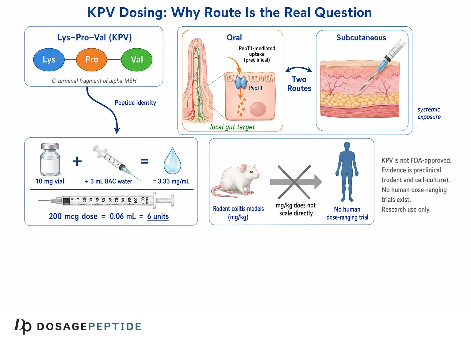
The question that brings most people to KPV peptide dosage is deceptively simple: how many micrograms, by which route, and how do you get there from a 10 mg vial? This article answers the arithmetic completely and transparently — 10 mg in 3 mL gives 3.33 mg/mL, and a 200 mcg dose is 0.06 mL, or 6 units on a U-100 insulin syringe — but it also answers the harder question underneath it: where those numbers come from, why KPV is one of the very few peptides for which the oral route is mechanistically defensible rather than vendor folklore, and why no honest source can give you a human milligram figure, because no human dose-ranging trial of KPV has ever been run.
What Is KPV, and Why Does Its Size Change the Dosing Question?
KPV is a tripeptide: lysine–proline–valine. Three amino acids. That is it. It is the C-terminal fragment of alpha-melanocyte-stimulating hormone (α-MSH), the thirteen-residue neuropeptide with sequence SYSMEHFRWGKPV. Cut off the last three residues — positions 11, 12 and 13 — and you have KPV, which is why the older immunology literature almost always calls it α-MSH(11–13) rather than KPV — the 2008 Muenster colitis paper, for instance, names the compound “α-MSH(11-13) (KPV)” in its own abstract.[2] If you are searching the literature and finding little, that naming convention is usually why.
Its molecular formula is C16H30N4O4, giving a molecular weight of approximately 342.4 g/mol for the free acid.[15] That number matters more than it looks. For comparison — using standard published reference values for each compound rather than any measurement of our own — BPC-157 is about 1419 g/mol, TB-500 (the acetylated fragment Ac-LKKTETQ) about 889 g/mol, and semaglutide about 4114 g/mol. KPV is roughly a quarter the mass of BPC-157 and about one-twelfth the mass of semaglutide. It is barely a peptide at all in pharmacokinetic terms — it sits closer to a small molecule than to a biologic.
Why size drives every dosing decision here
Three consequences follow from being that small, and all three shape the dosage conversation:
- Molar density per milligram is high. Because KPV is light, a given mass contains many more molecules than the same mass of a larger peptide. A 200 mcg dose of KPV is 0.58 micromoles. The same 200 mcg of BPC-157 is 0.14 micromoles — four times fewer molecules. Micrograms are not a species-neutral currency; molecules are. This is an arithmetic fact about molecular weight, not a statement about activity per molecule, and it is the reason the micromole column exists in the dosage chart below.
- It is a substrate for a real transporter. Di- and tripeptides are not degraded and absorbed like larger proteins — they have a dedicated intestinal uptake route, the proton-coupled oligopeptide transporter PepT1 (gene SLC15A1), which exists specifically to carry two- and three-residue fragments across the brush border intact.[11] That transporter is the entire reason the oral-route question is scientifically live for KPV and dead for almost everything else in a research fridge.
- It does not appear to signal primarily through melanocortin receptors. The parent hormone drives pigmentation through MC1R. KPV, in the studies that looked, does not appear to work primarily through melanocortin receptors — which is both mechanistically interesting and a reason the fragment attracted research interest over the parent peptide.[6] This is a reading of the pharmacology, not a safety claim: no human safety database exists for KPV, so nothing on this page should be taken as establishing the absence of any effect.
For a fuller structural and biological introduction, see our overview of what KPV is and how it fits into anti-inflammatory peptide research. This article assumes that background and focuses on the numbers.
Research Context
KPV entered the literature through dermatology and immunology in the 1990s and early 2000s, as researchers tried to work out which part of α-MSH carried its anti-inflammatory activity and which part carried its pigmenting activity. The answer, roughly, was that the core (His-Phe-Arg-Trp, positions 6–9) drives melanocortin receptor signalling and pigmentation, while the C-terminal tripeptide carries a substantial share of the anti-inflammatory effect through a different route.[6]
That structural dissection is what made KPV interesting: it looked like a way to get the anti-inflammatory arm of melanocortin biology without the pigment.
It is worth flagging immediately that the “different route” part of that sentence is contested in the primary literature rather than settled. Getting and colleagues, working in peritonitis, concluded that KPV does not act through melanocortin receptors.[6] The Muenster dermatology group, reviewing their own work, reported the opposite in one respect: that KPV — not only the parent hormone — was able to bind MC-1R and modulate antigen-presenting cell function.[9] Those two readings are in tension, and this article does not pretend otherwise. The receptor question for KPV is not fully resolved; what is consistent across both lineages is the downstream NF-κB result, not the receptor mechanism upstream of it.
The three research lineages
Practically all published KPV work falls into one of three streams, and it helps to know which stream a claim came from before you decide what it is worth.
- Melanocortin immunology (Muenster / London / Milan / Dundee, roughly 1999–2012). Skin, peritonitis, contact hypersensitivity, airway epithelium. Cell culture and mouse models. This lineage established that the tripeptide is anti-inflammatory and started to work out how.[9] The airway-epithelium strand is geographically and topically separate from the skin work — it is single-author research from Dundee, and it is where the most specific molecular mechanism in the whole KPV literature comes from.[7]
- Gut / PepT1 pharmacology (Emory and Georgia State, 2008–present). The Merlin group’s work is the single most important body of evidence for KPV specifically, because it connected the tripeptide to a named transporter that is upregulated in inflamed colon and demonstrated efficacy in mouse colitis via oral administration.[1]
- Drug-delivery engineering (2010–present, largely Chinese and US groups). A large and still-growing set of papers in which KPV is the payload inside nanoparticles, hydrogels, or self-assembled carriers.[3][4] These papers are frequently cited as evidence that “KPV works,” but read carefully, most of them are evidence that a specific delivery system works. That distinction turns out to matter enormously for dosing, and we return to it below.
The regulatory position, stated plainly
KPV is not an FDA-approved drug. It is not approved for inflammatory bowel disease, ulcerative colitis, Crohn’s disease, atopic dermatitis, wound healing, or any other indication, in the United States or, to our knowledge, in any major jurisdiction. There is no approved label, therefore no approved dose, no approved route, and no approved schedule.
Nor is KPV an investigational drug in the formal sense. A search of ClinicalTrials.gov for KPV, Lys-Pro-Val, or α-MSH(11–13) as an intervention returns no registered interventional trials. It has never, as far as the public trial registry shows, been through a Phase 1 in humans. It is a research chemical: material sold for laboratory investigation, outside the drug approval framework, with no human safety database behind it.
This is the single most important fact on this page, and everything that follows should be read through it. When this article gives you an arithmetic answer — and it will give you complete, checkable arithmetic — that arithmetic tells you what is in the syringe. It does not tell you that anything should be in the syringe.
Mechanisms Studied
KPV has an unusually well-characterised mechanism for a research peptide, which is worth appreciating precisely because the dose evidence is so thin. We know a fair amount about how it works and almost nothing about how much.
NF-κB: the central node
Across cell types — intestinal epithelium, T cells, bronchial epithelium, sebocytes — the recurring finding is inhibition of NF-κB signalling. In Caco2-BBE intestinal epithelial cells and Jurkat T cells stimulated with pro-inflammatory cytokines, KPV inhibited NF-κB activation and MAP kinase inflammatory signalling and reduced pro-inflammatory cytokine secretion, with the reported in-vitro work using concentrations around 10 nM.[1]
The mechanism at that node has been probed further. In immortalised human bronchial epithelial cells, KPV suppressed TNF-α- and virus-evoked NF-κB signalling in a dose-dependent way, and the effect was associated with KPV entering the nucleus, stabilising IκBα, and blocking nuclear translocation of the p65RelA subunit — with competition assays pointing to an interaction at the importin-α binding site on p65RelA.[7] That is a specific, intracellular, non-receptor mechanism: the tripeptide gets inside the cell and interferes with the nuclear import machinery of the master inflammatory transcription factor.
Not (mainly) a melanocortin receptor story
This is the point most vendor copy gets wrong. In a crystal-induced peritonitis model, systemic KPV significantly reduced neutrophil accumulation, and that effect was not blocked by the MC3/MC4 receptor antagonist SHU9119 and persisted in mice with a non-functional MC1R. KPV also failed to raise cAMP in macrophages, unlike the core melanocortin peptides. The authors concluded that KPV is unlikely to mediate its effects through melanocortin receptors and is more likely to act through inhibition of IL-1β function.[6]
The colitis work independently supports MC1R-independence: in mice expressing a non-functional MC1 receptor and given DSS, KPV treatment still produced a marked effect, leading the authors to conclude the anti-inflammatory action is at least partially independent of MC1R signalling.[2] A 2023 review of the melanocortin system in inflammatory bowel disease reaches a similar reading of the accumulated mouse-model evidence.[12]
The honest synthesis is therefore hedged rather than clean. The peritonitis and colitis lineages both point to melanocortin-receptor independence, and they are the studies that tested it directly with antagonists and receptor-defective animals. Against that, the Muenster review reports KPV binding MC-1R.[9] These are reconcilable — binding a receptor and requiring it for an effect are different claims, and a peptide can do the former without the latter — but nobody has done the experiment that reconciles them. So: KPV is a melanocortin fragment whose anti-inflammatory effects, in the models that tested the question directly, do not appear to require melanocortin receptor signalling. It is probably best understood as a small anti-inflammatory molecule with a melanocortin pedigree and an unfinished receptor story.
PepT1: the transporter that makes the route question real
The mechanism that matters most for dosing is transport. PepT1 (SLC15A1) is a proton-coupled symporter that carries di- and tripeptides — and peptide-like drugs including certain aminocephalosporins, ACE inhibitors and antiviral prodrugs — across cell membranes intact.[11] It is normally expressed in the small intestine and at low levels in the healthy colon, and is reported to be upregulated in the colon during intestinal inflammation.[5][11] It is worth being precise about the provenance of that statement, because a great deal rests on it. The experimental work establishing the colitis–PepT1–KPV chain is murine; the human end of it is thinner — the same group reports increased PepT1 expression in human colonic biopsies from colorectal cancer patients,[5] and the transporter literature treats induction in inflamed human colon as an accepted feature of the SLC15 family’s pathological biology.[11] But no experiment has demonstrated the full chain — induced colonic PepT1 taking up orally administered KPV to anti-inflammatory effect — in a human being. The oral route’s mechanistic case is built on a mouse chain plus a human expression observation, not on a human chain.
The 2008 Gastroenterology paper did the transport work directly, using tritiated KPV and competition experiments, and reported a Km for hPepT1-mediated KPV uptake of roughly 160 µM in Caco2-BBE epithelial cells and roughly 700 µM in Jurkat immune cells — higher affinity in the epithelium than in immune cells.[1] The functional payoff came from the knockout side: in the colitis-associated cancer work, KPV prevented carcinogenesis in wild-type mice, but when given to PepT1-knockout mice it produced none of the effects seen in wild-types.[5] That is about as clean a demonstration as preclinical work offers that the transporter is required for the effect.
The transporter has since been used as a targeting handle in its own right, with nanoparticle designs employing KPV both as payload and as a PepT1-seeking ligand aimed at colonic epithelial cells and macrophages.[4]
Barrier and tight-junction effects
A recurring secondary theme is epithelial barrier integrity. The closely related tripeptide KdPT — a derivative of the same C-terminal region — increased colonic epithelial proliferation, accelerated wound closure, improved transepithelial electrical resistance after interferon-γ/TNF-α challenge, and prevented loss of tight junction protein expression in mice.[10] Two details from that paper are worth carrying forward, because both are routinely mangled in secondary sources. First, KdPT was reported to act independently of IL-1 receptor type I in vivo — so the widely repeated claim that this tripeptide line works through an IL-1 receptor interaction is not what the barrier paper found; it found the opposite. Second, KdPT did not affect melanogenesis in vitro, which was rather the point of making it. KPV-loaded delivery systems in colitis models likewise report accelerated mucosal healing alongside cytokine suppression.[4] This is the mechanistic bridge to the broader peptide and gut barrier permeability research literature — though note carefully that a good deal of the barrier data is for KdPT, not KPV, and the two are not interchangeable.
Antimicrobial activity
Separately, α-MSH and its C-terminal tripeptide were reported to inhibit Staphylococcus aureus colony formation and to reduce viability and germ tube formation of Candida albicans, with effects reported across a broad concentration range including the picomolar, apparently mediated via increases in cellular cAMP in the pathogen.[8] This is a genuine and frequently cited finding, but it is a 2000-vintage in-vitro result in isolated organisms, in a paper whose primary subject was the parent hormone. It has not been replicated into any human evidence, and it is a long way from “KPV is an antimicrobial.”
How Do You Reconstitute a 10 mg KPV Vial?
KPV ships as a lyophilised (freeze-dried) powder in a sealed vial — typically a small, almost invisible white disc or film at the bottom. It has to be dissolved before any volumetric dose can be measured. The reconstitution diluent used in research settings is bacteriostatic water for injection (BAC water), which is sterile water containing 0.9% benzyl alcohol as a preservative, or plain sterile water for single-use work.
The arithmetic has exactly one step, and everyone should be able to do it without a calculator:
Concentration = total mass in the vial ÷ volume of diluent added.
The site-standard 10 mg / 3 mL configuration, worked through
Our catalog uses 3 mL of BAC water for a 10 mg KPV vial as the reference configuration. Here is every step of that calculation, spelled out:
- Concentration. 10 mg ÷ 3 mL = 3.333 mg/mL. Converting to micrograms, which is the unit KPV doses are actually discussed in: 3.333 mg/mL × 1000 = 3,333 mcg/mL.
- Volume for a 200 mcg dose. 200 mcg ÷ 3,333 mcg/mL = 0.06 mL.
- Syringe units. A U-100 insulin syringe is graduated so that 100 units = 1.0 mL, therefore 1 unit = 0.01 mL. So 0.06 mL × 100 = 6 units.
- Check the reverse direction. 6 units = 0.06 mL. 0.06 mL × 3,333 mcg/mL = 200 mcg. The arithmetic closes.
So the arithmetic answer for a 10 mg vial at 200 mcg is: with 3 mL of BAC water, 200 mcg corresponds to the 6-unit mark on a U-100 syringe. At that reference dose the vial contains 10 mg ÷ 0.2 mg = 50 doses of 200 mcg. That is a description of a conversion, not an instruction to anyone.
If you would rather not do this by hand for a configuration we have not tabulated, our peptide dosage calculator performs the same arithmetic for arbitrary vial sizes and diluent volumes, and the peptide reconstitution guide covers the physical technique — angling the diluent stream down the vial wall rather than onto the powder cake, letting it dissolve rather than shaking it, and so on. The U-100 unit conversion, which is where most arithmetic errors originate, is covered in detail in our insulin syringe units guide.
The vial must physically hold the water
One constraint that trips people up: the diluent has to fit. Peptide vials are commonly 2 mL, 3 mL or 5 mL nominal capacity, and the nominal capacity is not the same as the usable capacity — you need headspace, and the stopper displaces a little volume. A 3 mL vial will accept 3 mL only awkwardly and with no room for error. If the KPV vial in front of you is a small 2 mL vial, 3 mL of BAC water is not going in it, and the configuration has to change — which changes the concentration and therefore every unit figure on this page.
Always check the physical vial before committing to a reconstitution volume. Then recompute. The math is one division; there is no excuse for guessing.
Reconstitution comparison: same 10 mg, four different volumes
Because the diluent volume is a free choice, the same vial can be set up at any of several concentrations. Higher concentration means smaller, harder-to-measure volumes; lower concentration means larger, easier-to-measure volumes but a diluent load the vial may not accommodate. This table shows what a 200 mcg dose becomes under each option:
| BAC water added | Concentration | Concentration (mcg/mL) | Volume for 200 mcg | U-100 units for 200 mcg | Practical note |
|---|---|---|---|---|---|
| 1 mL | 10 mg/mL | 10,000 | 0.02 mL | 2 units | Very concentrated; a 1-unit misread is a 50% dose error |
| 2 mL | 5 mg/mL | 5,000 | 0.04 mL | 4 units | Workable; fits a 2 mL vial poorly |
| 3 mL | 3.33 mg/mL | 3,333 | 0.06 mL | 6 units | Site reference configuration |
| 5 mL | 2 mg/mL | 2,000 | 0.10 mL | 10 units | Easiest to measure; requires a 5 mL+ vial |
Note what does not change across that table: the vial still contains 10 mg, and still contains 50 doses of 200 mcg. Reconstitution volume changes the readability of the measurement, not the amount of peptide you have. Choosing 5 mL over 1 mL does not make the vial “weaker” — it makes each unit on the syringe correspond to less peptide, which is a precision decision, not a potency decision.
The rule of thumb that actually generalises
If you want one relationship to memorise instead of a table:
Units on a U-100 syringe = (desired dose in mcg × diluent volume in mL) ÷ (vial mass in mg × 10).
Check it against the reference case: (200 × 3) ÷ (10 × 10) = 600 ÷ 100 = 6 units. Correct. Check it against the 5 mL case: (200 × 5) ÷ 100 = 10 units. Correct. This formula holds for any peptide in a mg-labelled vial, not just KPV.
Micrograms to Syringe Units: The KPV Conversion Table
This is the table most people came here for. Every row is the same single division carried out at the site reference configuration of 10 mg reconstituted with 3 mL BAC water = 3.33 mg/mL = 3,333 mcg/mL.
Read this as a conversion table, not a recommendation table. It tells you what volume corresponds to what mass. It expresses no view on which row anyone should be interested in, and as the evidence sections of this article establish, no published human data exist that would let anyone form such a view.
| Dose (mcg) | Dose (mg) | Volume (mL) | U-100 units | Doses per 10 mg vial | Micromoles per dose |
|---|---|---|---|---|---|
| 50 mcg | 0.05 mg | 0.015 mL | 1.5 units | 200 | 0.15 µmol |
| 100 mcg | 0.10 mg | 0.03 mL | 3 units | 100 | 0.29 µmol |
| 150 mcg | 0.15 mg | 0.045 mL | 4.5 units | 66 | 0.44 µmol |
| 200 mcg | 0.20 mg | 0.06 mL | 6 units | 50 | 0.58 µmol |
| 250 mcg | 0.25 mg | 0.075 mL | 7.5 units | 40 | 0.73 µmol |
| 300 mcg | 0.30 mg | 0.09 mL | 9 units | 33 | 0.88 µmol |
| 400 mcg | 0.40 mg | 0.12 mL | 12 units | 25 | 1.17 µmol |
| 500 mcg | 0.50 mg | 0.15 mL | 15 units | 20 | 1.46 µmol |
| 1000 mcg (1 mg) | 1.0 mg | 0.30 mL | 30 units | 10 | 2.92 µmol |
The micromole column is included deliberately. It is the column that lets you compare KPV to anything else on a like-for-like basis, because it counts molecules rather than mass. Using the 342.4 g/mol molecular weight,[15] a 10 mg vial contains 10 mg ÷ 342.4 mg/mmol = 0.0292 mmol = 29.2 µmol of KPV. Dissolved in 3 mL, that is a 9.73 mM stock solution. Hold onto that number — it becomes important when we compare the vial to the concentrations that actually appear in the published research, and the comparison is not flattering to the folklore.
The half-unit problem
Notice that 50 mcg and 150 mcg land on 1.5 and 4.5 units. Standard U-100 insulin syringes are graduated in 1-unit or 2-unit increments; half-unit syringes exist but are less common. At the 3 mL configuration, sub-100 mcg doses require reading between the lines, and the relative error grows fast: misreading by half a unit at the 6-unit mark is an 8% error, while the same half-unit misread at the 1.5-unit mark is a 33% error.
If a research protocol calls for small KPV quantities, the correct response is to change the reconstitution volume upward — 5 mL puts 100 mcg at 5 units instead of 3 — not to squint at a 1.5-unit graduation. Precision is a function of your dilution choice, and dilution is free.
What the chart cannot tell you
The chart above is arithmetic, and arithmetic is always correct. That is exactly why it is dangerous. A perfectly accurate conversion table creates a strong impression that the underlying quantity is well established, when in KPV’s case the underlying quantity is a convention. The 200 mcg reference figure is not derived from a dose-finding study. It is a number that circulates. We tabulate it because it is what people are searching for and because a wrong table is worse than a right one — not because it is validated.
Oral vs Subcutaneous: Which Route Does the Research Actually Support?
This is where KPV genuinely differs from the rest of the research-peptide catalog, and it is worth being precise about why, because the honest answer is more interesting than either “oral peptides don’t work” or “KPV is orally bioavailable.”
Why oral peptides normally fail
The standing objection to swallowing a peptide is well founded. The gastrointestinal tract is a protein-destroying environment by design. Gastric acid denatures. Pepsin cleaves. Pancreatic trypsin, chymotrypsin and elastase cleave further. Then the intestinal brush border presents a dense array of membrane-bound peptidases — aminopeptidase N, dipeptidyl peptidase IV, and others — whose specific job is to reduce whatever survives to free amino acids before absorption. A 30-residue peptide entering that gauntlet does not arrive at the far end as a 30-residue peptide. This is why BPC-157’s oral claims are contested, why insulin is injected, and why oral semaglutide requires a dedicated absorption enhancer (SNAC) co-formulated in the tablet and still achieves a bioavailability on the order of 1% — a figure quoted here as a widely reported reference value for that product, not as a KPV finding.
Why a tripeptide is a different case
Three structural facts change the calculus for Lys-Pro-Val specifically:
- There is nothing left to cleave down to. A tripeptide is at the floor of the size distribution. The endopeptidases that do the bulk of the damage require internal peptide bonds flanked by recognisable sequence context; a three-residue substrate offers essentially two bonds and almost no context. The digestive machinery is optimised to produce fragments of this size, not to destroy them.
- Proline is a peptidase roadblock. The central residue is proline, whose rigid pyrrolidine ring imposes a conformational constraint that many peptidases cannot accommodate. Proline-containing bonds are a well-known point of enzymatic resistance in peptide chemistry — it is precisely why DPP-4, a proline-specific enzyme, exists as a separate specialist. A Lys-Pro-Val motif is far more resistant to generic brush-border peptidase attack than a comparable three-residue sequence without proline.
- There is a dedicated uptake transporter. This is the decisive one. PepT1 exists specifically to absorb di- and tripeptides intact, and it is the route by which peptide-like drugs including certain aminocephalosporins and ACE inhibitors achieve oral bioavailability.[11] KPV is a demonstrated PepT1 substrate, with measured uptake kinetics.[1]
So oral KPV is not a marketing conceit grafted onto an injectable. It is a mechanistically coherent proposition, and — crucially — the pivotal preclinical work was done that way. In the 2008 Gastroenterology study, KPV was added to the drinking water of mice at 100 µM, and oral administration reduced the incidence of both DSS- and TNBS-induced colitis, with decreased pro-inflammatory cytokine expression.[1] The independent Muenster group separately reported anti-inflammatory efficacy for KPV in the DSS and CD45RBhi transfer models, though its published abstract does not specify the route of administration, and we have not been able to confirm the route from a primary source.[2] So the accurate statement is narrower than the one usually made: the pivotal Dalmasso colitis work was oral,[1] and it is the paper that carries the oral case. Secondary sources that describe both foundational colitis papers as oral papers are asserting more than the published record supports — a small example of exactly the drift this article is trying to resist.
The part almost nobody says out loud: a local target changes everything
Here is the argument that actually matters, and it is not the one you usually see.
When people ask “is oral KPV bioavailable,” they are implicitly asking “does it reach the bloodstream.” For a gut target, that is the wrong question — and arguably the wrong goal.
PepT1 is upregulated in the inflamed colon.[5] The cells the colitis literature is interested in — colonic epithelium and lamina propria immune cells — are reachable from the lumen. If your target tissue is the wall of the colon, then delivering the compound to the colonic lumen and having it taken up locally by an inflammation-induced transporter is not a compromise route. It is the better route. Systemic exposure would be, at best, irrelevant and at worst a source of off-target effect.
This reframing has a sharp corollary that cuts against oral KPV as commonly practised. If the mechanism is local luminal uptake by colonic PepT1, then the peptide has to arrive at the colon. Free KPV swallowed on an empty stomach is a small, highly soluble molecule that will be substantially absorbed by small-intestinal PepT1 long before it reaches the colon — where PepT1 is abundantly expressed in health. The tripeptide’s greatest advantage over larger peptides (efficient proximal absorption) is, for a colonic target, its greatest liability.
The researchers knew this. It is the entire reason the field pivoted to delivery engineering. The 2008 authors explicitly chose a high drinking-water concentration to compensate for absorption losses before the compound reached the colon.[1] And when Laroui and colleagues loaded KPV into nanoparticles inside a colon-targeting alginate/chitosan hydrogel, they reported that KPV could be delivered at a concentration roughly 12,000-fold lower than free KPV in solution while producing similar therapeutic efficacy in the DSS model.[3]
Sit with that number. A four-order-of-magnitude dose reduction from packaging alone. It is the single most informative dosing datum in the entire KPV literature, and what it tells you is that the dose of free oral KPV is almost entirely a function of delivery inefficiency, not of pharmacological potency. The molecule is potent at nanomolar concentrations at the cell.[1] Everything above that is overhead paid to get it where it is going. Subsequent hyaluronic-acid-functionalised nanoparticle systems pursued the same logic, targeting colonic epithelial cells and macrophages directly.[4]
None of those delivery systems is what anyone has. They are laboratory constructs — engineered particles, characterised by zeta potential and swelling degree, made in a materials lab. A vial of lyophilised KPV powder is free peptide. Citing the nanoparticle papers as evidence for what free KPV does at what dose is citing the wrong variable: those papers largely measure the carrier.
Subcutaneous KPV: what is actually known
The subcutaneous route bypasses the gut entirely and produces systemic exposure. Is there a research basis for it?
Partially, and mostly by analogy. In the crystal-induced peritonitis model, systemically administered KPV reduced neutrophil accumulation — that is a KPV-specific parenteral result.[6] In the contact hypersensitivity model, the Muenster review reports that systemic and topical application of “α-MSH or KPV” inhibited both the sensitisation and elicitation phases.[9] Note the phrasing carefully: the tripeptide is named, so this is not a case of a parent-hormone result being silently reassigned — but the two molecules are reported together in a single grouped statement, which is weaker than a KPV-only arm reported on its own. So “injected KPV does something systemically in rodents” is a supportable statement, with the peritonitis work carrying more of that weight than the CHS work.
What is not supportable is any claim about human subcutaneous pharmacokinetics, because none have been published. We do not know KPV’s human plasma half-life. We do not know its subcutaneous bioavailability. We do not know its volume of distribution or its clearance route. For a molecule of 342 g/mol, both renal filtration and rapid plasma aminopeptidase degradation are plausible — and both point toward a short half-life — but “plausible” is not “measured,” and this article will not dress inference up as data.
Route comparison: an honest scorecard
| Route | Preclinical basis | Human data | Mechanistic coherence | Main unresolved problem |
|---|---|---|---|---|
| Oral (free peptide) | Strongest — the pivotal Dalmasso colitis paper is oral (100 µM in drinking water)[1] | None | High for a gut target: PepT1 substrate, proline-stabilised, luminal delivery to an inflammation-upregulated transporter | Proximal absorption may consume the dose before the colon; free peptide requires ~12,000× more than a targeted carrier[3] |
| Oral (targeted carrier) | Extensive and positive in rodents[4] | None | Very high — this is what the field actually pursues | Not available outside a materials laboratory; irrelevant to a powder vial |
| Subcutaneous | Indirect — parenteral systemic models in rodents[6] | None | Moderate — reaches circulation, but bypasses the luminal PepT1 mechanism the gut work depends on | No human PK whatsoever; unknown half-life |
| Topical | Weakest — contact hypersensitivity work included topical application, but reported for “α-MSH or KPV” as a group rather than a KPV-only arm[9] | None | Low to moderate — local delivery to a local target is coherent in principle, but the KPV-specific topical evidence is grouped with the parent hormone and the mechanism is unresolved | No vehicle standardisation; no penetration data; no KPV-only topical dose-response |
| Rectal / intracolonic | Used in several rodent hydrogel studies | None | High for distal colitis — delivers straight to the target | Studied only as engineered formulations, not free peptide |
The honest summary of that table: every route has zero human data, and the route with the best preclinical support is the one where the dose depends almost entirely on a delivery system nobody has. That is not a satisfying answer. It is the correct one.
Where Do the Commonly Quoted KPV Numbers Come From?
If there is no human trial, why does everyone say 200 mcg? This deserves a direct answer, because the provenance of a number is part of its meaning.
The 200 mcg figure
The 200 mcg reference dose — which our own KPV 10mg dosage chart catalogs, and which this article converts — is a convention of the research-supply market, not a finding. It is not traceable to a dose-ranging study, a pharmacokinetic model, or an allometric calculation from the rodent work. As far as we can determine, it emerged the way most research-peptide reference doses emerge:
- A vial size gets standardised, usually for manufacturing and shipping reasons. 10 mg is a convenient synthesis and fill quantity.
- A reconstitution volume gets standardised, usually to whatever the vial physically accepts. 3 mL for a 3–5 mL vial.
- A dose gets chosen that produces a clean, readable number on the resulting syringe. At 3.33 mg/mL, 200 mcg is exactly 6 units — an easy mark to hit, comfortably above the half-unit ambiguity zone, and it divides the vial into a round 50 doses.
- The number propagates, gets repeated, and acquires authority through repetition alone.
Notice that none of those four steps involves biology. The 200 mcg figure is downstream of vial economics and syringe legibility. That is not a scandal — it has to be some number, and a conventional reference figure is genuinely useful for comparing protocols and for the arithmetic on this page. But it should be called what it is. It is a unit of account, not a therapeutic dose.
The concentrations that appear in the actual papers
It is instructive to lay the published concentrations next to the vial and see how badly they fail to line up:
| Source | Concentration / amount | System | What it tells you about a human dose |
|---|---|---|---|
| In-vitro anti-inflammatory effect[1] | ~10 nM | Caco2-BBE cells, Jurkat T cells | Effects occur at nanomolar concentrations at the cell. Says nothing about the dose needed to produce that concentration at a tissue. |
| PepT1 uptake Km[1] | ~160 µM (epithelial); ~700 µM (immune) | Transport kinetics | Half-maximal transport requires micromolar — four orders of magnitude above the effect concentration. Transport, not potency, is rate-limiting. |
| Mouse colitis, drinking water[1] | 100 µM in drinking water, 8 days (DSS) / 48 h (TNBS) | Mouse, oral, free peptide | A luminal concentration, not a dose. Chosen to overcome proximal absorption losses. |
| Targeted nanoparticle delivery[3] | ~12,000× lower than free solution for similar efficacy | Mouse, oral, engineered carrier | The dose is a delivery artefact. Change the carrier, change the dose by four orders of magnitude. |
| The vial in your fridge | 10 mg in 3 mL = 9.73 mM stock | — | Roughly 100× more concentrated than the mouse drinking water; roughly a million-fold more concentrated than the in-vitro effect concentration. |
The last row is the point of the table. The reconstituted stock is not on the same scale as anything in the literature, in either direction, and there is no published function that maps between them. Anyone who tells you the mouse data “work out to” 200 mcg is performing a calculation that does not exist.
Current Evidence Level
Let us grade this precisely, because precision here is the whole value of the page.
What tier is KPV on?
| Evidence tier | Does KPV have it? | Detail |
|---|---|---|
| FDA-approved for any indication | No | No approval, no label, no approved dose or route. |
| Phase 3 human trial | No | None registered. |
| Phase 2 human trial | No | None registered. |
| Phase 1 / human dose-ranging | No | None registered. No published human pharmacokinetics of any kind. |
| Human case series or observational data | No | We are not aware of any peer-reviewed human data. |
| Animal efficacy (multiple models, independent groups) | Yes | Mouse DSS colitis, TNBS colitis, CD45RBhi transfer colitis, colitis-associated cancer, peritonitis, contact hypersensitivity. Replicated across at least three independent groups.[1][2][6] |
| Defined molecular mechanism | Yes | NF-κB inhibition via blocked p65RelA nuclear import; PepT1-dependent uptake confirmed by knockout.[7][5] |
| Measured transport kinetics | Yes | Km determined in two cell types with radiolabelled peptide.[1] |
Verdict: preclinical only. KPV sits at the top of the preclinical tier — genuinely good mechanistic characterisation, genuine independent replication in animals, a named transporter with knockout confirmation — and it has not taken a single step onto the human tier. Nearly two decades after the pivotal 2008 paper called it a possible “new therapeutic agent for IBD,”[1] no company or academic group has taken free KPV into a registered human trial.
The silence of the drug developers is itself information
This deserves a moment of honesty that most peptide content skips. When a compound has clean mechanism, replicated animal efficacy, a plausible oral route, an obvious commercial indication in a large market, no patent obstacle worth speaking of, and eighteen years of opportunity — and still nobody runs a Phase 1 — the reasons are worth considering. The candidates are unglamorous but real: an unmodified tripeptide is difficult to protect commercially; the free-peptide dose required is impractical without a carrier, and the carrier then becomes the actual product; and rodent colitis models have an unusually poor record of predicting human IBD outcomes.
Notably, the field’s own trajectory tells the story. The follow-on work moved to KdPT — a modified analogue[10] — and to nanoparticle carriers. Both moves are, implicitly, a judgement that free KPV was not the developable asset. A 2023 review of melanocortins in IBD still frames these molecules as candidates that “could represent new drugs,” which is the language of an open question, not a settled one.[12]
Why Rodent Colitis Doses Do Not Convert to a Human Milligram Figure
The obvious move, and the one many articles make, is to take the mouse data and scale it. We are not going to do that, and it is worth explaining exactly why rather than just declining.
What the naive conversion looks like
Here is the calculation someone would perform, stated openly so you can see where it fails — and we are going to stop it before it reaches the end, deliberately. The 2008 study used 100 µM KPV in drinking water.[1] At 342.4 g/mol, 100 µM is about 34 mcg/mL. A laboratory mouse drinks somewhere in the region of 4–6 mL of water per day and weighs roughly 25 g. Multiply: about 140–200 mcg per mouse per day, or roughly 6–8 mg/kg/day in the mouse.
That is where the worked example stops. The next step — dividing by a body-surface-area factor to produce a human-equivalent milligram figure[13] — is the step that every article performing this conversion takes and that we decline to take. It would yield a human-equivalent figure in the tens of milligrams per day, and we are not going to print the digits, because a number like that is precisely the kind of thing that gets lifted out of its context, quoted in a forum, and repeated until it looks like a finding. Every input to it is contaminated, and a quotable number does not survive the caveats that follow it. Here is why the conversion cannot be carried across the species line at all:
Failure 1: it is a concentration, not a dose
Drinking-water administration does not deliver a dose. It delivers a concentration to a variable-volume, variable-timing intake. Water consumption in a DSS-colitis mouse is not the water consumption of a healthy mouse — sick animals with colitis and diarrhoea drink differently, and the direction and magnitude of that change is exactly the sort of thing that makes drinking-water dosing notoriously imprecise. The “4–6 mL/day” assumption in the calculation above is not a measurement from that paper. It is a textbook number I supplied. The moment I supplied it, the calculation stopped being derived from the study.
Failure 2: allometric scaling assumes systemic exposure, and this is a local mechanism
Body-surface-area scaling is a heuristic for matching systemic exposure across species — it rests on the observation that basal metabolic rate, and therefore clearance of systemically distributed drugs, scales roughly with surface area rather than mass. The FDA’s guidance on estimating a maximum recommended starting dose describes the approach and is explicit that it is a conservative safety heuristic for first-in-human trials, not a way to predict an efficacious dose.[14]
But the KPV colitis mechanism is not systemic. It is luminal concentration meeting an upregulated colonic transporter. Surface-area scaling has no purchase on that at all. What would actually need to be matched between mouse and human is luminal concentration in the inflamed colonic segment, which depends on transit time, water volume, proximal absorption, colonic surface area, luminal pH (PepT1 is proton-coupled, so the pH gradient is part of the driving force), and the degree of transporter upregulation. Mouse and human differ on every one of those axes, and none of them is captured by a factor of 12.3.
The published review literature is candid that interspecies allometric dose conversion is one of the most controversial areas in clinical pharmacology even under favourable assumptions.[13] Applying it to a locally-acting, transporter-dependent compound is using the tool outside its stated domain.
Failure 3: saturable transport is non-linear by construction
PepT1 uptake follows Michaelis-Menten kinetics with a Km around 160 µM in epithelial cells.[1] Allometric scaling is a linear operation. Linear scaling of a saturable process is invalid by definition: below Km, uptake rises roughly proportionally with concentration; approaching and above Km, it flattens. Doubling the luminal concentration near saturation does not double uptake. There is no scalar you can multiply by to fix this — you would need the full kinetic model, and the human colonic parameters for it do not exist.
Worse, PepT1 is a shared transporter. It carries dietary di- and tripeptides — which arrive in gram quantities after a protein-containing meal — and peptide-like drugs. Any KPV in the small-intestinal lumen is competing for the transporter against the digestion products of whatever was last eaten.[11] That competition is a first-order determinant of uptake and is completely absent from any scaling calculation.
Failure 4: transporter expression is the dependent variable
The most subtle failure. Colonic PepT1 is low in health and induced by inflammation.[5] That means KPV’s uptake capacity at its target site is a function of how inflamed the target is. The effective dose is not a constant of the organism; it moves with disease state. A DSS mouse at day 6 is not a DSS mouse at day 2, and neither is a human colon.
Compounds whose delivery depends on a disease-induced transporter do not have a single dose. They have a dose-response surface with disease severity as an axis. Collapsing that to one milligram number for a human, from mouse data, from a concentration in drinking water, is not extrapolation. It is invention.
The conclusion, stated without hedging
There is no valid path from the published KPV research to a human dose. Not by allometry, not by molar equivalence, not by any published model. The 200 mcg reference figure this article converts is not the output of such a path. Anyone presenting a human KPV dose as derived from the science is either mistaken about what the science says or has done the arithmetic above without noticing that it is invalid.
Timing, Frequency and Cycle Length: What Is Known and What Is Assumed
Search demand for “KPV cycle length” and “how long to run KPV” is real, so here is a straight answer: there is no established cycle length for KPV, because there is no established dose to run for a length of time. But the published durations are worth knowing, because they are the only anchors that exist.
Durations actually used in the research
| Study | Model | Duration of KPV exposure | Route |
|---|---|---|---|
| Dalmasso 2008[1] | DSS colitis, mouse | 8 days | Oral (drinking water) |
| Dalmasso 2008[1] | TNBS colitis, mouse | 48 hours | Oral (drinking water) |
| Kannengiesser 2008[2] | DSS colitis and CD45RBhi transfer colitis, mouse | Not stated in the published abstract | Not stated in the published abstract |
Two observations. First, these are short. Eight days. Forty-eight hours. The KPV literature contains essentially no chronic exposure data — nothing resembling the weeks-to-months timeframes that circulate as “cycles.” Second, these durations were set by the model, not by the compound. DSS colitis runs about a week; that is why the KPV arm ran about a week. It says nothing about how long KPV should be given for any purpose.
The frequency question and the half-life you do not have
Dosing frequency is normally derived from half-life: you dose often enough to keep concentration in a useful window. For KPV, the input to that calculation is missing. No published human plasma half-life exists. No published rodent plasma half-life for the free peptide is readily available either. The drinking-water studies sidestepped the question entirely by making exposure continuous — which is itself a hint that the investigators did not expect a single daily bolus to maintain useful concentrations.
That is actually a substantive point about the shape of the evidence. The most successful published KPV administration was continuous low-level luminal exposure, not intermittent bolus. Anyone reasoning from those papers toward a once-daily injection is not extending the research — they are inverting its design.
What can honestly be said about scheduling
- Known: effective exposures in rodent colitis models were continuous and short-duration (48 hours to ~8 days).
- Known: the in-vitro anti-inflammatory effect appears at nanomolar concentrations, so a very low sustained concentration is mechanistically sufficient at the cell.[1]
- Unknown: half-life in any species by any route.
- Unknown: whether intermittent dosing reproduces continuous-exposure effects at all.
- Unknown: everything about chronic exposure — tolerance, accumulation, safety, transporter downregulation.
- Assumed, with no support: every once- or twice-daily schedule and every 4-to-8-week “cycle” you will find quoted. These are transplanted from the general research-peptide culture, not from KPV data.
How Does the Tri-Heal Blend Change the KPV Arithmetic?
KPV also appears as one component of the Tri-Heal blend, and the dosing arithmetic there is fundamentally different in a way that catches people out. Our dedicated page covers what the Tri-Heal TB-500 / BPC-157 / KPV blend is; here we deal only with the numbers.
The composition
A Tri-Heal vial in our catalog contains TB-500 25 mg + BPC-157 10 mg + KPV 10 mg — a total of 45 mg of peptide in a single vial. The KPV component is the same 10 mg as a standalone KPV vial. That coincidence is exactly what causes the confusion: people assume that because the KPV mass is identical, the KPV dosing is identical. It is not.
Worked comparison
Reconstitute both vials with the same 3 mL of BAC water and compare:
| Standalone KPV vial | Tri-Heal vial | |
|---|---|---|
| Total peptide mass | 10 mg | 45 mg |
| BAC water | 3 mL | 3 mL |
| Total concentration | 3.33 mg/mL | 15 mg/mL |
| KPV concentration | 3.33 mg/mL | 3.33 mg/mL |
| BPC-157 concentration | 0 | 3.33 mg/mL |
| TB-500 concentration | 0 | 8.33 mg/mL |
| What 6 units (0.06 mL) delivers | 200 mcg KPV | 200 mcg KPV + 200 mcg BPC-157 + 500 mcg TB-500 |
The KPV column is identical — at the same reconstitution volume, 6 units delivers 200 mcg of KPV from either vial. That part of the arithmetic genuinely does transfer. What does not transfer is everything else in the syringe.
The constraint nobody mentions: you cannot dose KPV independently in a blend
This is the point that matters. In a blend, the three peptides are locked in a fixed 2.5 : 1 : 1 ratio (TB-500 : BPC-157 : KPV) that was decided at the fill line. Every volumetric adjustment moves all three together. If a research protocol calls for more KPV, the only lever is volume, and pulling that lever also increases BPC-157 and TB-500 proportionally. There is no way to titrate one component of a premixed vial.
Three practical consequences follow:
- Dose-finding is impossible in a blend. If you observe an effect, you cannot attribute it. If you observe a problem, you cannot isolate it. A blend is, by construction, an experiment with three simultaneously confounded variables.
- The blend ratio is not derived from any comparative research. There is no published study establishing that 2.5 : 1 : 1 is a meaningful ratio for these three compounds. It is a product formulation decision.
- The three components have completely different evidence profiles. They are not three interchangeable healing peptides. KPV’s evidence base is the colitis/PepT1 literature described in this article; TB-500 and BPC-157 have entirely separate and separately limited literatures. Combining them does not combine their evidence — there is no published study of the three together at all, so the combination has less support than any individual component.
If the goal is to reason about KPV specifically — which is what a dosage article is for — the standalone vial is the only configuration where the arithmetic on this page means what it says.
Handling, Storage and Stability
Dosing arithmetic assumes the peptide in the vial is intact. That assumption is not free.
Before reconstitution
Lyophilised KPV is the stable form. Kept sealed, dry, cold and dark, lyophilised peptides are generally stable for extended periods — the powder state is what the freeze-drying was for. The enemies are moisture and heat. A vial that has been through an uncooled summer shipment has an unknown history, and no amount of correct arithmetic downstream repairs that.
After reconstitution
Once dissolved, the clock starts. Peptides in solution degrade — hydrolysis, oxidation, aggregation — and they do it faster warm than cold. The general research-handling convention is refrigeration at 2–8°C and a limited in-use window, commonly cited as around 28 days, which is inherited from the benzyl-alcohol preservative convention rather than from KPV-specific stability data.
There is a specific arithmetic problem hiding here. A 10 mg vial at 3 mL contains 50 doses of 200 mcg. At one dose per day, that vial takes 50 days to finish — comfortably beyond the conventional 28-day in-use window. The vial size and the reference dose are, in that sense, mutually inconsistent. Something has to give: a smaller reconstitution volume, a shorter usage period, discarding remainder, or accepting an unknown degradation state for the final third of the vial. Vendors do not flag this, because vial size is a manufacturing decision and dose is a folklore convention, and nobody reconciled the two.
It is worth being explicit about what is not known here, because this is a section where confident-sounding claims are easy to make and hard to check. We are not aware of a published stability study of free KPV in bacteriostatic water at refrigerator temperature — no measured degradation curve, no percentage remaining at thirty days, no identification of the primary degradation product. The 28-day convention is not a KPV finding; it is a preservative convention applied to KPV. Anyone quoting a specific KPV-in-solution stability figure is quoting something that, as far as we can establish, has not been measured and published. Our guide to storing peptides before and after reconstitution covers the general handling principles in detail.
Diluent choice
Bacteriostatic water contains 0.9% benzyl alcohol, which suppresses microbial growth and is what makes multi-dose use of a vial conventional at all. Sterile water without preservative offers no such protection and is a single-use proposition. This is a sterility distinction, not a peptide-chemistry one — both dissolve KPV identically, and the concentration arithmetic on this page is unaffected by which you use.
Limitations
This article has given precise arithmetic and imprecise biology, and the reader is entitled to a clear inventory of what that means.
Limitations of the evidence base
- No human data of any kind. No trials, no pharmacokinetics, no dose-ranging, no safety database. Every human-relevant statement about KPV dosing is inference. This is not a gap that careful reasoning fills.
- Model-specific efficacy. DSS and TNBS colitis are chemical injury models. They are useful, standardised, and reproducible; they are also not human inflammatory bowel disease, and the translational record of compounds that fixed DSS colitis and then failed in humans is long. The CD45RBhi transfer model[2] is immunologically richer, which strengthens the case somewhat, but the gap remains.
- The delivery-system confound. A large fraction of positive KPV literature tests engineered carriers.[3][4] When a nanoparticle formulation works at 1/12,000 the free-peptide concentration, the paper is substantially about the nanoparticle. Those results do not transfer to free powder.
- Analogue contamination. Several of the most encouraging findings — barrier repair, accelerated epithelial wound closure, tight junction preservation — are for KdPT, a modified tripeptide, not KPV.[10] KdPT was developed because it behaves differently. Attributing its results to KPV is a citation error that is extremely common in secondary sources.
- Parent-compound contamination. The same drift runs in the other direction, from α-MSH down to KPV. Much of the melanocortin immunology literature studies the parent hormone and its fragments together, reporting them in a single sentence — “α-MSH or KPV” — which makes it easy for a secondary source to quietly assign a parent-hormone result to the tripeptide. Where this article cites that lineage, the tripeptide is named in the source; but readers evaluating any other KPV claim from this literature should check which molecule the experiment actually used.
- Small and ageing literature. A PubMed search on
KPV[Title](July 2026) returns fourteen papers across two decades — a query anyone can re-run and date-stamp for themselves. Fourteen is a thin base by any standard, and the foundational transport work is now over fifteen years old with no human follow-up. Broadening the query to the full text or to α-MSH(11–13) returns more, but much of that additional yield is delivery-engineering work in which KPV is the payload rather than the object of study. - Publication bias. A field this small, with no failed clinical program to report, is exactly the shape of literature in which negative results are unpublished and effect sizes drift upward.
Limitations of the numbers on this page
- The 200 mcg reference is a convention. It is not derived from research. We convert it because it is the catalog reference and because people search for it, not because it is validated.
- The chart assumes exact reconstitution. Every unit figure assumes precisely 3.00 mL of diluent went in and precisely 10 mg of KPV was in the vial. Neither is guaranteed. Fill tolerances on research-grade vials are not pharmaceutical fill tolerances, and drawing 3 mL by eye into a small vial is not a volumetric measurement.
- Purity is unverified. The whole chart rests on the vial containing 10 mg of KPV. Research-chemical supply is not subject to the identity, purity and content-uniformity requirements that apply to approved drugs. A third-party certificate of analysis addresses this partially; nothing addresses it fully.
- Salt form is unstated. Peptides are frequently supplied as acetate or TFA salts, in which case the labelled mass may or may not be the mass of the free peptide. A 10–20% discrepancy from this source alone is entirely possible and is invisible on a syringe.
- The micromole column assumes the free acid. KPV is sometimes made as the C-terminal amide (H-KPV-NH2), which shifts the molecular weight by about 1 g/mol — negligible — but the same is not true of salt counterions.
The honest bottom line
KPV is a mechanistically well-characterised anti-inflammatory tripeptide with genuine, independently replicated preclinical efficacy in rodent colitis, a demonstrated and knockout-confirmed dependence on the PepT1 transporter, and an unusually credible mechanistic case for the oral route when the target is the gut. It is also a compound with zero human evidence, whose most impressive published results belong to delivery systems rather than to the peptide, whose commonly quoted dose is a product of vial economics, and which no drug developer has taken into a Phase 1 in eighteen years of opportunity.
Both halves of that sentence are true. Any source giving you only one of them is selling something.
Frequently Asked Questions
What is the standard KPV peptide dosage?
There is no standard dose in any regulatory or clinical sense, because KPV has never been through a human dose-ranging trial. The figure that circulates in research-supply catalogs, and which this site uses as a reference for conversion purposes, is 200 mcg. That figure is a market convention derived from vial size and syringe legibility — 200 mcg is exactly 6 units when a 10 mg vial is reconstituted with 3 mL — not from published dose-finding research.
How do you reconstitute a KPV 10mg vial?
The site reference configuration is 10 mg of KPV with 3 mL of bacteriostatic water, giving 10 ÷ 3 = 3.33 mg/mL, or 3,333 mcg/mL. Add the diluent slowly down the inside wall of the vial rather than directly onto the powder, and allow it to dissolve without shaking. Confirm the vial physically holds 3 mL before committing — a 2 mL vial requires a different volume and therefore different arithmetic throughout.
How many units is 200 mcg of KPV on an insulin syringe?
At the 10 mg / 3 mL configuration: 200 mcg ÷ 3,333 mcg/mL = 0.06 mL. A U-100 insulin syringe is graduated so 100 units equals 1 mL, so 0.06 mL is 6 units. At 5 mL of diluent (2 mg/mL), the same 200 mcg would be 0.10 mL, or 10 units. The reconstitution volume, not the peptide, determines the unit figure.
What is the KPV oral dosage?
No human oral dose has been established. The mechanistic case for the oral route is real — KPV is a confirmed substrate of the PepT1 di/tripeptide transporter and the pivotal mouse colitis studies were oral — but the rodent work used 100 µM KPV in drinking water, which is a luminal concentration under continuous exposure, not a dose. No published method converts that to a human milligram figure, and naive body-surface-area scaling is invalid here because the mechanism is local and transporter-saturable.
Is KPV better taken orally or subcutaneously?
The research does not answer this, but it frames it usefully. The strongest preclinical evidence is oral, and for a gut target that is mechanistically appropriate rather than a compromise — colonic PepT1 is upregulated by inflammation and reachable from the lumen. Subcutaneous administration produces systemic exposure but bypasses that mechanism entirely. Neither route has any human pharmacokinetic data, so any confident preference is opinion.
How many doses are in a 10mg KPV vial?
At the 200 mcg reference dose, 10 mg ÷ 0.2 mg = 50 doses, regardless of reconstitution volume — the diluent changes concentration, not peptide quantity. Note the practical tension: at one dose per day, 50 doses take 50 days, which exceeds the conventional ~28-day post-reconstitution window for a refrigerated multi-dose vial.
Does KPV in the Tri-Heal blend dose the same as a standalone vial?
The KPV component does. A Tri-Heal vial contains 10 mg of KPV alongside 25 mg TB-500 and 10 mg BPC-157 (45 mg total), so at 3 mL the KPV concentration is still 3.33 mg/mL and 6 units still delivers 200 mcg of KPV. But those same 6 units also deliver 200 mcg of BPC-157 and 500 mcg of TB-500. The three are locked in a fixed ratio, so KPV cannot be adjusted independently.
Is KPV FDA-approved for IBD or colitis?
No. KPV is not FDA-approved for inflammatory bowel disease, ulcerative colitis, Crohn’s disease, any skin condition, or any other indication. It has no approved label anywhere we are aware of, and no registered interventional clinical trial. It is a research chemical. The colitis evidence is entirely preclinical — rodent models and cell culture.
How long does a KPV cycle last?
No cycle length is established, because no dose is established. For reference, the published rodent exposures were short and continuous: eight days of drinking-water administration in the DSS colitis model and 48 hours in the TNBS model. Those durations were set by how long the disease model runs, not by any property of the peptide. Multi-week “cycles” quoted online are imported from general research-peptide culture and have no KPV-specific basis.
References
- Dalmasso G, Charrier-Hisamuddin L, Nguyen HTT, Yan Y, Sitaraman S, Merlin D. PepT1-mediated tripeptide KPV uptake reduces intestinal inflammation. Gastroenterology. 2008;134(1):166–178. https://pmc.ncbi.nlm.nih.gov/articles/PMC2431115/ PMID: 18061177.
- Kannengiesser K, Maaser C, Heidemann J, et al. Melanocortin-derived tripeptide KPV has anti-inflammatory potential in murine models of inflammatory bowel disease. Inflamm Bowel Dis. 2008;14(3):324–331. https://doi.org/10.1002/ibd.20334 PMID: 18092346.
- Laroui H, Dalmasso G, Nguyen HTT, Yan Y, Sitaraman SV, Merlin D. Drug-loaded nanoparticles targeted to the colon with polysaccharide hydrogel reduce colitis in a mouse model. Gastroenterology. 2010;138(3):843–853. https://doi.org/10.1053/j.gastro.2009.11.003 PMID: 19909746.
- Xiao B, Xu Z, Viennois E, et al. Orally targeted delivery of tripeptide KPV via hyaluronic acid-functionalized nanoparticles efficiently alleviates ulcerative colitis. Mol Ther. 2017;25(7):1628–1640. https://pmc.ncbi.nlm.nih.gov/articles/PMC5498804/ PMID: 28143741.
- Viennois E, Ingersoll SA, Ayyadurai S, et al. Critical role of PepT1 in promoting colitis-associated cancer and therapeutic benefits of the anti-inflammatory PepT1-mediated tripeptide KPV in a murine model. Cell Mol Gastroenterol Hepatol. 2016;2(3):340–357. https://pmc.ncbi.nlm.nih.gov/articles/PMC4957955/ PMID: 27458604.
- Getting SJ, Schiöth HB, Perretti M. Dissection of the anti-inflammatory effect of the core and C-terminal (KPV) alpha-melanocyte-stimulating hormone peptides. J Pharmacol Exp Ther. 2003;306(2):631–637. https://doi.org/10.1124/jpet.103.051623 PMID: 12750433.
- Land SC. Inhibition of cellular and systemic inflammation cues in human bronchial epithelial cells by melanocortin-related peptides: mechanism of KPV action and a role for MC3R agonists. Int J Physiol Pathophysiol Pharmacol. 2012;4(2):59–73. https://pmc.ncbi.nlm.nih.gov/articles/PMC3403564/ PMID: 22837805.
- Cutuli M, Cristiani S, Lipton JM, Catania A. Antimicrobial effects of alpha-MSH peptides. J Leukoc Biol. 2000;67(2):233–239. https://doi.org/10.1002/jlb.67.2.233 PMID: 10670585.
- Luger TA, Scholzen TE, Brzoska T, Böhm M. New insights into the functions of alpha-MSH and related peptides in the immune system. Ann N Y Acad Sci. 2003;994:133–140. https://doi.org/10.1111/j.1749-6632.2003.tb03172.x PMID: 12851308.
- Bettenworth D, Buyse M, Böhm M, et al. The tripeptide KdPT protects from intestinal inflammation and maintains intestinal barrier function. Am J Pathol. 2011;179(3):1230–1242. https://pmc.ncbi.nlm.nih.gov/articles/PMC3157275/ PMID: 21741932.
- Smith DE, Clémençon B, Hediger MA. Proton-coupled oligopeptide transporter family SLC15: physiological, pharmacological and pathological implications. Mol Aspects Med. 2013;34(2–3):323–336. https://pmc.ncbi.nlm.nih.gov/articles/PMC3602806/ PMID: 23506874.
- Gravina AG, Pellegrino R, Durante T, et al. The melanocortin system in inflammatory bowel diseases: insights into its mechanisms and therapeutic potentials. Cells. 2023;12(14):1889. https://pmc.ncbi.nlm.nih.gov/articles/PMC10378568/ PMID: 37508552.
- Nair AB, Jacob S. A simple practice guide for dose conversion between animals and human. J Basic Clin Pharm. 2016;7(2):27–31. https://pmc.ncbi.nlm.nih.gov/articles/PMC4804402/ PMID: 27057123.
- U.S. Food and Drug Administration. Guidance for Industry: Estimating the Maximum Safe Starting Dose in Initial Clinical Trials for Therapeutics in Adult Healthy Volunteers. 2005. https://www.fda.gov/regulatory-information/search-fda-guidance-documents/estimating-maximum-safe-starting-dose-initial-clinical-trials-therapeutics-adult-healthy-volunteers
- National Center for Biotechnology Information. PubChem Compound Summary for CID 125672, MSH (11-13) [Lys-Pro-Val]. https://pubchem.ncbi.nlm.nih.gov/compound/125672
Research use only. KPV is not an FDA-approved drug and is not approved for the diagnosis, treatment, cure or prevention of any disease in humans or animals. It has not been evaluated in any registered human clinical trial, and no human dosing, safety or pharmacokinetic data exist. Every quantity, concentration, volume and syringe-unit figure on this page is presented for laboratory reference and arithmetic verification only, and describes what has been reported in published research settings or what a stated reconstitution produces — not a recommendation, protocol or instruction for use by any person. Nothing here is medical advice, and dosagepeptide.com is an independent reference library, not a seller, clinic or prescriber. Consult a qualified healthcare professional for any question concerning a medical condition.