Short answer: yes — and then it stops working. In diet-induced obese mice, Melanotan II sharply suppressed feeding for roughly the first four days of continuous dosing, after which food intake climbed back toward control levels by about day five even though dosing continued.9 That fade, called tachyphylaxis, is the single most consequential thing the appetite literature has established about this molecule, and it is the part vendors never mention.
The suppression itself is real: MT-II is a potent agonist at MC3R and MC4R, so injecting it is functionally like flooding the hypothalamic circuit where the decision to eat is computed with an indestructible satiety signal. But that circuit is homeostatically defended and adapts — receptor signalling down-regulates and the opposing AgRP arm compensates. Intermittent dosing preserves the response better than constant infusion, without abolishing the fade.10 Melanotan II has never been approved for any use. The appetite lesson it taught was cashed in by setmelanotide, an MC4R-selective drug approved in 2020 for specific rare genetic forms of obesity.11
What Melanotan II Is, and Why Its Lack of Selectivity Defines Everything
Melanotan II is a cyclic synthetic heptapeptide derived from α-melanocyte-stimulating hormone (α-MSH), one of the peptides cleaved from the pro-opiomelanocortin (POMC) precursor. Its origin traces to the University of Arizona in the late 1980s, where Al-Obeidi, Hadley, Hruby, and colleagues set out to build a more stable, more potent analog of α-MSH. They truncated the native hormone to its minimal active core — the His-Phe-Arg-Trp sequence at positions 6 through 9 that carries most of the receptor-binding information — substituted a D-phenylalanine at position 7 to resist enzymatic degradation, and cyclized the molecule with a lactam bridge between an aspartate and a lysine side chain. The result, conventionally written Ac-Nle⁴-c[Asp⁵-His⁶-D-Phe⁷-Arg⁸-Trp⁹-Lys¹⁰]-NH₂, is a rigidified ring that resists breakdown and binds its targets with markedly higher affinity and duration of action than the parent hormone.1
That engineering succeeded almost too well. The same structural features that make MT-II potent also make it promiscuous: it is a high-affinity agonist at four of the five known melanocortin receptor subtypes — MC1R, MC3R, MC4R, and MC5R — with no meaningful ability to distinguish among them.1 This nonselectivity is the single most important fact about MT-II as a research tool, and it cuts both ways. On one hand it is what makes the molecule a clean, powerful activator of the appetite-relevant receptors (MC3R and MC4R) without the researcher having to worry that the compound is a weak or partial agonist. On the other hand it means that any effect observed after MT-II administration is, by default, a composite of actions at every melanocortin receptor the drug can reach: pigmentation and inflammation via MC1R, feeding and energy balance via MC3R and MC4R, sexual and cardiovascular effects via MC4R, and exocrine functions via MC5R. Attributing an appetite finding specifically to MC4R therefore requires additional experimental controls — selective antagonists, receptor knockouts, or site-restricted delivery — rather than the agonist alone.
It helps to separate three concepts that popular writing blurs. There is the endogenous system: POMC-derived α-MSH acting on a family of receptors with distinct tissue distributions. There is the probe: MT-II, a stabilized, nonselective super-agonist built to activate that system hard and long. And there is the question: which receptor, in which brain region, produces a given behavioral output. MT-II answers the third question only when paired with tools that restore the selectivity the molecule itself lacks. Researchers interested in how the same probe is used to dissect the pigmentation arm of this biology can compare the appetite work described here with the MC1R pigmentation-signaling literature, where the identical molecule is deployed against an entirely different receptor and readout.
One further point of identity is worth fixing early, because it recurs. MT-II is the parent compound of bremelanotide (marketed as Vyleesi), an approved drug for a wholly unrelated indication. Bremelanotide is essentially a metabolite-derived analog in which the C-terminal amide of MT-II is replaced by a hydroxyl group, a change that shifts the pharmacology toward central sexual-arousal effects.8 The relationship is genealogical, not interchangeable: the fact that a descendant molecule became a licensed medicine says nothing about MT-II’s own safety or approval status, which remain, respectively, concerning and nonexistent.
The Central Melanocortin System: Where Appetite Is Actually Decided
To understand what MT-II characterizes, you have to understand the circuit it plugs into. The central melanocortin system is arguably the best-mapped appetite-regulating pathway in the mammalian brain, and MT-II is one of the instruments that mapped it.
The system’s hub sits in the arcuate nucleus of the hypothalamus, where two opposing neuron populations sense the body’s energy state. One population expresses POMC and, when activated, releases α-MSH, which acts on downstream melanocortin-4 receptors to suppress feeding and raise energy expenditure — an anorexigenic (appetite-reducing) signal. The opposing population expresses agouti-related peptide (AgRP) and neuropeptide Y (NPY); it is orexigenic (appetite-promoting), and it works in part by releasing AgRP, an inverse agonist at MC4R that actively drives the receptor below its baseline activity. Downstream MC4R-expressing neurons, concentrated in the paraventricular nucleus of the hypothalamus and scattered through the brainstem, integrate these opposing inputs. The balance of α-MSH agonism against AgRP inverse agonism at MC4R is, in a real sense, where the decision to eat or stop eating is computed.1
MT-II enters this circuit as an exogenous, long-acting stand-in for α-MSH. Because it is a potent agonist at MC3R and MC4R, injecting it centrally is functionally like flooding the system with an indestructible satiety signal. The elegance of the tool is that it lets researchers ask a causal question the endogenous system cannot easily answer on its own: if you force melanocortin signaling up, does feeding go down, and by how much, and through which node? The reciprocal experiment — blocking the system with the antagonist/inverse-agonist SHU9119 or with AgRP and watching feeding rise — completes the logic.
The foundational demonstration came from Fan and colleagues in 1997. Working across four different mouse models of overeating — fasted normal mice, genetically obese ob/ob mice, agouti (Aµ) mice, and mice given appetite-stimulating NPY — they showed that intracerebroventricular MT-II inhibited feeding in every case, and that co-administering SHU9119 completely blocked that inhibition. Crucially, giving SHU9119 alone increased feeding, demonstrating that the melanocortin system exerts a tonic, ongoing brake on appetite rather than merely being available to apply one on demand.2 That single study reframed obesity genetics: the agouti obesity syndrome could now be understood as chronic antagonism of a normally active MC4R brake, and MT-II was the key that turned the causal argument from correlation into demonstration.
| Melanocortin receptor | Principal tissue / site | Relevance to appetite research |
|---|---|---|
| MC1R | Melanocytes, immune cells | Pigmentation and inflammation; a confound, not an appetite target |
| MC2R | Adrenal cortex | ACTH/cortisol; MT-II does not activate it (ACTH-selective) |
| MC3R | Hypothalamus, limbic regions | Modulates feeding efficiency and energy partitioning |
| MC4R | PVN, brainstem, wider CNS | Primary anorexigenic receptor; dominant appetite target2 |
| MC5R | Exocrine glands | Sebaceous/exocrine function; not appetite-relevant |
The table makes the interpretive challenge concrete. MT-II hits MC1R, MC3R, MC4R, and MC5R indiscriminately, yet only two of those rows are appetite-relevant, and one of those two (MC4R) does most of the work. So when a study reports that MT-II reduced food intake, the honest reading is “the melanocortin system was activated and feeding fell”; pinning the effect to MC4R specifically is an additional inferential step that the best studies earn with knockouts or selective antagonists and that weaker studies simply assume.
What “Characterize” Means: MT-II as a Pharmacological Probe
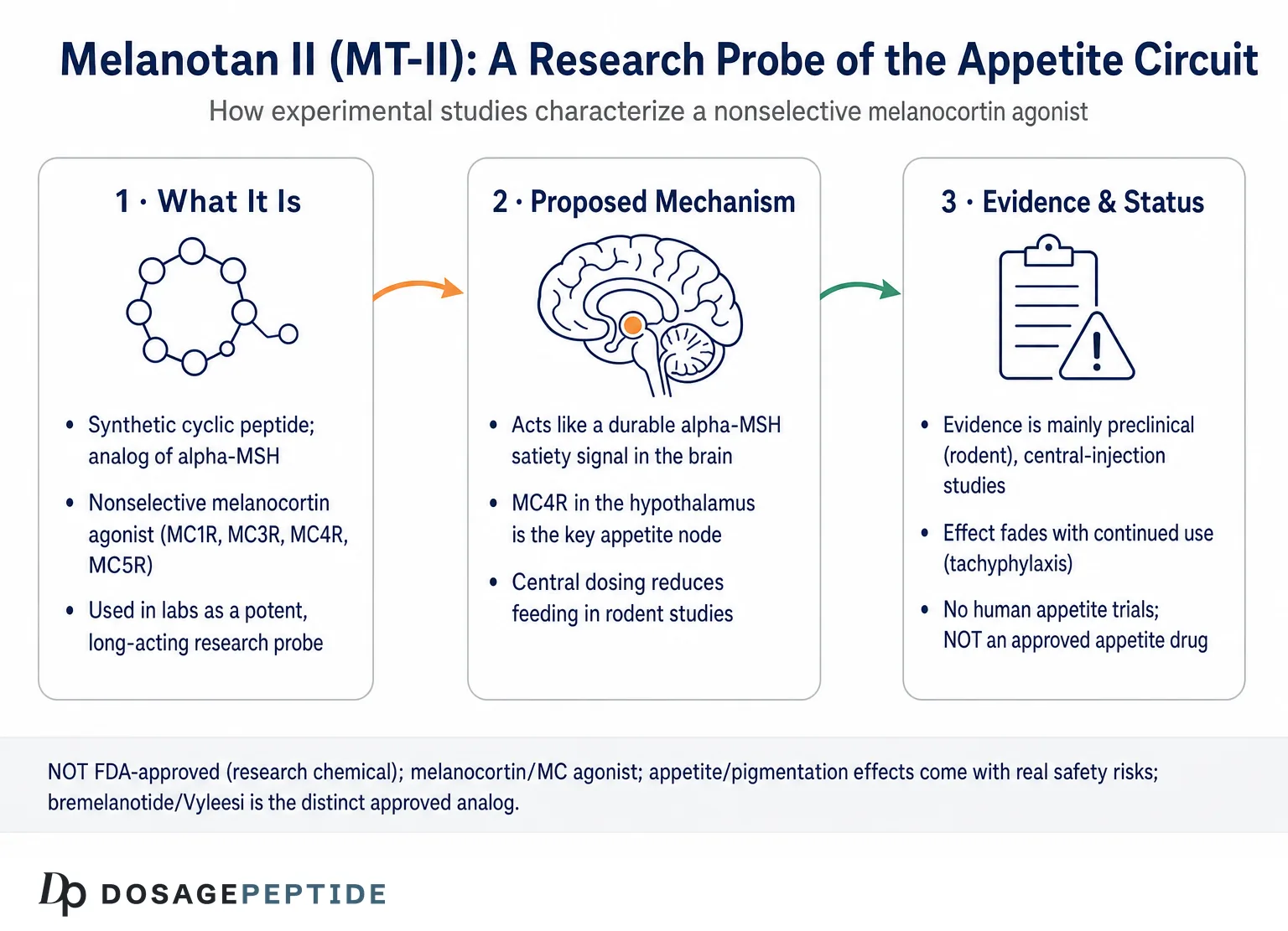
The word characterize deserves unpacking, because it describes a specific kind of scientific work that is different from testing a therapy. When researchers characterize a compound in appetite research, they are typically doing one of several things: establishing a dose–response relationship, mapping the anatomical site where an effect originates, defining the receptor subtype responsible, distinguishing a genuine appetite effect from a nonspecific one such as malaise, or measuring how the effect changes over time with repeated exposure. MT-II has been used for all of these, and the resulting body of work is a case study in how a nonselective agonist can still yield selective conclusions when the surrounding experimental design is rigorous.
The core methodological toolkit for MT-II appetite characterization has four pillars. The first is route of administration. Because the appetite-relevant receptors sit inside the blood-brain barrier, the cleanest experiments deliver MT-II directly into the brain — intracerebroventricular (ICV) infusion into the ventricular system, or, more precisely, stereotaxic microinjection into a specific nucleus. Comparing central with peripheral (subcutaneous or intravenous) delivery then reveals whether an effect is truly central or partly mediated outside the brain. The second pillar is pharmacological subtraction: co-administering a selective antagonist such as SHU9119 to see whether it abolishes the MT-II effect, thereby confirming that the effect ran through melanocortin receptors rather than off-target actions. The third is genetic dissection: giving MT-II to animals lacking a specific receptor (MC4R- or MC3R-knockout mice) and asking whether the response disappears. The fourth is behavioral discrimination: distinguishing reduced feeding caused by genuine satiety from reduced feeding caused by nausea or aversion, using paradigms such as conditioned taste aversion, pica, or kaolin intake.
| Paradigm | What it manipulates | What it characterizes | Representative finding |
|---|---|---|---|
| ICV infusion | Global central melanocortin tone | Whether & how strongly central MC activation suppresses feeding | Dose-dependent anorexia across hyperphagia models23 |
| Site-specific microinjection | MC signaling in one nucleus | Anatomical origin of the effect | PVN, brainstem (NTS), and nucleus accumbens each reduce intake6 |
| Antagonist co-infusion (SHU9119) | Receptor blockade | Whether effect is melanocortin-mediated | SHU9119 abolishes MT-II anorexia; alone increases feeding2 |
| Receptor knockout | Deletes MC3R or MC4R | Which subtype carries the effect | Loss of MC4R attenuates the anorectic response |
| Meal-pattern analysis | Fine-grained feeding microstructure | Satiety vs malaise; meal size vs frequency | MT-II reduces meal size specifically4 |
| Free-choice / macronutrient diets | Diet composition and palatability | Context-dependence of the anorectic signal | Effect depends on dietary fat, not obesity per se7 |
| Aversion controls (CTA, pica) | Nausea-like states | Whether “anorexia” is confounded by malaise | Some, though not all, sites show aversion |
This is what characterization looks like in practice: no single MT-II experiment proves anything definitive about appetite, but the convergence of these paradigms builds a picture whose confidence exceeds any one study. It is also why casual claims — “MT-II suppresses appetite via MC4R” — are only as good as the controls behind them. The statement is broadly supported by the field, but the support comes from the full apparatus in the table, not from the mere observation that hungry animals ate less after an injection.
The Core Findings: Dose, Duration, and the Anatomy of Anorexia
Beyond the foundational Fan study, a series of experiments through the late 1990s and 2000s characterized MT-II’s feeding effects in detail. Central administration of MT-II reliably and dose-dependently reduces food intake and body weight in rats and mice, and the effect is potent: in careful dose-ranging work, centrally administered MT-II reduced feeding across a wide range of doses, with the anorectic action separable, at the low end, from changes in drinking, body temperature, and locomotor activity.3 That separability matters. A crude appetite suppressant might reduce eating simply by making an animal sick or sedated; the finding that low-dose MT-II curtails feeding without dragging temperature and activity along with it argues for engagement of a genuine satiety circuit rather than a blunt malaise.
Meal-pattern analysis sharpened the picture further. When Azzara and colleagues examined the microstructure of feeding, central melanocortin receptor agonism reduced the size of spontaneous and scheduled meals rather than simply abolishing eating, and it did so without augmenting the feeding inhibition produced by a gastric preload.4 The interpretation is that melanocortin signaling acts on the central controllers of meal termination — nudging the brain toward “enough” earlier within a meal — rather than overriding the whole feeding system. This is precisely the kind of mechanistic granularity that a nonselective probe can still deliver when paired with a sensitive behavioral readout.
The anatomical dissection is where MT-II earned its scientific keep. Because the peptide can be microinjected stereotaxically into a single nucleus, researchers used it to ask where in the brain the anorectic signal originates. Three answers emerged, and together they revealed that appetite is not governed by a single site but by a distributed melanocortin network:
- Hypothalamus (PVN). The paraventricular nucleus, dense with MC4R, is the classical satiety node; melanocortin agonism here reduces intake and is a canonical target of the POMC–AgRP balance.
- Brainstem (nucleus of the solitary tract). Melanocortin ligands applied to the hindbrain produce long-lasting effects on feeding and body weight, showing that the caudal brainstem — which receives vagal satiety signals from the gut — is an independent melanocortin appetite site, not merely a relay for hypothalamic commands.
- Nucleus accumbens (reward). In a 2022 study, bilateral microinjection of MT-II into the nucleus accumbens decreased both free feeding and operant responding for food — that is, it reduced not only consumption but the motivated work animals would perform to obtain food — and did so without inducing an aversive state or altering metabolic rate.6
That last finding is conceptually important. It extends the melanocortin appetite story out of the pure homeostatic (energy-balance) domain and into the reward domain: MT-II in the accumbens dampened the incentive value of food, the wanting rather than just the eating, and it did so cleanly enough that the authors could rule out malaise as the driver. Appetite, these MT-II studies collectively argue, is regulated at multiple anatomical levels — homeostatic hypothalamic circuits, gut-linked brainstem circuits, and reward-linked forebrain circuits — each of which the same probe can be used to interrogate in isolation. Readers assembling the terminology of these circuits and receptor conventions may find the site’s peptide research glossary a useful companion for the anatomical and pharmacological vocabulary used throughout this literature.
It is worth pausing on why the anatomical distribution is more than a curiosity of neuroanatomy. If appetite were controlled by a single melanocortin node, then activating that node would produce a uniform, predictable effect, and a drug developer could target it in isolation. Because the system is distributed — and because the different nodes appear to habituate at different rates, with brainstem stimulation producing more durable weight loss than hypothalamic stimulation — the practical implication is that the site of melanocortin engagement shapes both the magnitude and the persistence of the appetite effect. MT-II was the instrument that made this distributed architecture visible, because it could be delivered with surgical precision to one region at a time. A systemically injected agonist, by contrast, activates all of these nodes simultaneously and in unknown proportion, which is one more reason the whole-body pharmacology of MT-II is so much harder to interpret than its site-restricted research use. The distributed map is a scientific gift; it is also a warning that the appetite response to the compound in an intact organism is a sum of partially opposing, differently-adapting signals rather than a single clean readout.
Central Versus Peripheral: The Blood-Brain Barrier Question
A recurring and honestly-contested question in the MT-II literature is whether the peptide’s appetite effect requires it to reach the brain, or whether peripheral administration — the route relevant to real-world use — can produce anorexia on its own. This is not a pedantic distinction; it determines whether findings from ICV experiments have any bearing on what happens when the compound is injected subcutaneously.
Trivedi and colleagues addressed the site of anorectic action of peripherally administered MT-II directly. Studying intravenous delivery in rats, they found that peripheral MT-II could suppress feeding, and they set out to determine whether this required brain penetration.5 The general picture that emerges from this and related work is that MT-II given peripherally can access appetite-relevant sites — likely including circumventricular regions and the caudal brainstem where the blood-brain barrier is relatively permeable, as well as some penetration into the CNS — but that central delivery is far more potent per unit dose. In other words, the anorectic effect is real from the periphery but is fundamentally a central-melanocortin phenomenon; the periphery is simply a less efficient route to the same receptors.
The interpretive caution here is substantial. Peripheral studies are the most externally relevant to how the compound is actually (mis)used, yet they are also the hardest to interpret cleanly, because a systemically distributed nonselective agonist engages MC1R in the skin, MC4R in the cardiovascular system, and MC3R/MC4R in the brain all at once. Some fraction of a peripheral “appetite” effect could in principle reflect nausea or cardiovascular activation rather than pure central satiety. This is one reason the field trusts the central microinjection work more than the peripheral work for mechanistic claims: the closer the delivery is to the target receptor, the smaller the space for confounds. It is also a reason to be skeptical of extrapolating from a clean ICV result to a prediction about what a subcutaneous injection does to a whole organism.
Context Dependence: Diet, Palatability, and the Limits of the Signal
One of the more nuanced characterizations of MT-II concerns how its anorectic effect depends on what is on the menu. If MT-II simply clamped down on a global “eat” command, its effect should be roughly constant regardless of diet. It is not.
Van den Heuvel and colleagues studied MT-II in rats on free-choice diets, where animals could self-select among standard chow and palatable fat and sugar components. Their central finding was that the inhibitory effect of MT-II on feeding depended on the dietary fat content of the diet and not on whether the animal was obese.7 Put differently, the melanocortin brake bit differently depending on the macronutrient composition of what was being eaten, and the animal’s adiposity per se was not the determining variable. This complicates any simple story in which MT-II “reduces appetite” as a fixed quantity; the effect is modulated by the palatability and composition of available food, consistent with the accumbens-reward findings that place melanocortin signaling partly within the circuitry that assigns value to specific foods.
This context dependence is a genuinely useful characterization because it tells us something about the physiology, not just about the drug. It suggests that the central melanocortin system is not a master volume knob on all eating but a modulator that interacts with the incentive and macronutrient properties of food. For anyone tempted to read the animal appetite literature as a promise that a melanocortin agonist would produce uniform, dependable appetite suppression in a person eating a varied human diet, the free-choice data are a corrective: the effect is real but conditional, and its magnitude in a naturalistic feeding environment is harder to predict than a single-diet laboratory result implies. Researchers comparing how different peptide classes handle this same conditionality can contrast the melanocortin evidence with the amylin-analog literature summarized in the discussion of cagrilintide’s role in appetite regulation, where satiety signaling operates through an entirely separate receptor system.
Tachyphylaxis: The Finding That Reframes MT-II’s Whole Story
If there is one experimental result that most sharply separates “MT-II characterizes appetite biology” from “MT-II is a viable appetite drug,” it is tachyphylaxis — the rapid waning of the anorectic effect with continued exposure. This is not a footnote; it is arguably the single most consequential thing the MT-II appetite literature has established about sustained melanocortin agonism.
Pierroz and colleagues gave both acute and chronic MT-II to diet-induced obese mice and observed the pattern in stark form. MT-II markedly suppressed feeding during roughly the first four days of continuous treatment, with progressive weight loss over that window — and then food intake climbed back toward control levels by around day five, even as dosing continued.9 The anorectic response, in other words, defeated itself: the system adapted, and the appetite suppression faded despite ongoing receptor stimulation. Weight loss plateaued at a level below baseline, but the driving force behind it had largely dissipated. The proposed mechanisms include down-regulation of melanocortin receptor function with sustained agonism and compensatory up-regulation of the opposing AgRP signal.
Subsequent work probed whether the dosing schedule could outrun this adaptation. Zhang and colleagues showed that intermittent MT-II application — giving the drug, withdrawing it, and reintroducing it — could evoke repeated bouts of anorexia and produced robust fat and weight loss, preserving the anorectic response better than constant infusion, though it did not abolish tachyphylaxis entirely.10 Others found that targeting the brainstem nucleus of the solitary tract, rather than the hypothalamus, produced more prolonged weight loss with less anorectic tachyphylaxis, suggesting that where and how the melanocortin system is stimulated shapes how quickly it habituates.
The scientific lesson is layered. First, tachyphylaxis is a property of the biology, not merely of the molecule: it reveals that the central melanocortin appetite circuit is homeostatically defended, engineered to resist sustained suppression, which is exactly what one would expect of a system whose job is to defend energy stores. Second, and honestly, it is a major reason a simple, chronically-dosed melanocortin super-agonist was never going to be a straightforward obesity therapy. A drug whose flagship effect disappears within a week of steady use is, whatever its acute potency, a poor therapeutic bet in that form. The value of MT-II here was diagnostic — it exposed the adaptive resilience of the circuit — and that diagnosis directly shaped how successful melanocortin drugs were later designed and deployed. The distinction between a probe that reveals a problem and a therapy that solves one could hardly be clearer.
From Probe to Approved Drug: What MT-II Taught, and What Actually Reached Patients
The most honest way to convey MT-II’s real-world significance is to trace what the melanocortin appetite research it enabled ultimately produced — because the answer is not “MT-II as a weight-loss drug.” It is two other things.
The first is setmelanotide, a selective MC4R agonist approved by the FDA (in 2020) for chronic weight management in patients with specific, rare genetic defects in the melanocortin pathway — POMC deficiency, PCSK1 deficiency, and leptin-receptor deficiency, later extended to Bardet-Biedl syndrome. Setmelanotide is the therapeutic vindication of the circuit that MT-II helped map: it works by restoring appetite control in patients whose melanocortin signaling is genetically broken upstream of the receptor, reducing hunger and body weight where the pathway can still be driven at the MC4R level.11 The contrast with MT-II is instructive on two axes. Setmelanotide is selective for MC4R, avoiding much of the off-target pigmentary and other activity that makes nonselective MT-II hazardous; and it is deployed in defined genetic populations where the mechanism is known to be rate-limiting, rather than as a blunt appetite suppressant for general obesity. The lesson MT-II taught — that MC4R is the pivotal appetite receptor — is precisely the lesson setmelanotide monetized, but only after stripping away the nonselectivity and narrowing the indication.
The second descendant is bremelanotide (Vyleesi), approved by the FDA in 2019 for hypoactive sexual desire disorder in premenopausal women. Bremelanotide is a close chemical relative of MT-II — effectively a metabolite-derived analog — but it was developed for a completely different melanocortin function, central sexual arousal, and its appetite effects are not its therapeutic purpose.8 The existence of Vyleesi is often cited, misleadingly, as evidence that “MT-II-type peptides are FDA-approved.” The accurate statement is narrower: a chemically modified relative of MT-II, optimized for a different receptor-mediated behavior and shorn of MT-II’s tanning and other liabilities, cleared regulatory review for sexual dysfunction. Readers exploring that parallel branch of the melanocortin story can follow it through the discussion of whether Melanotan II could play a role in hypoactive sexual desire disorder, which examines the same pleiotropy from the sexual-function angle.
| Attribute | Melanotan II (MT-II) | Setmelanotide | Bremelanotide (Vyleesi) |
|---|---|---|---|
| Receptor selectivity | Nonselective (MC1R/3R/4R/5R) | MC4R-selective | MC4R-preferring analog |
| Primary research/clinical use | Laboratory appetite probe | Rare genetic obesity (appetite) | Hypoactive sexual desire disorder |
| Regulatory status | Not approved for any use | FDA-approved (2020), defined genetic indications11 | FDA-approved (2019)8 |
| Appetite as intended effect | No (research readout only) | Yes, in target populations | No |
| Relationship to MT-II | — | Conceptual heir (same receptor lesson) | Chemical descendant |
The through-line is that MT-II’s contribution to appetite science was real and lasting, but it was contributed as a tool. The drugs that emerged from the melanocortin appetite program are selective, targeted, and indicated for specific populations. None of them is MT-II. Anyone reasoning from “MT-II suppresses appetite in animals” to “MT-II is a legitimate weight-loss agent” is skipping the entire history in which the field deliberately engineered away from MT-II to get to something usable.
Reading Appetite Data Honestly: Confounds, Aversion, and Pleiotropy
A responsible account of how MT-II is characterized in appetite research has to spend real time on the ways such data can mislead, because the confounds here are unusually severe and unusually easy to overlook.
The first is the satiety-versus-malaise problem. When an animal eats less after a drug, that reduction can reflect genuine satiety (the drug engaged the “I’m full” circuit) or it can reflect nausea, discomfort, or a general sickness state (the animal eats less because it feels ill). These are behaviorally similar but scientifically opposite: only the first supports a claim about appetite regulation. Melanocortin agonists sit uncomfortably in this space because nausea is a known feature of melanocortin activation. The field addresses this with dedicated aversion controls — conditioned taste aversion tests, measurement of pica (eating of non-nutritive substances like kaolin as a nausea proxy), and metabolic-rate controls. The nucleus accumbens study is notable precisely because it demonstrated reduced feeding without an aversive state, which is what licenses its appetite-specific interpretation.6 Studies that report anorexia without such controls should be read more cautiously, because some fraction of the “appetite” effect at higher doses or via peripheral routes may be malaise.
The second confound is pleiotropy from nonselectivity. As established, MT-II activates every melanocortin receptor it reaches. In a whole-animal experiment, an observed change in feeding coexists with changes in pigmentation, cardiovascular tone, sexual behavior, and grooming. If any of these secondary effects indirectly influences feeding — for instance, if cardiovascular activation contributes to a stress-like anorexia — then the “appetite” readout is contaminated. This is why the most trustworthy MT-II appetite conclusions come from experiments that constrain the drug’s reach, whether anatomically (microinjection into one nucleus) or genetically (knockout of one receptor), rather than from whole-body dosing.
The third is the acute-versus-chronic gap, already met in the tachyphylaxis section but worth restating as an interpretive rule: an acute anorectic result says nothing reliable about a sustained one. Much of the striking MT-II appetite data is acute, and the chronic data show the effect eroding. Any characterization that quotes the dramatic acute suppression without the chronic adaptation is telling half the story.
The fourth is species and model specificity. Nearly all mechanistic MT-II appetite work is in rodents. The central melanocortin system is well conserved, and human genetics (MC4R mutations are among the most common monogenic obesity causes) strongly support its relevance to human appetite. But the quantitative translation — how much a given melanocortin drive changes human feeding, and how quickly humans habituate — is not settled by rodent data alone. The early human experience with MT-II was in tiny pilot studies aimed at tanning and sexual function, not appetite, and those studies are far too small to characterize a human appetite effect. Researchers cataloging which model systems best isolate these receptor-specific effects can consult the companion analysis of the experimental models used to assess Melanotan II, whose methodological framework applies with equal force to the appetite-focused MC3R/MC4R work.
Holding all four confounds together yields a disciplined summary: MT-II has robustly demonstrated, across well-controlled central studies, that activating the melanocortin system suppresses feeding through identifiable nodes; it has done so most convincingly when the experiments neutralized malaise, constrained the drug’s receptor reach, and looked beyond the acute window; and its findings translate to human appetite biology by inference and genetics rather than by direct human appetite trials.
Safety, Legality, and the Gap Between Tool and Product
No honest article on MT-II can treat it purely as an elegant probe, because the same molecule is widely and dangerously misused. The safety literature is not hypothetical; it is a catalog of real harms in real people who bought the peptide online.
Documented serious adverse effects associated with MT-II use include rhabdomyolysis and systemic sympathomimetic toxicity, renal infarction, priapism, and dermatologic complications including the appearance or change of pigmented lesions and reports of melanoma. In one representative case, a man who injected 6 mg of internet-sourced MT-II developed systemic toxicity with rhabdomyolysis;12 in another, six months of subcutaneous use preceded a renal infarction affecting roughly half of one kidney, with the authors implicating both a possible thrombotic mechanism and direct renal toxicity.13 The melanoma concern is mechanistically coherent rather than merely alarmist: MT-II is a potent MC1R agonist that drives melanocyte activity, and unchecked melanocyte stimulation is a plausible route to malignant transformation or to the darkening and proliferation of moles that has been repeatedly reported after MT-II use. None of these harms is exotic or theoretical; each appears in the published clinical record from people who obtained the peptide through unregulated channels and injected it without supervision, precisely the pattern that consumer marketing of the appetite and tanning effects encourages.
Several structural reasons make these harms worse than the pharmacology alone would predict. The compound is unregulated, so material sold as MT-II varies in purity, concentration, and sterility, introducing contaminants and dosing errors that have nothing to do with the molecule’s intrinsic effects. It is nonselective, so a user seeking one effect (tanning, appetite suppression) unavoidably receives all the others (cardiovascular activation, sexual effects, melanocyte stimulation). And it is typically self-administered by injection without medical oversight, in populations and at doses never studied for safety. The appetite-suppressing property that consumer marketing frames as a perk is, in this light, one facet of a systemically active agent with a genuinely concerning adverse-event record.
On legality and regulation: Melanotan II is not approved by the FDA, the EMA, or any comparable regulator for appetite suppression, obesity, tanning, or any other indication. Regulatory agencies in multiple countries have issued warnings against its sale and use, and it is not a lawful medicine. Its appearance in research-chemical and cosmetic-injectable markets does not confer any legitimacy. This is the practical endpoint of everything above: a molecule can be a superb scientific instrument — MT-II genuinely is — and simultaneously an unapproved, hazardous product that no framework endorses for human appetite control. For orientation to how the broader family of metabolic and appetite-relevant research peptides is organized and distinguished, the site’s central dosage reference index catalogs these compounds by their studied mechanisms rather than by any marketing claim.
The synthesis, then, mirrors the article’s opening distinction. Experimental studies characterize MT-II as a potent, nonselective melanocortin agonist that has been invaluable for mapping how the brain’s melanocortin system regulates appetite — establishing MC4R as the pivotal node, revealing a distributed hypothalamic-brainstem-reward architecture, and exposing the tachyphylaxis that defends the circuit. That characterization is a scientific achievement. It is emphatically not a clinical endorsement, and the drugs that appetite research ultimately delivered were selective, targeted successors, not MT-II itself.
Frequently Asked Questions
Does Melanotan II actually suppress appetite, or is that a myth?
It genuinely does, in the sense that central administration of MT-II reliably and dose-dependently reduces food intake in rodents, an effect demonstrated across multiple hyperphagia models and abolished by melanocortin antagonists.23 That is a real pharmacological property. What is a myth is the leap from that laboratory finding to the idea that MT-II is a safe or sanctioned appetite suppressant for people. The animal effect is central, potent, and mechanistically informative; it is also subject to tachyphylaxis, entangled with the compound’s many off-target actions, and never developed into an approved appetite drug in this molecule.
Which receptor is responsible for the appetite effect?
The melanocortin-4 receptor (MC4R) is the primary anorexigenic receptor and does most of the appetite work, with MC3R playing a modulatory role in energy partitioning.2 Because MT-II is nonselective and activates MC1R, MC3R, MC4R, and MC5R indiscriminately, attributing a feeding effect specifically to MC4R requires additional evidence — selective antagonists such as SHU9119 or receptor-knockout animals — rather than the agonist alone.
Why does the appetite-suppressing effect fade with continued use?
This is tachyphylaxis, and it is one of the most important findings in the MT-II appetite literature. In diet-induced obese mice, chronic MT-II suppressed feeding for roughly four days, after which intake returned toward normal despite continued dosing.9 The likely mechanisms are down-regulation of melanocortin receptor function and compensatory increases in the opposing AgRP signal. Intermittent dosing preserves the response better than continuous dosing but does not eliminate the adaptation.10 This self-limiting behavior is a major reason a simple chronic melanocortin super-agonist was never a viable obesity therapy.
Is Melanotan II the same as the approved drugs setmelanotide or Vyleesi?
No. Setmelanotide is a selective MC4R agonist approved for rare genetic obesity syndromes,11 and bremelanotide (Vyleesi) is an MT-II-derived analog approved for hypoactive sexual desire disorder.8 Both are chemically and pharmacologically distinct from nonselective MT-II, and both were deliberately engineered to shed MT-II’s liabilities and to target defined indications. MT-II itself is not approved for anything.
Do the appetite effects require the drug to reach the brain?
The effect is fundamentally central. The appetite-relevant receptors (MC3R and MC4R) sit inside the blood-brain barrier, and direct central administration is far more potent per dose than peripheral. Peripheral MT-II can suppress feeding, likely by reaching circumventricular and brainstem sites and by some CNS penetration,5 but peripheral results are harder to interpret because a systemically distributed nonselective agonist also activates skin, cardiovascular, and other melanocortin receptors that can indirectly influence eating.
How do researchers know MT-II reduces feeding through satiety and not nausea?
They use dedicated controls. Conditioned taste aversion tests, pica (kaolin intake) as a nausea proxy, and metabolic-rate measurements help distinguish genuine satiety from malaise. The nucleus accumbens microinjection study is a strong example: it reduced both feeding and food-motivated operant responding without inducing an aversive state or changing metabolic rate, which supports an appetite-specific interpretation.6 Studies lacking such controls, especially at high doses or via peripheral routes, warrant more caution.
Does diet composition change how well MT-II works?
Yes, and this is a genuinely informative characterization. In rats on free-choice diets, the inhibitory effect of MT-II on feeding depended on the dietary fat content rather than on whether the animal was obese.7 This context dependence suggests the central melanocortin system is not a fixed appetite volume knob but a modulator that interacts with the macronutrient and reward properties of available food — consistent with the finding that melanocortin signaling in reward regions dampens the incentive value of food.
Is it legal or safe to use Melanotan II to control appetite?
No. Melanotan II is not approved by the FDA, EMA, or comparable regulators for appetite suppression or any other use, and regulatory agencies have warned against it. Documented harms in people who used it include rhabdomyolysis and systemic toxicity, renal infarction, priapism, and pigmented-lesion changes including melanoma reports.1213 Its nonselectivity, unregulated supply, and typical unsupervised self-injection compound these risks. Its scientific value as a research probe does not translate into any endorsement for human appetite control.
References
- Dorr RT, Lines R, Levine N, et al. Evaluation of melanotan-II, a superpotent cyclic melanotropic peptide in a pilot phase-I clinical study. Life Sci. 1996;58(20):1777-1784. PMID: 8637402. https://pubmed.ncbi.nlm.nih.gov/8637402/
- Fan W, Boston BA, Kesterson RA, Hruby VJ, Cone RD. Role of melanocortinergic neurons in feeding and the agouti obesity syndrome. Nature. 1997;385(6612):165-168. PMID: 8990120. https://pubmed.ncbi.nlm.nih.gov/8990120/
- Murphy B, Nunes CN, Ronan JJ, Hanaway M, Fairhurst AM, Mellin TN. Centrally administered MTII affects feeding, drinking, temperature, and activity in the Sprague-Dawley rat. J Appl Physiol. 2000;89(1):273-282. PMID: 10904062. https://pubmed.ncbi.nlm.nih.gov/10904062/
- Azzara AV, Sokolnicki JP, Schwartz GJ. Central melanocortin receptor agonist reduces spontaneous and scheduled meal size but does not augment duodenal preload-induced feeding inhibition. Physiol Behav. 2002;77(2-3):411-416. PMID: 12419417. https://pubmed.ncbi.nlm.nih.gov/12419417/
- Trivedi P, Jiang M, Tamvakopoulos CC, et al. Exploring the site of anorectic action of peripherally administered synthetic melanocortin peptide MT-II in rats. Brain Res. 2003;977(2):221-230. PMID: 12834882. https://pubmed.ncbi.nlm.nih.gov/12834882/
- Eliason NL, Martin L, Low MJ, Sharpe AL. Melanocortin receptor agonist melanotan-II microinjected in the nucleus accumbens decreases appetitive and consumptive responding for food. Neuropeptides. 2022;96:102289. PMID: 36155088. PMCID: PMC10152796. https://pmc.ncbi.nlm.nih.gov/articles/PMC10152796/
- van den Heuvel JK, Eggels L, van Rozen AJ, et al. Inhibitory effect of the melanocortin receptor agonist melanotan-II (MTII) on feeding depends on dietary fat content and not obesity in rats on free-choice diets. Front Behav Neurosci. 2015;9:358. PMID: 26733840. PMCID: PMC4689860. https://pmc.ncbi.nlm.nih.gov/articles/PMC4689860/
- Cai M, Nyberg J, Hruby VJ. Melanotropins as drugs for the treatment of obesity and other feeding disorders: potential and problems. Curr Top Med Chem. 2009;9(6):554-563. PMID: 19689365. PMCID: PMC4608742. https://pmc.ncbi.nlm.nih.gov/articles/PMC4608742/
- Pierroz DD, Ziotopoulou M, Ungsunan L, Moschos S, Flier JS, Mantzoros CS. Effects of acute and chronic administration of the melanocortin agonist MTII in mice with diet-induced obesity. Diabetes. 2002;51(5):1337-1345. PMID: 11978628. https://pubmed.ncbi.nlm.nih.gov/11978628/
- Zhang Y, Collazo R, Gao Y, Li G, Scarpace PJ. Intermittent MTII application evokes repeated anorexia and robust fat and weight loss. Peptides. 2010;31(4):639-643. PMID: 20034526. PMCID: PMC2860181. https://pmc.ncbi.nlm.nih.gov/articles/PMC2860181/
- Collet TH, Dubern B, Mokrosinski J, et al. Evaluation of a melanocortin-4 receptor (MC4R) agonist (setmelanotide) in MC4R deficiency. Mol Metab. 2017;6(10):1321-1329. PMID: 29031731. PMCID: PMC5641599. https://pmc.ncbi.nlm.nih.gov/articles/PMC5641599/
- Nelson ME, Bryant SM, Aks SE. Melanotan II injection resulting in systemic toxicity and rhabdomyolysis. Clin Toxicol (Phila). 2012;50(10):1169-1173. PMID: 23121206. https://pubmed.ncbi.nlm.nih.gov/23121206/
- Peters B, Hadimeri H, Wahlberg R, Afghahi H. Melanotan II: a possible cause of renal infarction: review of the literature and case report. CEN Case Rep. 2020;9(2):159-161. PMID: 31953620. PMCID: PMC7148395. https://pmc.ncbi.nlm.nih.gov/articles/PMC7148395/
Educational and research-use disclaimer: This article is provided solely for scientific and educational purposes. Melanotan II is not approved by the FDA, EMA, or any comparable regulator for appetite suppression, obesity, tanning, sexual function, or any other use, and multiple regulatory agencies have issued warnings against its sale and use. It is a nonselective melanocortin agonist with a documented record of serious adverse effects, including rhabdomyolysis, systemic toxicity, renal infarction, priapism, and pigmented-lesion changes including melanoma. Nothing here is medical advice or an endorsement of human use; the compound’s value described above is strictly as a laboratory research probe for characterizing melanocortin appetite biology. The distinct approved melanocortin drugs setmelanotide and bremelanotide are separate, selective agents and are not interchangeable with Melanotan II. Readers should consult qualified professionals and applicable laws and regulations before making any decisions.